A New Approach To Metabolic Syndrome

The Greatest Healthcare/Financial Crisis
 
:dropcap_open:M:dropcap_close:etabolic Syndrome (aka Syndrome X) with its four hallmark symptoms of obesity, hypertension, dyslipidemia and hyperglycemia is devastating our country as well as the whole of North America.  In March of 2005 the National Institutes of Health and the New England Journal of Medicine published a paper stating that because of this epidemic the current generation is projected to have a shorter life expectancy then the previous one…for the first time in recorded history!  Since that paper things have become much worse.  Worse, despite the fact we have changed the USDA “Food Pyramid”, developed many new classes of pharmaceutical agents (especially ones for pre-diabetes and diabetes Type II), have taken soda machines out of schools, and even the First Lady’s top priority is the obesity epidemic.  This syndrome, with all of its comorbidities (cardiovascular disease, stroke, many cancers, kidney failure, blindness, amputations, etc.), accounts for the majority of healthcare dollars spent.  If the tide is not turned, Metabolic Syndrome will bankrupt our country. This is a fact.
 
obesitycrisisThe Pathophysiology of Metabolic Syndrome
In 1987 the late Gerald Reaven, MD, Professor of Medicine at Stanford University’s College of Medicine, first demonstrated that the four hallmark symptoms shared a commonality: hyper-insulinemia coupled with insulin resistance.  He coined the term “Syndrome X” to illustrate the point: the four legs of the “X” represent the symptoms (hypertension, central obesity, hyperglycemia and dyslipidemia) and the nexus of the “X” represents the causal agents of too much insulin along with insulin resistance (the cells do not respond to normal physiologic amounts of insulin).  This is the standard, accepted medical model of this disease.
 
The Failure of Current Treatments
We are being ravaged by this syndrome due to the simple fact that we have ignored the model!  Instead of focusing our attention on the root cause we have decided to treat each of the symptoms as separate, unrelated diseases. Thus we have new dietary recommendations and “diets d’Jour”, as well as a plethora of exercise regimens prescribed for obesity and, of course, “diet pills”.  There too are the myriad of prescription drugs to ‘control’ the other three symptoms.  If our attention is on ‘controlling symptoms’ we have admitted, by default, that there is NOTHING WE CAN DO FOR THE CAUSATIVE FACTORS and we will just have to LEARN TO LIVE WITH OUR DISEASE (i.e. ‘it will always be with us, we’ll just control it’).  This attitude of acceptance is bad enough and unaffordable in the long run, but that’s the least of it.  If we understand the pathophysiology of this syndrome we readily can see why many of these treatments actually make the other symptoms much worse!  Hyper-insulinemia means the patient’s pancreas is secreting an exaggerated amount of insulin in response to rises in blood glucose. 
 
This can easily be confirmed by doing a fasting insulin level OR the standard glucose challenge test and ordering insulin levels along with glucose levels at time zero, one hour and two hour intervals post challenge glucose administration. Sadly, the vast majority of practitioners do not even think about such an important marker. So we dwell on just the glucose level or Hemoglobin A1c (merely symptoms) and prescribe drugs such as the sulfonylureas (i.e. glyburide, glipizide, glimperide) which cause the pancreas to secrete EVEN MORE INSULIN, or we actually give them INSULIN ITSELF in an aggressive attempt to control a symptom.  If the model is correct then this therapy should make the syndrome worse……and it does!  This is the fundamental reason why we have failed to stem the tide (or actually reverse) this seemingly insidious malady.
 
The Concept of Homeostasis
If insulin just mediated glucose uptake by our cells and did nothing else, we probably would not have this problem. However, this is not the case, and when the amount of insulin remains consistently elevated it does other things…..and these things are NOT good.  Before discussing the effects of hyper-insulinemia, a review of the fundamental concept of homeostasis should be addressed.  The body is an organism that strives to maintain a constant internal environment in the face of constantly changing, often hostile, external factors.  Blood pressure, blood glucose, body temperature, acid/base balance, etc. must remain within a relatively narrow range in order to survive.  It does so by means of the action/reaction principle, or mechanisms that exert opposite effects so that a balance may be achieved.  Examples are: vasodilation/vasoconstriction, oxidation/reduction, anabolism/catabolism, assimilation/elimination, etc.  These systems are exquisitely regulated primarily by the nervous system and the endocrine (hormonal) system.  So if the environmental temperature is 125 degrees, our internal temperature remains 98.6 degrees.  

Likewise if the temperature drops to 20 degrees, certain mechanisms are in place to make certain our internal temperature remains a constant 98.6.  Glucose homeostasis is essential for life, as certain cells in the body can only use glucose as an energy source (certain brain cells, the adrenal medulla, red blood cells, etc.).  Whether in times of feast or famine, blood glucose must remain in a certain range and insulin and glucagon are the master hormones that control this process (forget about ghrelin, leptin, incretins and all these ‘new mini-hormones’ that are in the literature today…these are subservient to the two masters). The body needs BOTH of these “master hormones” to maintain balance (as they have exactly opposite physiological functions….if you know what insulin does, then you automatically know what glucagon does…the exact opposite!) and if an imbalance occurs, dysfunction or “disease” will arise.
 
The Physiological Effects of Insulin
:dropcap_open:Likewise if the temperature drops to 20 degrees, certain mechanisms are in place to make certain our internal temperature remains a constant 98.6.:quoteleft_close:
Insulin’s primary function is mediating glucose uptake to muscle cells, and in this way it helps regulate blood glucose homeostasis.  However, insulin binds to many other receptors in the body and affects MANY other physiological parameters.  And here’s the “rub”.  If insulin receptors on the muscle cells become resistant to insulin’s effect (and do not uptake glucose in an effective manner) the pancreas will produce more to ensure glucose uptake will occur.  But if we increase insulin levels, what happens to OTHER receptors that are not “resistant” yet and modulate other bodily functions?  This scenario becomes way more complicated, in that these receptors become ‘insulin resistant’ at different times.  So a ‘typical Syndrome Xer’ presents to the physician with some central obesity, slightly elevated blood pressure, slightly elevated blood glucose and a less than stellar lipid panel.  He is told to lose some weight by eating more fruits and vegetables, cutting down on fats and cholesterol (have oatmeal instead of bacon and eggs) and doing some light exercise.  This is standard, first line therapy of lifestyle changes and sounds very reasonable.  This compliant patient makes these changes and returns in two months, shocked and disappointed that his symptoms have become worse!  Now he is given a low dose ACE inhibitor coupled with a diuretic for his hypertension and placed on metformin and glyburide to help control hyperglycemia.  
 
The glyburide tells the pancreas to secrete even more insulin and he gains more weight.  Insulin also “ramps up” the enzyme HMG-Co A reductase, which basically tells the body to produce even more cholesterol.  Excess insulin also drives the kidneys to retain sodium and waste magnesium, which is an essential element for insulin receptor sensitivity.   Hypertension and insulin resistance worsen.  Usually at this point (if not sooner) a statin is added along with niacin and another oral hypoglycemic and we ‘start the march’ to insulin therapy. This is why many of these patients will find themselves on six to nine prescription drugs, and this is the current “Standard of Care” for this syndrome.
 
Let Your Food Be Your Medicine
Let us now suppose that the above patient visited a Chiropractic physician first.  This particular physician is skilled in the use of a ‘muscle sparing’ protein diet, not a hyper protein diet ala Atkins.  This diet is also low in fat, particularly saturated fat and is very carbohydrate restrictive (providing about 40 grams of carbohydrates daily mainly from fibrous vegetables).  The physician explains the “medical model” of Syndrome X and relates how the overproduction of insulin can contribute to all his symptoms.  Correcting hyperinsulinemia is very straightforward: all carbohydrates (with the exception of fiber) will eventually be turned into glucose….sometimes quickly, sometimes slowly.  As the glucose is absorbed the pancreas begins to secrete insulin (in this case, too much insulin).  By restricting the carbohydrates the production of insulin is immediately reduced.  
 
The patient is interested but confides that he can be hypoglycemic at times and is afraid of such a restrictive protocol. The physician relates that hypoglycemia is usually the consequence of an overproduction of insulin, not a lack of carbohydrates.  He further explains the body has “three tanks of energy” from which to draw.  Glycogen (or our stored glucose), muscle, which can be broken down via gluconeogenesis to supply glucose and fat (triglycerides), which can be turned into glucose (from the glycerol) and ketonic bodies that most of the cells in the body can use for fuel.  But the body draws on these compartments in a very specific order.  It will always use the glycogen first and only when ‘that tank’ is empty will it begin to simultaneously burn muscle and fat. 
 
The physician tells the patient if he keeps “putting fuel in the glycogen tank”, he will never be able to access his fat reserves, thus the restriction of carbohydrates.  He also says that we never want to lose muscle, thus the inclusion of the adequate amount of protein to replenish what is lost to gluconeogenesis.  
 
During the first three days of this protocol the patient may feel a little tired or weak (as the body depletes its glycogen) but once this is gone and the body ‘switches over to muscle and fat’ he will have plenty of energy and hypoglycemic episodes will be a thing of the past.  His patient is interested but asks: “ketonic bodies, does that mean ketosis…I thought that was bad?”  Again the physician explains that ketoacidosis is bad and that is why a Type I diabetic would never be placed on this program.  In this case ketosis just means ‘living off your reserves’ and is the reason human beings are able to survive times of famine.  His concerns allayed, the patient begins the program.
 
Under The Influence of Glucagon
Six weeks later the elated patient returns to his Chiropractor.  He is thirty pounds lighter and says that his medical doctor told him his blood work was fantastic!   In layman’s terms the physician tells him: “Well you have actually reset your pancreas, it no longer is pumping out too much insulin and now you can start to put fruits, grains and dairy back into your diet.”  After this patient’s glycogen reserves were depleted and carbohydrates continued to be restricted, the body had to ensure proper blood glucose levels were maintained.  Under these conditions the pancreas produces more glucagon (which raises blood sugar) and much less insulin (whose primary function is to lower blood sugar).  But there is more to glucagon than this primary function.  Glucagon stimulates two adipocyte (fat cell) enzymes (HSL and ATGL) and inhibits a third (Lipoprotein lipase).  
 
The result is the release of triglycerides from the fat cell (to be used as fuel) as opposed to insulin’s effect, which is to store fat.  Glucagon enhances the entry of free fatty acids across the mitochondrial membranes so they can be used as fuel (insulin inhibits this).  Glucagon also greatly inhibits the action of HMG-CoA reductase (along with all the other enzymes necessary for cholesterol synthesis) and forces cells to pull cholesterol from the blood stream via ‘ramping-up’ LDL receptors (1983 Nobel Prize in Medicine).  This is why this patient’s lipid panel came back stellar.  Finally, in the kidneys the retention of sodium (caused by excess insulin) has now been corrected and his hypertension has resolved.  The pathophysiology of Syndrome X is predictable.  The reversal of this syndrome is also predictable and repeatable!  As a matter of fact this exact method is being employed by over 700 chiropractic practices in the United States and Canada as well as many medical practices.  Tens of thousands of patients have experienced the same benefits described here.  
 
The Chiropractic physician (because of his/her training and philosophy) can become a leading force in helping to reverse this terrible syndrome.  Let this article be a call to action!

Michael P. Ciell, R.Ph., is the Chief Science Officer and Vice-President of Clinic Operations, Ideal Protein of America. Website: www.idealprotein.com

For more information, all of the in-depth science and an opportunity to speak with one of your fellow practitioners, please contact Ideal Protein @ 1-866-314-4447

Genetics, Diet, and Ankylosing Spondylitis

lowbackpainankylosingspond:dropcap_open:S:dropcap_close:ome health problems are purely genetic. Others have nothing to do with genetics but are purely the result of environmental influences, such as diet, infection, or trauma. Then, there are many conditions in which some genetic predisposition interacts with the environment to produce the disease. The classic example is celiac disease, which is tightly linked to genetics but can be controlled by eliminating wheat, rye, and barley products from the diet. Considerable evidence suggests that many of the inflammatory arthritides, including ankylosing spondylitis, could also respond to dietary interventions, including a low-fat, purely plant-based (vegan) diet or sometimes a gluten-free diet.

Ankylosing spondylitis is an inflammatory disease that can lead to fusion of the vertebrae. Ankylosis means stiffening and refers to fusion of the joints. Spondylitis means inflammation of the vertebrae. In severe cases, ankylosing spondylitis can lead to a permanent deformity called bamboo spine. Ankylosing spondylitis is more common in males than in females. It typically arises between the ages of 20 and 40 years but can begin as early as age 10.

Ankylosing spondylitis is the prototype and best studied subtype of a group of related but phenotypically distinct disorders that are collectively called spondyloarthritis.1 Other subtypes of spondyloarthritis include psoriatic arthritis, arthritis related to inflammatory bowel disease, reactive arthritis, and a form of juvenile idiopathic arthritis. Cases of spondyloarthritis are often classified according to whether the problem is manifested mainly in the joints of the spine (axial spondyloarthritis) or elsewhere in the body, including other joints and organs such as the eye (peripheral spondyloarthritis).

The hallmark of ankylosing spondylitis is involvement of the sacroiliac joint.2 The earliest symptom is typically tenderness in the sacroiliac joint or pain upon hyperextension of the hip. These symptoms are easily mistaken for evidence of sciatic nerve irritation. As the disease progresses, it may produce flattening of the normal lordosis of the lumbar spine, along with restriction of motion of the lumbar spine. Involvement of the thoracic vertebrae can produce costovertebral joint fusion resulting in restriction of the chest. Accentuation of kyphosis of the dorsal spine can produce a stooped appearance.

The Assessment of SpondyloArthritis international Society (ASAS) (formerly ASsessment in Ankylosing Spondylitis) has published a useful handbook for the assessment of axial spondyloarthritis in general and ankylosing spondylitis in particular.3 The handbook’s stated purpose is to standardize not only patient care but the design of clinical studies. It provides an overview of the classification systems used for inflammatory back pain. The handbook makes it clear that both genetics and the intestinal tract could play an important role in causing inflammatory back pain.

:dropcap_open:Involvement of the thoracic vertebrae can produce costovertebral joint fusion resulting in restriction of the chest.:quoteleft_close:

Ankylosing spondylitis is particularly common in people who carry one or two of the HLA-B*27 subtypes of the HLA-B gene of the major histocompatibility complex. Inflammatory back pain has also been linked to intestinal diseases, including Crohn’s disease. Chiropractors can’t alter their patients’ major histocompatibility complex. That’s the genetic hand of cards that the patient was dealt at conception. However, chiropractors can encourage patients to make the kind of dietary changes that could improve the health of their gastrointestinal tract and consequently the health of their joints.

The major histocompatibility complex (MHC) accounts for nearly half of the predisposition to ankylosing spondylitis, and HLA-B*27 subtypes seem to be the most important genetic influence.4 The MHC is a set of human leukocyte antigens (HLA) that are displayed on the surface of cells and play an important role in the immune system.

In humans, the genes for the MHC are found on chromosome 6. Each person has two copies of chromosome 6. Thus, each person carries two versions of the HLA-B gene: one from the father and the other from the mother. Thus, any given person could have one of the HLA-B*27 subtypes, or two different HLA-B*27 subtypes, or no HLA-B*27 subtypes of the HLA-B gene. So far, dozens of subtypes of HLA-B*27 have been identified.

Inheriting an HLA-B*27 subtype from one parent increases the risk of having ankylosing spondylitis. Inheriting a double dose of HLA-B*27 (an HLA-B*27 from each parent) increases the risk of ankylosing spondylitis even further. Yet not everyone who got a double dose of HLA-B*27 has ankylosing spondylitis. Other genetic and environmental factors could alter the likelihood and severity of the disease.

The important question for chiropractors to consider is whether the patient has any control over any of the factors that could contribute to spondyloarthropathy. One largely overlooked possibility is that diet could make a difference. After all, intestinal disorders such as colitis and Crohn’s disease are prominent in the diagnostic criteria for spondyloarthropathy,3 and subclinical gut inflammation has also been found in patients with ankylosing spondylitis.5 If the problem starts in the gut, the cause might be something that the patient has been eating. If so, then the patient might be able to alter the course of a potentially disabling and disfiguring disease by making a simple change in diet.

healthyfoodveggiesAn intriguing case report from Germany suggests that a vegan diet (i.e., a diet that excludes foods of animal origin) holds promise for patients with ankylosing spondylitis.6 The report describes a 33-year-old patient who inherited an HLA-B*27 from both parents and had a 10-year history of ankylosing spondylitis that had responded poorly to conventional medical treatment and physiotherapy. The authors recommended that the patient switch temporarily to a purely vegan diet, avoiding all animal fats and proteins. The patient’s symptoms started to improve within 3 to 4 days after the switch to a vegan diet. After the patient resumed eating meat 6 weeks later, the condition started to worsen again. The patient then resumed the vegan diet. At 3 months’ follow-up, the patient had quit taking tramadol and ibuprofen, had cut his meloxicam dose in half, and was “almost completely free of complaints.”

Normally, the lining of the intestine serves as a barrier that prevents potentially dangerous substances and infective agents from gaining access to the bloodstream. If this barrier is breached, then many harmful substances could enter the bloodstream. This includes not only bacteria and viruses but food proteins that haven’t been completely digested. Proteins from other animals might be particularly hazardous in this regard. They are different enough from human proteins that the human immune system will recognize them as foreign and make antibodies against them. Yet they might still be similar enough to human proteins (a phenomenon called molecular mimicry) that those antibodies will “cross-react” with human proteins and attack the body’s own tissues.7 This cross-reaction helps to explain why eating animal protein seems to predispose people to autoimmune disease.

Sadly, the current “evidence-based” guidelines for managing ankylosing spondylitis focus entirely on drug treatments, exercise, and physiotherapy but completely ignore the dietary angle.8 That doesn’t mean that diet isn’t important. It merely reflects the neglect of the dietary angle in the existing research on ankylosing spondylitis.

The anti-inflammatory and disease-modifying drugs used in the management of ankylosing spondylitis are outside the scope of chiropractic practice. Nevertheless, chiropractors may be involved in helping ankylosing spondylitis patients with exercise and physiotherapy. Given the lack of data on dietary interventions for spondyloarthropathy, what is a chiropractor to do? The first thing is simply to alert patients to the fact that inflammatory back pain has been linked to intestinal problems that could respond to a change in diet. The second thing would be to develop some simple guidelines that could help patients eliminate the foods that are likely to contribute to gut problems and inflammatory arthritis.  A switch from the standard American diet to a low-fat vegan diet is beneficial to a patient’s overall health and might be sufficient to arrest the inflammatory disease. Some other patients may also have to eliminate other troublesome foods. For example, people with gluten intolerance would have to eliminate wheat, rye, and barley. A registered dietitian, or qualified nutritionist can help patients identify and eliminate the foods that are contributing to their back problems.


Laurie Endicott Thomas has worked as a medical editor and writer for more than 20 years. She is the author of the upcoming book Where Do Gorillas Get Their Protein? Her Web site is www.gorillaprotein.com and she blogs at www.wheredogorillasgettheirprotein.blogspot.com.


REFERENCE LIST

Dougados M, Baeten D. Spondyloarthritis. Lancet 2011;377:2127-2137.

Khan MA. Ankylosing spondylitis: clinical features.3rd ed. St Louis, MO: Mosby; 2003.

Sieper J, Rudwaleit M, Baraliakos X et al. The Assessment of Spon dyloArthritis international Society (ASAS) handbook: a guide to assess spondyloarthritis. Ann Rheum Dis 2009;68 Suppl 2:ii1-44. http://www.asas-group.org/pdf/ASAS-Handbook.pdf

Reveille JD. The genetic basis of spondyloarthritis. Ann Rheum Dis 2011;70 Suppl 1:i44-i50.

Ciccia F, Bombardieri M, Rizzo A et al. Over-expression of paneth cell-derived anti-microbial peptides in the gut of patients with ankylosing spondylitis and subclinical intestinal inflammation. Rheuma- tology (Oxford) 2010;49:2076-2083.

Huber R, Herdrich A, Rostock M, Vogel T. [Clinical remission of an HLA B27-positive sacroiliitis on vegan diet]. Forsch Komplemen tarmed Klass Naturheilkd 2001;8:228-231.

Pérez-Maceda B, Lopez-Bote JP, Langa C, Bernabeu C. Antibodies to dietary antigens in rheumatoid arthritis–possible molecular mimicry mechanism. Clin Chim Acta 1991;203:153-165.

Braun J, van den Berg R, Baraliakos X et al. 2010 update of the ASAS/ EULAR recommendations for the management of ankylosing spondylitis. Ann Rheum Dis 2011;70:896-904.

Calcium Supplementation Without a Proper Balance of Magnesium Can Increase Risk of Heart Attack, Osteoporosis and Kidney Stones

ORANGE, Calif., Sept. 14, 2011 /PRNewswire/ — The British Medical Journal recently published a large analysis based on the results of 5 clinical trials conducted in the U.S., Great Britain and New Zealand that involved more than 8,000 people. The analysis concluded that, “Calcium supplements … increase the risk of cardiovascular events, especially myocardial infarction (heart attack) … A reassessment of the role of calcium supplements in osteoporosis management is warranted.”(1)

elementchartMgCaAccording to Dr. Carolyn Dean, MD, ND, magnesium expert and Medical Director of the nonprofit Nutritional Magnesium Association (http://www.nutritionalmagnesium.org), approximately 30 to 40 years ago doctors began prescribing calcium on a routine basis to many men and almost all women over the age of 40 to counter the effects of bone loss due to aging. The conventional wisdom was that bone loss is due to calcium deficiency. “After 40 years, it has become clear that taking calcium alone does not stop or even slow bone loss and does not prevent osteoporosis.”(2)

The new wisdom now emerging is that magnesium is actually the key to the body’s proper assimilation and use of calcium as well as vitamin D. Dr. Dean says, “If we consume too much calcium without sufficient magnesium, the excess calcium is not utilized correctly and may actually become toxic, causing painful conditions such as some forms of arthritis, kidney stones, osteoporosis and calcification of the arteries leading to heart attack and cardiovascular disease. The effectiveness and benefits of calcium with respect to bone health and the prevention of osteoporosis are enormously impaired in the absence of adequate levels of magnesium in the body.”

Many medical doctors, research scientists and nutritionists now believe magnesium supplementation is more important than calcium in order to maintain healthy bones as well as a healthy heart.

“Magnesium keeps calcium dissolved in the blood. Without the proper balance of magnesium to calcium, about a 2:1 ratio, calcium ends up depositing in kidneys and can create kidney stones, in coronary arteries where it can lead to clogged arteries, and in joint cartilage, rather than in bones where we need it most. The more calcium taken without the balancing effect of magnesium, the more symptoms of magnesium deficiency and calcium excess you are liable to experience,” Dr. Dean says.

New York Times best-selling author Dr. Joseph Mercola concurs, “If you decide to supplement with magnesium it is important to understand that its complementary partner is calcium. So you should use both. Typically you would use twice as much elemental magnesium relative to the elemental calcium. That ratio works out quite well for most.”

Dr. Guy Abraham, M.D., a research gynecologist and endocrinologist specializing in premenstrual syndrome and osteoporosis, has found strong evidence to suggest that women with osteoporosis have a deficiency of the hormone calcitonin that is only made when they take twice as much magnesium as calcium. In fact, he has found that when calcium intake is decreased, it is better utilized than when it is high. Dr. Abraham is one of many doctors and biochemists who advocate taking more magnesium to correct calcium-deficiency diseases.

Dr. Dean adds, “Adequate levels of magnesium in the body are essential for the absorption and metabolism not only of calcium but of Vitamin D, because magnesium converts Vitamin D into its active form so that it can help calcium absorption. Magnesium also stimulates a particular hormone, calcitonin, which helps to preserve bone structure and draws calcium out of the blood and soft tissues back into the bones, lowering the likelihood of osteoporosis, some forms of arthritis, heart attack and kidney stones.”

A 32-page guide to the benefits of magnesium and how to avoid osteoporosis, strengthen bones naturally and support a healthy heart is available as a free download at http://www.nutritionalmagnesium.org.


Carolyn Dean, MD, ND, Medical Director – Nutritional Magnesium Association at www.nutritionalmagnesium.org and author of 22 books including, “The Magnesium Miracle”, “Future Health Now Encyclopedia” and “IBS For Dummies”.


REFERENCES

1. BMJ. 2011;342:d2040. Epub 2011 Apr 19. PMID: 21505219

2. Feskanich D; Willett WC; Colditz GA. Calcium, vitamin D, milk consumption, and hip fractures: a prospective study among postmenopausal women. Am J Clin Nutr 2003 Feb;77(2):504-11

Food Factors and Athletic Performance

Want to improve athletic performance?  Find your personalized eating plan. Avoid the foods you do not tolerate. 

FOOD INTOLERANCE IS A RAPIDLY INCREASING PROBLEM THAT DIFFERS FROM FOOD “ALLERGY” IN THAT SYMPTOMS ARE USUALLY CHRONIC AND DELAYED, PERHAPS BY SEVERAL HOURS OR EVEN DAYS.  HENCE THEY HAVE RECEIVED RELATIVELY LITTLE ATTENTION. NONETHELESS, KNOWING WHAT FOODS YOU DO NOT TOLERATE MAY BE THE MOST POWERFUL TOOL FOR IMPROVING ATHLETIC PERFORMANCE AND WELL BEING.

Ask many of the top professional and amateur athletes what their training secret is and they will respond by saying, “Personalized eating!”

saladgirlEating healthful food is one thing, but eating in accordance with your own unique, genetically determined biochemical makeup is quite another thing.  One man’s meat is another man’s poison.  Finding out what foods are right (and which are wrong) for you is the key to health and performance.

Foods that are compatible with your make up will increase your strength and energy.  Consumption of foods or added chemicals that YOUR body perceives as harmful will result in intolerance reactions.  Food intolerance induces the excessive generation of toxic free radicals and inflammatory chemicals. The damage can result in a wide range of health problems.  The following are some of the effects of food intolerance that are of strong interest to athletes:

1. Direct damage to the mitochondria in our cells.  The mitochondria are the sites where energy is produced.  The wrong food activates the immune system and free radicals and attacking molecules produced can cause damage to the DNA, and other structures within the mitochondria, resulting in lower energy production.  This is of special significance to athletes because intense exercise alone will generate damaging free radicals from the normal metabolic process, sped up somewhat by the intensity of the exercise.  Usually, this is not a problem if your anti-oxidant enzymes and nutritional anti-oxidants are sufficient.  Complicate it with food intolerance and the generation of free radicals will overload your systems.

2. Related in some ways to this is that inflammation will also cause a shortening of the ends of the strands of your DNA, called telomeres.  Telomeres are like the plastic pieces at the end of a shoe lace, holding the DNA strands together.  Whenever the cell divides, and immune cells divide frequently, the telomeres are shortened.  When they become too short the cell can no longer reproduce; it becomes old and dies.  This could explain cancer and generalized weakened immunity in old age.  Avoid this by avoiding inflammation.  We can choose what we eat.

3. Auto-immunity can also arise from too much inflammation.  Some immune cells die in the process of trying to neutralize a food.  The DNA from the dead cells release their own genetic material, which, if the amount is too great, may be perceived as “foreign” bodies the immune system must attack.  However, this time you are attacking the blueprint for your own bodily proteins (structures) and auto-immune reacts may occur.  Again, it’s better to avoid this if you can.

4.  Certain immune chemicals with names like Interleukin 6 and tumor necrosis factor alpha will block insulin receptors on your muscle (and brain and liver) cells.  This is a recently discovered finding made by medical scientists that explains why food intolerance makes people fat.  The muscle cells are starved of important energy material which is instead stored as fat in fat cells.

5. Food intolerance can cause inflammation in the gut.  An extreme form is celiac disease.  This reduces the absorption of nutrients that are needed for proper energy production as well as all other biological processes.  It also consumes serotonin in the gut.  Serotonin deficits are related to sugar craving.

6. Toxic immune chemicals such as elastase damage muscle tissue.

7. Food intolerance can cause inflammation in the upper and lower airways.  Decreased air intake limits aerobic metabolic function.

8. Chronic inflammation also makes you fatigued.  Ever wonder why you feel so tired when you get the flu?  It’s because the same immune chemicals that are intended to fight viruses cause fatigue, so you are forced to rest when you are sick.  Your body wants you to rest, not exercise, so that your energy can be used to fight the invader.  The immune system that’s inappropriately activated by the wrong food will thus cause you to be chronically fatigued to a lesser or greater extent.  The immune system is now mistaking a food particle for a virus or bacterium.

In contrast with “True” allergy, whereby a few molecules of peanut may, for example, induce anaphylaxis, the sheer magnitude of exposures to intolerogenic foods, despite it’s less dramatic flare (pun intended) render FOOD INTOLERANCE FAR MORE COMMON.

Allergy vs. Intolerance

The biological cause of true allergy has been known since 1967.  An immune system structure called IgE plays a central role.  A small amount of allergen, interacting with IgE, can trigger the release of chemicals that create allergic symptoms.  Many people react to animals, pollens, and, in rare, unfortunate cases, foods.

Food allergy symptoms are quite dramatic, and intentionally so, as this pathway represents our natural defense against large parasites, like helminths.  Unlike other pathogens (i.e. viruses and bacterium) these parasitic worms are significantly larger than the cells that protect against them.  Hence, our immune defense must be very strong, which is why allergies produce such dramatic symptoms.

In contrast, intolerances to foods follow different pathways, and symptoms are dose related, chronic and delayed.  Consequently, most people are unaware of their food intolerances.

:dropcap_open:When chemicals in foods (naturally occurring or otherwise) cannot be adequately detoxified by the liver, the immune system is called to action.:quoteleft_close:

Offending food may be tolerable until such time as a chemical naturally occurring within the food, or that has been added, exceeds a certain threshold.  Many athletes are consuming energy bars that are complex.  The modern diet is, in general, very complex and certainly not what our ancestors had been exposed to for sufficient time for us to adapt.  Hence, even “normal” foods may be simply unfamiliar to us, from a genetic point of view, and aggravate the immune system.

Another consequence of the modern diet is that nutritional cofactors required for hepatic biotransformation are lower in commercially-grown produce.  When chemicals in foods (naturally occurring or otherwise) cannot be adequately detoxified by the liver, the immune system is called to action.

This “total load” is further impacted by the integrity of the gut membrane which, under normal circumstances, forms a natural barrier.  Infection, an imbalance in gut flora, antibiotics, cortisone (exogenous or endogenously produced by excessive stress) and hormones used in birth control, compromises the gut barrier.

Classical, or IgE, allergy to food has been recognized for centuries.  The first recorded anaphylactic reaction to eggs occurred in the sixteenth century (1) and a fish-induced allergy was reported in the seventeenth century.  However, the more recent development of other non-allergic adverse reactions to foods, including food intolerance, only began receiving recognition following the work of Chicago-based allergist Theron Randolf in the 1950s (3).

Modern agriculture causes modern diseases, inflammation

The link between food intolerance, chemical sensitivity and the dramatic increase in degenerative diseases is clear; coinciding, as they do, with consumption of junk food.  When low quality and non-compatible foods are avoided, inflammation resolves, weight normalizes, and a number of other inflammatory-based health problems subside.

As seen from this necessarily simplified analysis, adverse food reactions may be toxic or nontoxic reactions. Toxic reactions occur in anyone, given sufficient exposure. Nontoxic reactions occur in susceptible individuals and may result from chemicals occurring in aged cheese or chocolate; and may involve either immune mechanisms (allergy or hypersensitivity) or non- immune mechanisms.  The former are referred to as “hypersensitivities”, the latter, “intolerances”.

Food intolerances are the most common.  They are most likely caused by the pharmacologic activities of chemicals that naturally occur in the food, or those that are added to the food.

However, some intolerance results from inherited enzyme deficiencies, and thus remain fixed.   Some reactions are exacerbated by poor digestion related to intestinal disorders, or the overwhelming of specific detoxification pathways, that are rate limited (4).  Hence, addressing these underlying issues can result in tolerance of moderate quantities of the food.

Because numerous and varied mechanisms play a role in the pathogenesis of adverse reactions to foods, definitive identification of offending foods relies upon provocation of symptoms following oral challenge under double-blind conditions—not always a convenient option for a training athlete. Various serum tests exist but are of questionable value.  Whereas testing serum levels of allergen-specific IgE is a useful test for classical allergy, it is of limited value for identification of foods and chemicals associated with intolerance that are not IgE mediated.  Rather, a useful test for intolerances would have to measure the direct effect of the food substance on the very immune system cells that are responsible for these intolerances.  It should show a good correlation with clinical symptoms, as confirmed by double-blinded oral challenges.  The only test meeting these criteria is the Alcat test.  The Alcat test was developed by scientists working in the US in the mid ‘80s.  It has since spread throughout the world and is now available in Germany.  It has begun to start a revolutionary breakthrough in sports and general medicine that many have not yet come to terms with.  However, its many benefits are well documented.diabetesblsug

Other Manifestations

Previously, childhood diabetes was exclusively of the Type 1, auto-immune based type.  The consequence: high blood sugar levels and tissue degeneration.  Perhaps this is just the tip of the iceberg.  Now, due to over activation of the innate immune system, due to food intolerance, so called “adult onset” diabetes occurs even in children. Adult onset diabetes is not auto-immune per se, but occurs when insulin receptors on muscle, the liver and the brain lose effectiveness.  Insulin resistance is the hallmark of metabolic syndrome.  Initially, insulin is produced, but it cannot sufficiently facilitate the uptake of glucose because of the insensitivity of the insulin receptors.  The pancreas then produces increasing quantities of insulin but of lower quality. Blood sugar levels increase.

Interleukin 6 and tumor necrosis factor alpha block insulin receptors.  Glucose is stored in adipocytes (fat cells) which in turn produce these very same mediators perpetuating the cycle of inflammation, muscle degeneration and inefficient metabolism.

Solutions

The foremost approach to achieving health, leanness and improved energy and strength should be dietary; emphasizing healthy, natural, nutritious food along with exercise, stress management, intestinal health and adequate nutrition.

Foods that act as triggers require proper identification and avoidance. Testing of white blood cell reactions is the best approach.  It reflects pathological responses to foods that are mediated by immunologic, non-immunologic, pharmacologic as well as toxic pathways.   The Alcat test is a scientifically validated approach that exhibits the highest degree of correlation with blinded challenges and is the most accurate (5). Symptom resolution, normalization of weight and broad clinical correlation affirm this as a most beneficial tool to be added to any health and sports regimen (6).


Roger Davis Deutsch founded Science Systems Corp (CSS), located in Deerfield Beach,  FL. CSS developed The ALCAT Test to determine intolerances to foods, additives, environmental chemicals, molds, drugs and other substances.  He began his involvement of development in food intolerance and other Alcat technology-based tests in 1986. He may be reached at Cell Science  Systems, Corp., 852 S. Military Trail, Deerfield Beach, FL 33442 or [email protected].


REFERENCES

1. Cohen SG, Saavedra-Delgado AM. Through the centuries with food and drink, for better or worse II. Allergy Proc 1989; 10:363-73.

2. Harper DS. Egg?—Ugh! In: Avenberg KM, editor. Footnotes on Allergy. Uppsala: Upplands Grafiska AB; 1980. p. 52.

3. Bruijnzeel-Koomen C, Ortolani C, Aas K, et al. Adverse reactions to food allergy. 1995; 50:623-35.

4. Deutsch, R. The Right Stuff: Use of Alcat testing for determining dietary factors effecting immune balance, health and longevity.  Anti-Aging Therapeutics. Chapt. 9, Vol. 10, 2007

5. Høj, L.  Diagnostic value of ALCAT test in intolerance to food additives compared with double blind placebo controlled (DBPC) oral challenges. Alleg Clin Immun 1996: No 1, part 3.

6. Brostoff, J., et. al., 45th An. Congress, Am. Col. of Allergy & Imm.

INTERNAL HEALTH: A Chiropractic Specialty The Lower Extremities—Part III


lower-extremities:dropcap_open:T:dropcap_close:his article is the conclusion of a series on the short leg syndrome. Previously, I have discussed the importance of proper alignment of the hip, knee, and ankle joints to each other.  This month, we move into the pelvis and summarize the role of proper lower extremity mechanics on walking and maintaining proper spinal alignment. 

Prone hip extension to verify pelvic misalignment

Ask the patient,while lying prone,  to bend one knee to 90 degrees and raise that leg off the table as far as possible.  Check the height.  Return the leg to the table and straighten the knee. Now, have the patient similarly raise the opposite leg, and determine the side of RESTRICTED hip extension. The following muscles are involved with extending the hip with the knee flexed:

• Adductor Magnus (posterior part)

• Gluteus Maximus

• Semitendinosus and Semimembranosus

Lombard’s Paradox

There is an interesting relationship involving the quadriceps and the hamstring muscles. Despite being antagonists to each other, both the hamstrings and quadriceps contract when one is rising to stand from a sitting or squatting position. Their combined effort allows for efficient walking.

• The hamstrings (L4 to S3) pull harder on the hip than the rectus femoris, resulting in hip extension. Hip extension also adds a passive stretch component to the rectus femoris, which results in a knee extension force.

• The rectus femoris (L2 to L4) pulls greater on the knee than the hamstrings, resulting in knee extension.

Minor’s Sign

This is a useful clinical sign for determining acute problems in the lumbar spine that also involve the quadriceps muscle, which is innervated by the femoral nerve (L2 to L4). Patients with low back pain will rise from a seated position by using their hands to “walk up” their thighs.

I have been describing the importance of finding the exact cause of the short leg, so that continual stress can be removed and your corrections can have lasting benefit. Now, it is time to put it all together and describe exactly why we, as chiropractors, can become known as healers who truly restore normalcy and function and maintain health.

During normal walking, where opposite arm and leg movements occur simultaneously, tradition holds that the legs propel the body, and the trunk is carried along as a passive passenger. However, modern studies show that there is a rotational component that is essential for optimal movement and to maintain the upright posture against gravity. Loss of coordinated motion between the pelvis and the lumbar during gait is clearly involved in any low back or hip pain condition.

It may be convenient to think of this as an “anti-gravity spiral spring mechanism.” The mechanism involves the lateral side-bending of the lumbar spine causing or driving an axial torque in the pelvis. This torqueing occurs when the legs move, but the motion is actually driven by the interaction of the global muscles involved in movement.  This movement is best observed and described as follows:

• The left heel strike produces a downward motion of the trunk and spinal rotation with convexity of the curve to the left. At the same time, the sacrum side bends to the right.

• The erector spinae muscles are partly responsible for this side bending. But the kinetic energy chain begins in the tibialis anterior and Peroneus L & B muscles. We examined these muscles when we tested ankle plantar flexion and dorsiflexion. Think of these muscles as the foot’s stirrup muscles. Keep in mind that stress points located within these muscles are often implicated in disruption of cranial-sacral respiration and coccygeal fixations.

• The kinetic chain continues up through the knee to the biceps femoris muscle, and after reaching the ischial tuberosity, the energy is transferred to the sacrotuberous ligament. The lower border of the ligament is often continuous with the tendon of origin of the long head of the biceps femoris. The biceps femoris, therefore, acts to stabilize the sacroiliac joint via the sacrotuberous ligament.

• The sacrotuberous (great or posterior sacrosciatic) ligament then runs upward from the tuberosity of the ischium to the sacrum and coccyx—specifically, to the inferior margins of the sacrum and the upper coccyx and to the lower transverse sacral tubercles. It is flat, and triangular in form; narrower in the middle than at the ends.

I would like to close this account of the short leg syndrome by pointing out that the dura mater and the sympathetic chain also originate from the sacro-coccygeal articulation. The implications are profound and easily account for the success of our profession in the past. I believe an appreciation for the material presented in these articles will take you a long way toward professional success in the 21st century.

Five Steps to Overcoming a Food Addiction

hamburgeraddiction:dropcap_open:Y:dropcap_close:ou know the food you’re eating is bad for you—you just can’t stop eating it. You have a food addiction and you’re not alone. Millions of people are suffering as well.

This is the area of health where most of us have our greatest challenge. We want to eat healthier, we want to be healthier, but when it comes to eating right, we just can’t seem to do it.

If We Know Better, Why Do We Still Eat Unhealthy Food?

What we eat has less to do with our intellect and more to do with the associations we have to food. An association is a link in your mind between an emotion and an experience, person, place, or thing.

An example of an association is a fear of dogs. A fear of dogs is often the result of a person having been attacked or frightened severely by a dog. That event creates an association in the person’s mind, linking dogs with fear. Now every time this person sees a dog, even if it’s a friendly one with a big smile on its face, he or she will experience fear.

Food is another area where we’ve created associations. Once upon a time, you ate a food—a cake, a piece of chocolate, ice cream—and you liked the way it tasted. At that moment you created an association in your mind linking this food to feeling good. And each time you ate this food, it continued to make you feel good, further strengthening the association.

The result is you have an association to food that is extremely strong. As strong, if not more so, than the person who has a fear of dogs. If I tell that person that the dog approaching is friendly it won’t matter because associations generally override intellect and that person will still be afraid of the dog. If I tell you that a food is bad for you, it also won’t matter because your associations will override your intellect and you will eat that food anyway.

This is why it’s so hard for you to give up certain foods. Your intellect is telling you that a food is bad for you, but your emotions, which are actually your associations, are telling you this food will make you feel good. How can you give up something that makes you feel good? See the conflict?

How Do We Overcome a Food Addiction?

The first and most important step to overcoming a food addiction is motivation. You have to want to make the change. Your motivation can come from different sources, such as a doctor’s diagnosis of a health condition that will worsen unless you make a change to your weight.

:dropcap_open:How can you give up something that makes you feel good? See the conflict?:quoteleft_close:

It can come from your family and your desire to be healthy enough to participate with your kids in activities and sports. Your motivation can also come from the fact that you want to be around to see your children graduate high school or college, or to see them have families of their own.

Or maybe your motivation comes from the fact that you deserve it. You deserve to feel good, you deserve to feel healthy, and you deserve to live a long and healthy life.

The second step to overcoming a food addiction is identifying your current associations to the food that you want to stop eating. Answer the following question: Why do I eat this food? Be honest. Does it make you feel good? Does it give you pleasure? Does it satisfy you? Does it relax you?

The third step is to create new powerful negative associations to the food that you want to stop eating. Remember, it’s very difficult to give up something that makes you feel good and gives you pleasure. Therefore, you need to change the way you feel about the food from a positive to a negative.

The fourth step is to create new positive associations to not eating the food. The goal is that every time you resist the urge to eat the unhealthy food, you will feel good and feel pleasure. And each time, thereafter, that you resist the urge, you will feel even more pleasure, thereby reinforcing the association as well as building your strength and confidence.

The fifth and final step to overcoming a food addiction is to anchor your new associations. Anchoring an association is a way of reinforcing it, or making the link in your mind stronger. An association is anchored when a strong emotion is involved, as in the case of the fear of dogs, or through repetition, as is often the case with food.

Keep in mind that you may have to do this process more than once. Overcoming a food addiction is not easy, but if you’re motivated and committed to doing it, it can be done.


Hedley Turk, a former personal trainer, has overcome his food addiction and today is the author of Why Intelligent People Are Overweight: A Guide to a Healthier Life. He received his Bachelor of Science from the State University of New York at Albany School of Business and currently lives in Great Neck, NY where he is passionate about helping others overcome their food addictions and lead healthier lives. For more information, please visit www.WhyAreWeOverweight.com.

INTERNAL HEALTH: A Chiropractic Specialty The Lower Extremities—Part II

shortlegexam:dropcap_open:L:dropcap_close:ast month, I started a three-part series on the short leg syndrome and the absolute necessity of not only properly identifying it, but finding the exact cause, and tracing the effects up the leg into the pelvis and onto the spine.
I discussed the frequency of short leg syndrome in the population and its relevance to low back pain. We described the Allis Test for determining when the short leg was produced by structural misalignment above the knee, in the hip, and when the problem was below the knee, in the ankle. Either way, restricted joint range of motion was the result and, unfortunately, often goes undetected.

This month, we move down the leg to the feet and ankles. I’ll begin by describing what I believe is the most overlooked and underrated stress responsible for prolonging recurring and chronic structural problems.

Morton’s Syndrome—Long 2nd Metatarsal

Morton’s Toe is the presence of a second toe being longer than the first toe. This may occur in one or both feet and is not an unusual finding, since it is estimated that 40% of the population has a 2nd toe that is longer than the big toe.

This condition causes the weight bearing surface of the foot to shift laterally from the 1st metatarsal to the 2nd metatarsal. This creates a knife-edge rolling effect and can be seen as the lateral heel and medial sole wear out on shoes. Since the big toe is designed to bear weight when walking, there is a profound instability in weight bearing. The only possible answer is to support the 1st toe. The patient should be fitted with orthotics to correct this mechanical problem and failure to do so will result in a continued major mechanical stress for the patient and render your therapeutic efforts ineffective.

Ankle Fixations

Have the patient straighten their legs and examine for limited plantar flexion and dorsiflexion.

Ankle Dorsiflexion

Examiner stands at the patient’s feet: Passively dorsiflex and slightly evert the patient’s feet. Notice if there is restriction, especially on one side. When positive, search the Gastrocnemius muscle for evidence of muscle contraction.

Ankle Plantarflexion

Passively plantar flex and slightly invert the patient’s feet. Notice if there is restriction, especially on one side more than the other. When positive, palpate the reflex point for the anterior tibial muscle.

There are a number of adjusting techniques available for correction of these restrictions; however, because of the stress of weight-bearing, orthotics are often necessary.

The Prone Examination

With the patient in the prone position, we examine for restricted knee flexion, restricted dorsiflexion (heel tension), and restricted hip extension.

Prone Knee Flexion

This test is conducted by lifting the lower leg, and gently pushing the heels towards the pelvis. You are looking for a restriction in one or both legs. It is very important to place your hands closer to the knee than ankles, as you gently test for equal “springing” in the knees and do not force the legs toward the buttocks. In other words, we are checking unequal range of motion in the two legs and muscle contraction in the Quadriceps muscle.

  • Restriction may also indicate cartilage degeneration damage in the knee joint.
  • Very often when conducting the test, the patient complains of pain or soreness in the lumbo-sacral area. This is caused by the stretching of the ilo-lumbar ligament that runs from the Ilium to the 5th lumbar and the increase of the A-P lumbar curvature.
  • When this occurs there will be muscle contraction and subluxation in the dorso-lumbar area, especially involving T11 to L1. Correction here will relieve the pain in the lumbo-sacral area much to the patient’s delight.

Ankle Dorsiflexion with Knee Flexion

Bend the lower legs off the table to 90o. Press downward on the balls of the feet pushing the toes toward the floor. The test is positive when there is unequal resistance. This indicates the presence of a stress point in the Soleus muscle. The Soleus and Gastrocnemius muscles are used to plantar flex the ankle joint. However, they can be examined separately because the Gastrocnemius is disabled when the knee is flexed to 90 degrees.

The Soleus is critical for running and jumping. When it is contracted, the patient often complains of painful heels and difficulty walking up and down stairs, as well as sacroiliac pain on the same side of the body.  The Soleus is often referred to as the “2nd heart” because of its role in returning blood up the legs. Large venous sinuses are found in the calves covered by a tough fascia with large veins above that.

Next month, I conclude this series by describing restricted hip extension and its significance. Then, we’ll conclude by summarizing the flow of kinetic energy when walking, from the feet up the leg to the pelvis and lower back, and its importance to our profession.

INTERNAL HEALTH: A Chiropractic Specialty The Lower Extremities—Part I

lower-extremities

:dropcap_open:S:dropcap_close:uccessful doctors treat the cause of the problem, not the symptoms.  They are able to quickly and accurately determine the source of thepatient’s stress, devise a plan of treatment, and confidently convey their findings to the patient. They specialize in helping problem cases—the ones no one else can help. They solve these cases by identifying the specific cause(s) of the patients’ symptoms that have not been identified elsewhere.

So allow me to ask you, what tests do you perform in your office that other practitioners do not? Oddly enough in my years of lecturing, I have discovered that very often the quickest and simplest physical tests are not routinely used. Yet it is these very procedures that reveal hidden sources of stress that prevent correction.

One incredibly important area of concern in our profession is the finding of unequal leg length not related to fracture or surgery. Such a finding is the source of continual stress on the body whether standing or sitting. It is imperative that the cause of this inequality is found and corrected if we are to expect satisfactory therapeutic results. So with that preamble in mind, I will begin the first of a three-part series on a convenient screening examination you will find useful regardless of your office procedure.

The Short Leg Syndrome

Inequality in the length of the legs is a continual stress to the body and undermines virtually any other condition the patient may have.

Studies have consistently and repeatedly shown the following statistics for the prevalence of short legs in an asymptomatic population:measuringtapemay

71 percent of the population has a leg deficiency of 1/16”

33 percent of the population has a leg deficiency of 3/16”

4 percent of the population has a leg deficiency of 7/16”

3/16” to 1/4” of leg deficiency is considered to be sufficient to produce low back pain

However, it should not be assumed that any leg deficiency is permanent. Unless there is a past history of surgery or fracture in the lower extremities, these inequalities should be considered as functional and can be corrected with specific exercises.

The Supine Examination

Standing at the patient’s feet, we can quickly determine structural stress coming upward from the legs. We will describe, in order, the Allis Test for determining the short leg, examination for a long 2nd toe or Morton’s Syndrome, internal and external hip rotation fixations, and ankle fixations. All of these problems, if overlooked, will render your therapy ineffective for lasting correction.

While lying supine, have the patient bend their knees and place the soles of their feet flat upon the table. Determine that the feet are properly aligned to each other at the heels.

Problems Above the Knee

Place your hands on the front of the knees, over the patella and compare. The hand closest to the pelvis indicates that the cause of the short leg probably is in the hip. We then check for limited hip range of motion, in particular, limited internal and external fixations.

:quoteright_open:So allow me to ask you, what tests do you perform in your office that other practitioners do not?:quoteright_close:

Have the patient straighten their legs. Stand at their feet and cup the heels in the palm of your hands. Raise the legs off the table slightly and rotate the legs first inward and then outward. Compare the rotation of one side and then the other. Restriction indicates muscle and ligamentous contraction and also indicates probable rotation of the ilium on the sacrum.

Limited Internal Rotation indicates the ilium is fixated in internal rotation on the sacrum. I recommend palpating the medial thigh, medial surface of the tibia (where the deep fascia attaches) and the medial surface of the calcaneus to locate a possible source of stress.

Limited External Rotation indicates the ilium is fixated in external rotation. I recommend palpating the lateral thigh, the lateral fibula, and the lateral surface of the calcaneus to locate a possible source of stress.

The astute clinician will recognize that any leg restriction described above is a function of the sacral base angle and side of weakness that we examined in earlier articles. This test is used to ascertain chronicity and involvement of the hip joints. The knee is not being tested specifically here, because there is no rotation in the knee joint when the knee is locked in extension, as it is when we perform this test. Nevertheless, this test is useful in determining a prognosis of knee and hip degeneration:

Prolonged fixation in internal rotation leads to knee degeneration due to compression stress on the knee.

Prolonged fixation in external rotation leads to hip degeneration due to shearing stress on the hip.

Problems Below the Knee

Next, place your hands on top of the knees and compare the height. The low knee indicates the cause of the short leg is probably in the ankle. While we still have the patient with their knees bent, we move our attention to their toes, looking for the presence of a bunion, but also the length of the toes.

Next month we will continue our examination of the lower extremities and look at the feet and ankles.

INTERNAL HEALTH: A Chiropractic Specialty Parasympathetic Dominance Sympathetic Weakness

:dropcap_open:T:dropcap_close:his is my third article in a series designed to highlight visceral dysfunctions that are perpetuating muscle contractions, loss of joint range of motion, and reoccurring structural misalignments that have become chronic and defy permanent correction. It is exactly these problems that cause patients to seek alternatives to continuing chiropractic care. I believe our profession must, in addition to specializing in structural disorders, begin identifying these underlying visceral problems and specialize in restoring normal function before disease entities can be identified.

Throughout 2010, my articles identified a series of tests for screening structural problems that may have an underlying visceral cause. I ended 2010 by discussing spinal flexibility tests and then began in February discussing the effects the autonomic nervous system has on the organs that share the same spinal innervation as the joints involved and the muscles that move them. More precisely, I am attempting to outline a means of quickly discerning possible lifestyle and dietary patterns responsible for visceral dysfunctions that produce muscle contractions responsible for loss of joint range of motion, pain and discomfort. That leads me to this month’s article and the correlation between excessive dietary alkalinity (protein and calcium deficiency) and the effects of parasympathetic stimulation.

:dropcap_open:We must always bear in mind that, because of the stress cascade, sympathetic stimulation always trumps parasympathetic stimulation.:quoteleft_close:

Last month, I discussed the effects of sympathetic stimulation and the stress cascade—a specific cascade of physiological events initiated by the hypothalamus. Both the endocrine system and the sympathetic system must respond in a specific manner. We must always bear in mind that, because of the stress cascade, sympathetic stimulation always trumps parasympathetic stimulation. It is precisely this that causes so many seemingly indefinable symptomatic patterns.

The effects of autonomic stimulation are well known and possible causes easily recognized. But what is often overlooked is the possibility that, while the central nervous system may be calling for increased parasympathetic activity on the part of one or more organs, the organ or tissue may not be able to respond adequately due to a relative calcium deficiency. Let me say the same thing only differently; the hypothalamus may be signaling parasympathetic stimulation to an organ or tissue, but the cells may not be able to respond appropriately. Let’s examine what is required nutritionally for organs to respond appropriately to parasympathetic stimulation.

circlek

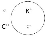
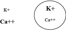
Just as an antagonistic relationship exists between H+ (acid) and OH- (alkaline), so does an antagonism exist between potassium and calcium with in the cells. The ability or inability of the organ/tissue to respond appropriately to autonomic stimulation rests on the balance between those minerals. Normally, the concentration of calcium ions is higher in the extracellular fluids while the concentration of potassium ions is higher within the cells.

symptomchart

Adequate calcium in the extracellular fluids is necessary for maintenance of a lesser amount inside the cells; otherwise intracellular calcium must begiven up to the extracellular fluid to maintain homeostasis. That would prevent the cells from responding appropriately to sympathetic stimulation and the patient would exhibit symptoms of parasympathetic dominance. Generally speaking, the patient would complain of excessive symptoms of an inappropriate need for rest, the inability to heal or recuperate, and reproductive problems.

Of course, the above has incredibly far-reaching influences and sometimes confusing consequences, not only for patients with chronic structural problems but for those who cannot respond appropriately to stress.

Lets’ carry that scenario a little further into a patient’s life and recognize the need for chiropractic as a profession to become recognized as the profession that restores normal function and maintains health.

The following is a list of symptoms associated with a deficiency of protein and calcium that causes the body to struggle maintaining homeostasis in the extracellular fluids. Obviously this stress, if long continued, results in necessary compensations and the appearance of chronic degenerative conditions. How many of your patients do you recognize from the following list of symptoms related to increased dietary alkalinity?

That is quite an extensive list of symptoms that you see frequently in your office. Picture in your mind a few patients, one at a time, which fit into a symptom pattern above. Now, superimpose over those symptoms the structural problems that you repeatedly see in these patients and understand that you cannot separate structure and function. Their dietary choices, with excesses and deficiencies, may very well be causing and perpetuating the chronic structural problems you seek to correct.

INTERNAL HEALTH: A Chiropractic Specialty Sympathetic Dominance, Parasympathetic Weakness

chart1loomismarch:dropcap_open:T:dropcap_close:his month, I begin a series of 24 articles each designed to highlight a set of symptoms that collectively point at a specific visceral problem that is perpetuating muscle contractions, loss of joint range of motion, and reoccurring structural misalignments that have become chronic and defy permanent correction. It is exactly these problems that cause patients to seek alternatives to continuing chiropractic care. I believe our profession must, in addition to specializing in structural disorders, begin identifying these underlying visceral problems and specialize in restoring normal function before disease entities can be identified.

Throughout 2010, my articles identified a series of tests for screening structural problems that may have an underlying visceral cause. I ended 2010 by discussing spinal flexibility tests and then began last month discussing the effects of sympathetic and parasympathetic stimulation. That leads me to this month’s article.

The effects of autonomic stimulation are well known and possible causes easily recognized. But what is often overlooked is the possibility that, while the central nervous system may be calling for increased sympathetic activity on the part of one or more organs, the organ or tissue may not be able to respond adequately due to a relative potassium deficiency. Let’s examine what is required nutritionally for organs to respond appropriately to autonomic stimulation. I’ll discuss sympathetic dominance this month and parasympathetic dominance next month.chart2loomismarch

Just as an antagonistic relationship exists between H+ (acid) and OH- (alkaline), so does an antagonism exist between potassium and calcium within the cells. The ability or inability of the organ/tissue to respond appropriately to autonomic stimulation rests on the balance between those minerals. Normally, the concentration of calcium ions is higher in the extracellular fluids, while the concentration of potassium ions is higher within the cells.

Adequate potassium within the cells is necessary for the cell to respond appropriately to parasympathetic stimulation.

A diet with adequate alkaline minerals, provided by fresh fruits and vegetables, is needed to supply those minerals including potassium.

A diet high in simple sugars depletes alkaline minerals and is largely responsible for the potassium deficiencies seen in our offices.

The symptoms of a potassium deficiency are stiff sore joints, constipation, inability to concentrate or think clearly, and cardiac arrhythmias. Also sodium is, of course, an alkaline mineral whose ions are needed in the extracellular fluid. While the body produces aldosterone and vasopressin to maintain sodium, the dietary stress placed on the body can still produce a mild deficiency. The symptoms of a mild sodium deficiency are almost insignificant, but do result in movement of potassium out of the cell into the extracellular fluid to maintain homeostasis. This causes a compensatory movement of calcium into the cells.

chart3loomismarchWhile all of this may seem inconsequential in a structural practice, it does result in an inability to respond appropriately to parasympathetic stimulation with an apparent sympathetic dominance and the symptoms of Fright-Fight-Flight are well documented. Every day, we see patients who are irritable and argumentative, or cannot adequately tolerate stress.

Let’s carry that scenario just a little further into a patient’s life and acknowledge the need for Chiropractic to become recognized as the profession that restores normal function and maintains health.

During sympathetic dominance, there is an increase of intracellular calcium accompanied by a deficiency of intracellular potassium. Excessive sympathetic stimulation increases the heart rate and force of contraction, as well as constricting blood vessels and elevating blood pressure. Ultimately, this results in heart disease. Pharmaceutically, calcium channel blockers are used to blunt the sympathetic nervous system and slow the progression of congestive heart failure.

A prudent nutritional approach would be early recognition of the symptoms of potassium deficiency and sympathetic dominance as seen with:

The appearance of stiff sore joints and constipation accompanied by the muscle contractions, loss of joint range of motion

The structural misalignments associated with the various visceral dysfunctions.

:dropcap_open:Let’s examine what is required nutritionally for organs to respond appropriately to autonomic stimulation.:quoteleft_close:

In addition to chiropractic care, dietary modification should be suggested by increasing the ingestion and digestion of foods high in alkaline minerals, such as fresh fruits and vegetables and reducing the consumption of simple sugars.

The following is a list of symptoms associated with a deficiency of alkaline minerals that causes the body to struggle with maintaining homeostasis in the extracellular fluids. Obviously this stress, if long continued, results in necessary compensations and the appearance of chronic degenerative conditions. How many of your patients do you recognize from the following list of symptoms related to increased dietary acidity?

Next month I will take up the 2nd article in this series of 24 – the other side of the autonomic nervous system – parasympathetic dominance.