Hip Pain in a Soccer Star

History and Presenting Symptoms

The patient is a 14-year-old soccer player who reports frequent pain in her right hip and posterior thigh region for the past several months.  This is most noticeable the day after a strenuous soccer competition or scrimmage when she has obvious tightness and tension in the back of her upper right thigh.  She denies any specific injury but admits that she has been playing especially hard since being named team captain.  She finds that lying down and putting warm towels around her hip helps the most.

Exam Findings

Vitals. This athletic girl weighs 120 lbs., which, at 5’4″, results in a BMI of 21—she is very active and fit.  She is a non-smoker, and her blood pressure and pulse rate are well within the normal range.

Posture and gait. Standing postural evaluation finds generally good alignment with intact spinal curves and no evidence of scoliosis.  Closer inspection identifies a higher left iliac crest, mild bilateral knee valgus, and static pronation of the right foot (calcaneal eversion with low medial arch).  The navicular drop test (Brody’s) finds 7 mm of excursion of the right navicular prominence between sitting and standing, compared to 3 mm of drop on the left.  Gait screening is negative for limp or noticeable asymmetry.

Chiropractic evaluation. Motion palpation identifies a right sacroiliac fixation, with moderate tenderness and loss of endrange mobility.  Straight leg raise is limited to 80° on the right by pain at the hamstring origin.

Primary complaint. The right hamstring is weaker than the left on manual muscle testing, and palpation finds tenderness at the right ischial tuberosity and increased tension in the proximal hamstring muscle.  All knee and ankle ranges of motion are full and pain free.

Imaging

Standing AP lumbopelvic view shows a leg length discrepancy with the right femur head 6 mm lower.  Frog-leg views of both hips are negative for ischemic necrosis (Legg-Calve-Perthes) and slipped femoral capital epiphysis.

Clinical Impression

Chronic hamstring strain, with leg length discrepancy (right short leg) and asymmetric foot pronation.

Treatment

Adjustments. Adjustments of the right SI joint and right foot and ankle were provided as needed.  The adjustments were supplemented by contract-relax stretches for the right hamstring muscle.

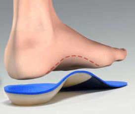
Stabilization. Flexible, shock-attenuating orthotics were fitted into her soccer shoes, and another pair was provided for daily wear.  Both were custom made for her individual postural needs.

Rehabilitation. Daily strengthening exercises for the right hamstring were progressed from light to strenuous resistance using elastic exercise tubing in a standing position.

Response to Care

This young athlete responded rapidly to the adjustments and strengthening exercises. She adapted to the custom-made stabilizing orthotics with little difficulty and reported that her ankles and knees felt more secure when on the field. Within three weeks of receiving the orthotics, she had no post-exercise pain or tenderness. She was released from care after a total of eight visits over two months.

Discussion

Hip, upper leg and, even, knee pain in a young person with an immature skeleton always raises concerns of ischemic necrosis (Legg-Calve-Perthes) and slipped femoral capital epiphysis. This star athlete had no X-ray evidence of either condition, but did have a biomechanical asymmetry in the lower extremities which caused a functional short leg.  Appropriate, focused treatment consisting of adjustments and stabilizing orthotics, along with stretching and strengthening exercises, brought about a rapid response.

While this patient had initial concerns about wearing orthotics in her well-fitting soccer shoes, she found them to be effective in helping reduce her hip symptoms and enhancing her athletic performance.  To get the right fit in her specialized athletic shoes, tracings of the inside of her soccer shoes were sent to the orthotics lab along with her foam-casted weightbearing foot images.

Studies have found a significant decrease in electromyographic activity of the hamstring muscles during running while wearing orthotics.  This is thought to be due to the increased stability of the ankles and knee joints, which allows greater relaxation of the hamstrings during gait, especially when running. 

Dr. John J. Danchik is the seventh inductee to the American Chiropractic Association Sports Hall of Fame.  He is the current chairperson of the United States Olympic Committee’s Chiropractic Selection Program.  He lectures extensively in the United States and abroad on current trends in sports chiropractic and rehabilitation.  Dr. Danchik is an associate editor of the Journal of the Neuromusculoskeletal System.  He has been in private practice in Massachusetts for 29 years.  He can be reached by e-mail at [email protected].

Morning Heel Pain

History and Presenting Symptoms

The patient is a 56-year-old male who reports severe pain on the bottom of his right foot when he begins to walk in the morning.  He also notices pain under his right heel when he has been standing for a long time (greater than one-half hour).  Over-the-counter pain medications help somewhat; but his condition does not seem to be improving, even though he has been avoiding extensive walking and standing.  He has not played racquetball for the past couple of months, due to his heel pain.  There is no history of prior injury to his right foot or ankle.

Exam Findings

orthoticblueVitals: This 5’11’’ financial consultant weighs 195 lbs. which means that he is overweight (BMI of 27).  He demonstrates a thickened waist (43 in.), confirming that his excess weight is due to abdominal fat deposition.  He is a non-smoker, and his blood pressure and pulse rate are within the normal range, probably due to his history of regular vigorous exercise during racquetball.

Posture and gait: Standing postural evaluation finds generally good alignment, but a decreased lumbar lordosis.  He has bilateral pes planus (flat foot), with no medial arches and bilateral calcaneal eversion.  These findings are somewhat more pronounced on the right side.  Both feet toe out during walking.

Chiropractic evaluation: The lumbar spine is moderately tender throughout, and he demonstrates a generalized loss of vertebral mobility, with few specific subluxations.  Orthopedic and neurological provocative testing of the spine and pelvis is negative.

Primary complaint: Examination of the right foot finds exquisite tenderness to palpation over the antero-medial aspect of the calcaneus.  All right foot ranges of motion are full and pain-free, and manual muscle testing finds no evidence of weakness when compared to the left side.

Imaging

A lateral X-ray of the right foot demonstrates a small bony outgrowth from the anterior aspect of the calcaneus.

Clinical Impression

Chronic irritation of the point of insertion of the plantar fascia into the calcaneus, with radiographic evidence of a heel spur.  This appears to be secondary to long-standing biomechanical stress associated with poor foot function, and excessive loading from strenuous exercise activity and too much body weight.

Treatment Plan

Adjustments: Mobilization and adjustments were provided to the lumbopelvic region.  The right calcaneus was adjusted anteriorly and both navicular bones were adjusted superiorly.

Stabilization: Orthotics with viscoelastic, shock-absorbing materials were custom-made and fitted to support his arches and to reduce calcaneal eversion.  In addition, a calcaneal “divot” was ordered for the area under the right heel, in order to decrease the pressure on the bone spur.

Rehabilitation: The patient was shown a series of foot exercises (marble pick-up and towel-scrunching) to improve the coordination and strength of his foot intrinsic muscles.  Once he had his orthotics, he also performed standing Achilles tendon stretches, keeping his feet in forward alignment. 

Response to Care

While his heel pain was initially somewhat slow in responding, this patient was diligent with his exercises and, after five weeks, he was able to walk in the morning with no foot pain.  At that point, he was advised to return to his regular racquetball exercise program and he had no recurrence of heel pain.  He was released to a self-directed maintenance program after a total of sixteen visits over three months.

Discussion

Radiographic evidence of a heel spur does not always correlate with heel pain.  However, it is frequently an indication of chronic biomechanical stress to the insertion of the plantar fascia.  Symptomatic heel spurs are difficult case presentations, and they require appropriate patient education.

If this overweight 56-year-old man had been less active (or had been a swimmer), or if he had inherited feet with better arches, he would have been less susceptible.  And, if he is able to follow through on his decision to drop twenty pounds of abdominal adipose tissue (which is necessary for him to be considered in the normal weight range for his height), he will be less likely to suffer future recurrences.

Dr. John J. Danchik is the seventh inductee to the American Chiropractic Association Sports Hall of Fame.  He is the current chairperson of the United States Olympic Committee’s Chiropractic Selection Program.  He lectures extensively in the United States and abroad on current trends in sports chiropractic and rehabilitation.  Dr. Danchik is an associate editor of the Journal of the Neuromusculoskeletal System.  He has been in private practice in Massachusetts for 29 years.  He can be reached by e-mail at [email protected].

Case Study: Functional Scoliosis

History and presenting symptoms

The patient is a 12 year-old girl, who was identified at a school screening as having a discrepancy in her shoulder heights.  Her parents were advised to seek further evaluation by an orthopedic surgeon.  They reported that the orthopedist found evidence of a scoliosis, but recommended a “watch and wait” approach.  He offered no treatment, but said that, if her spinal curve increased, he would be available to perform spinal corrective surgery.

The girl’s parents are requesting a second opinion, and any recommendations for conservative care.  The patient has no back symptoms, and she recalls no back injury.  Her delivery was relatively easy and uncomplicated, as reported by the parents.  She is regularly active in several physical activities, including soccer and softball.

Exam Findings

Vitals.  This 5’0’’ tall, athletic 12 year-old girl weighs 105 lbs, which results in a BMI of 20—she is not overweight.

Postural examination.  Standing postural evaluation identifies a right low pelvis, and a left low shoulder.  Her knees are well-aligned, but she has an obvious medial bowing of the right Achilles tendon, with a lower medial arch on the right foot.

Chiropractic evaluation.  Motion palpation identifies several mild limitations in spinal motion: The right SI joint, the lumbosacral junction, T11/12, and at the cervicothoracic junction.  Palpation finds no local tenderness in these regions, and she has full and pain-free active spinal ranges of motion.  Thoracolumbar lateral bending is equal to both sides, and Adams test finds no evidence of rib hump or persisting curve.

Lower extremities.  Closer examination finds that the right medial arch of the foot is lower than the left when standing.  When she is seated and non-weightbearing, the right arch appears equal to the left.  And when she performs a toe-raise while standing, the right arch returns.  Manual muscle testing finds no evidence of muscle weakness in the peroneal or anterior tibial muscles.

Imaging

A P-A full-spine film demonstrates a C-curve scoliosis, which encompasses the lumbar and thoracic regions.  The sacral base is lower on the right by 3 mm, and the Cobb angle is 12°.  A collimated pelvis view with the femur heads centered finds a difference of 6 mm in the heights of the femur heads, with the right side lower.

Clinical Impression

This is a classic case of a functional scoliosis associated with a unilateral flexible flat foot (pes planus).  By definition, this eliminates the concern of a progressive idiopathic scoliosis, which had caused the parents so much concern.  The condition is accompanied by multiple areas of mild joint motion restriction and compensatory spinal subluxations.

Treatment Plan

Adjustments.  Specific adjustments for the lumbopelvic and thoracolumbar spinal regions were provided as needed.  Manipulation of the right foot, including the navicular and cuboid bones, was performed.

Support.  Custom-made stabilizing orthotics were provided to ensure balanced support for both arches and to reduce weight-bearing asymmetry.  Particular emphasis was placed on wearing the supports in her athletic shoes.

Rehabilitation.  Because of her age and athletic pursuits, no specific rehabilitation exercises were provided.  She was able to continue in her sports activities without difficulty.

Response to Care

The spinal and foot adjustments were well tolerated, since she was young and symptom-free.  The orthotics improved her postural alignment and eliminated the shoulder discrepancy.  After three months of care, repeat full-spine X-rays with her orthotics in place found only a minimal (3 mm) leg length discrepancy, a level sacral base, and a 5° Cobb angle (which is considered non-scoliotic).  She was released to a self-directed maintenance program after a total of 10 treatment sessions over three months.

Discussion

This active 12 year-old girl responded well to a combination of spinal adjustments and custom-made orthotics.  Although she was asymptomatic, her parents considered her at risk for spinal surgery, based on the specialist’s opinion.  Chiropractic evaluation found her scoliosis to be functional, and her flat foot was found to be flexible.  Appropriate conservative care was initiated, and was ultimately very successful.  In most cases, a functional scoliosis responds well to chiropractic care, and is unlikely to require surgery.

Dr. John J. Danchik is the seventh inductee to the American Chiropractic Association Sports Hall of Fame.  He is the current chairperson of the United States Olympic Committee’s Chiropractic Selection Program.  He lectures extensively in the United States and abroad on current trends in sports chiropractic and rehabilitation.  Dr. Danchik is an associate editor of the Journal of the Neuromusculoskeletal System.  He has been in private practice in Massachusetts for 29 years.  He can be reached by e-mail at [email protected].

Case Study: Knee Pain in a Tennis Player

History and Presenting Symptoms

The patient is a 48-year-old real estate broker, who is also the president of the local Chamber of Commerce.  She reports pain in the front of her right knee, over the past several months, especially when playing tennis.  She denies any specific injury, and has no obvious swelling or discoloration.  She reports that she takes ibuprofen for relief, but is worried that she has to take this drug in order to play tennis.

Exam Findings

Vitals.  This active, middle-aged woman weighs 140 lbs, which, at 5’5’’, results in a body mass index (BMI) of 23—she is not overweight.  She is a non-smoker, and her blood pressure and pulse rate are both at the lower end of normal range.

Posture and gait.  Standing postural evaluation finds generally good alignment throughout her pelvis and spine.  She has a mild bilateral knee valgus and moderate calcaneal eversion and hyperpronation on the right side.  Measurement finds a standing Q-angle of 28° on the right and 24° on the left (20° is normal for women).

Chiropractic evaluation.  Motion palpation identifies a limitation in lumbosacral motion, with moderate tenderness and loss of endrange mobility to the right at L5/S1.  A compensatory subluxation is also noted at T10/T11.  Neurologic testing is negative.

Primary complaint.  Examination of the right knee finds no ligament instability, but there is a positive patellar grinding test.  All knee ranges of motion are full and pain free, bilaterally.  Manual muscle testing finds no evidence of muscle weakness.

Imaging

No X-rays or other forms of musculoskeletal imaging were requested.

Clinical Impression

Patello-femoral arthralgia (previously chondromalacia patellae) on the right, associated with an elevated Q-angle and foot pronation.  This is accompanied by lumbosacral joint motion restriction and compensatory lower thoracic subluxation.

Treatment Plan

Adjustments.  Specific, corrective adjustments for the lumbosacral joint and the lower thoracic region were provided as needed.  Manipulation of the right knee into external rotation was performed to decrease the internal rotation associated with hyperpronation and her elevated Q-angle.

Support.  Custom-made stabilizing orthotics were provided to support the arches and decrease the Q-angles.  Two pairs of stabilizing orthotics were ordered, one designed specifically for her tennis shoes and the other for her job-related dress shoes.

Rehabilitation.  Due to her active lifestyle, no specific rehabilitation exercises were provided.  She continued with her frequent tennis playing.

Response to Care

The spinal and knee adjustments were well tolerated, and she reported a rapid decrease in symptoms. Once she began wearing her orthotics regularly, she noted a substantial decrease in knee irritation with use, and a firmer foot plant during tennis playing.  Within two weeks of receiving her orthotics, she related that she had successfully completed a round-robin tournament with absolutely no knee pain or limitation.  She was released to a self-directed maintenance program after a total of eight treatment sessions over two months.

Discussion

Several interesting factors are present in this case.  This high-powered businesswoman used her tennis games as both a form of business interaction and a recreational relaxation.  As her knee began to bother her more, she was driven to using anti-inflammatory drugs.  She was wearing supportive shoes, but the underlying biomechanical problem had not been sufficiently addressed.

When women are physically active, their naturally higher Q-angles are frequently a source of patello-femoral pain.  This condition was previously called chondromalacia patellae, but it has been recognized that it is actually a biomechanical “tracking” disorder of the kneecap in the femoral groove.  The best treatment is a conservative approach, with a combination of chiropractic adjustments, flexible stabilizing orthotic support, and—when indicated—rehabilitative strengthening sessions using elastic exercise tubing.

Dr. John J. Danchik is the seventh inductee to the American Chiropractic Association Sports Hall of Fame.  He is the current chairperson of the United States Olympic Committee’s Chiropractic Selection Program.  He lectures extensively in the United States and abroad on current trends in sports chiropractic and rehabilitation.  Dr. Danchik is an associate editor of the Journal of the Neuromusculoskeletal System.  He has been in private practice in Massachusetts for 29 years.  He can be reached by e-mail at [email protected].

Case Study: Ankle Pain in a Young Athlete

orthoticblueHistory and presenting symptoms:

The patient is a 16-year-old athlete who plays football and baseball.  He reports recurrent pain in his left ankle over the past year, especially after lengthy running practices.  Occasionally, the ankle will swell, which he treats with an ice pack and over-the-counter anti-inflammatory drugs.  He recalls no specific injury and has no significant disability, but is concerned for his future sports performance.  Parental advice prompted him to seek chiropractic care.

Exam findings:

Vitals:  The patient weighs 180 lbs, which, at 5’10’’, results in a body mass index (BMI) of 26—at his age, he is considered overweight.  His appearance, however, confirms that excess weight is due to muscular development, not excess fat mass.  He doesn’t smoke, and his blood pressure and pulse rate are well within normal range.

Posture and gait:  Standing postural evaluation finds generally good alignment, with intact spinal curves, and no lateral listing of pelvis or spine.  He demonstrates a mild bilateral knee valgus (more prominent on left), with bilateral pes planus and calcaneal eversion.  These findings cause medial bowing of the Achilles tendons when standing, especially on the left.  A toeing-out tendency (foot flare) is seen during gait screening; this is also somewhat more prominent on left.

Chiropractic evaluation:  Motion palpation identifies a limitation in left sacroiliac motion, with moderate tenderness and loss of end range mobility.  Several compensatory subluxations are identified throughout the thoracolumbar region.  Orthopedic and neurological testing is negative.

Primary complaint:  Left ankle examination reveals slight swelling of entire Achilles tendon, which is moderately tender to palpation. All left ankle ranges of motion are full and pain-free, except dorsiflexion, which is slightly limited by tightness, not pain. Manual muscle testing finds slight weakness in plantar flexion when compared to right side.

Imaging:

No X-rays or other forms of musculoskeletal imaging were requested.

Clinical impression:

Chronic, recurrent Achilles tendinosis secondary to lower extremity biomechanical stress.  This is accompanied by sacroiliac joint motion restriction and compensatory thoracolumbar subluxations.

Treatment

Adjustment:  Specific, corrective adjustments for the SI joints and thoracolumbar region were provided, as needed.  Manipulation of the left talus during traction was performed to increase the range of ankle dorsiflexion motion.

Stabilization:  Custom-made stabilizing orthotics were provided to support the arches, decrease calcaneal eversion, and reduce the asymmetrical biomechanical forces being transmitted to the spine and pelvis.

Rehabilitation:  Initially, was taught to perform daily self-mobilization to increase left ankle dorsiflexion motion, along with towel-scrunching exercises.  After two weeks, daily strengthening of plantar flexion was introduced, using elastic exercise tubing.

Response to care:

The spinal and pelvic adjustments were well-tolerated, and this young, in-shape athlete required few re-adjustments.  His compliance with the stabilization and exercise recommendations was excellent, since he was motivated to improve his performance and prevent potential sports injuries.  He adapted to and wore the orthotics without difficulty.  He filled out and brought in his exercise log at every visit, which provided an opportunity to support his home-based efforts.

Within four weeks of receiving his orthotics, the patient was able to complete several long running practices without symptoms or swelling in the ankle.  He described a noticeable improvement overall in his athletic performance.  He was released from acute care to a self-directed maintenance program after a total of 12 visits over 2 months.

Discussion:

Using pediatric BMI calculations, this patient was classified as overweight.  This and his high activity level are likely to be significant factors in his ankle problem.  However, it is important to recognize that, in the case of athletes, overweight may be due to greater than usual amounts of lean body mass (muscle and bone), not fatty deposits.  This was readily apparent during examination.

While the traditional term for this condition is “Achilles tendinitis,” the more accurate and contemporary description is “tendinosis.”  As was apparent in this case, the problem is usually not an acute inflammatory event, but rather a long-standing biomechanical irritation that needs to have the underlying problem correctly addressed.

This case demonstrates that young athletes are superior patients.  Most have a great healing potential and are willingly to follow self-care recommendations—particularly when they include the use of orthotics for stabilization, specific exercises for rehabilitation, and a rapid return to full sports function.

Dr. John J. Danchik is the seventh inductee to the American Chiropractic Association Sports Hall of Fame.  He is the current chairperson of the United States Olympic Committee’s Chiropractic Selection Program.  He lectures extensively in the United States and abroad on current trends in sports chiropractic and rehabilitation.  Dr. Danchik is an associate editor of the Journal of the Neuromusculoskeletal System.  He has been in private practice in Massachusetts for 29 years.  He can be reached by e-mail at [email protected].

Case Study: Pedal Imbalance With Associated Conditions

History and subjective complaints

The patient has suffered from a postural imbalance most of her life, causing stress and strain to the pelvic and spinal areas.  The patient states that she has received chiropractic care “on and off” most of her life.

As the patient has grown older, she has experienced more frequent and intense pain in her lower back, right leg, and feet.  She was 52, when she learned that some of the problems she was experiencing when standing or walking were the result of flat feet (pes planus), lack of arch support, heel spurs, and a neuroma.  There are other contributing factors to her overall health and well-being: fibromyalgia, diverticulitis, and excess weight.

Upon her first office visit, the patient was experiencing problems walking at a slow, strolling pace.  She reported having constant lower back pain and a great deal of right leg and right thigh pain.  She particularly noticed the pain in her right thigh when she climbed stairs, bearing weight on that leg.  She also had severe pain in her feet that included numbness in her toes, at times.

Objective findings

The patient is a medium-framed, slightly overweight Caucasian female, 53 years of age.  She is a full-time customer service representative, a position which requires her to be seated most of her workday.  In addition to working full-time, she is also a certified massage therapist and is on her feet for long periods during evenings and on weekends, to treat her clients.

Vital index

    Height: 5 ft. 2 in.
    Weight: 146 pounds
    Blood pressure: Systolic, 132; Diastolic, 86

Objective and neurological findings

  • Gaenslen’s test positive with right leg.
  • Nachlas’ test positive, with radiation down the right thigh.
  • Morton’s squeeze test positive for chronic onset of interdigital neuroma in both feet.
  • Positive pain indicators in 13 of the 18 tender points for fibromyalgia.

Clinical impression and working diagnosis

The patient’s lower back pain was rooted in the lower lumbar area, where she has a curve.  She also experienced pain from the ilium, torquing forward.  Additionally, her right leg is shorter than the left, and the tightening of the muscles that is part of fibromyalgia also intensified her pain.  The patient had a neuroma, or entrapment of the nerve, in her left metatarsal.

Postural imbalances in the pedal foundation were also contributing to her musculoskeletal conditions, as well as obstructing the efficacy of chiropractic adjustments received prior to current care.

Treatment

The patient received a program of specific lumbosacral and lower extremity adjustments.  She also regularly received routine checks of her leg length, routine manipulation of vertebrae, and did hip rotator exercises—hip flexors and extenders.  Muscle trigger point therapy was also initiated to help manage her fibromyalgia symptoms.

The patient was also fitted for custom-made stabilizing orthotics, to address her spinal/pelvic instability and foot conditions.  She had begun wearing well-known name brand sandals at the time she learned of her flat feet, lack of arch support, heel spurs, and neuroma.  The sandals offered some stability to her low back, but did not correct her postural imbalances.  They also didn’t provide adequate arch support.

The patient currently receives chiropractic care every two weeks, minimum, gets a massage every two weeks, and wears her custom-made stabilizing orthotics every day.

Results

After only eight weeks of receiving adjustments and wearing the orthotics, the patient was no longer experiencing pain in the SI joint area.  In addition, the neuroma was corrected without surgery.  She no longer has numbness in her toes.  And, within a few days of doing hip rotator exercises, she found relief from her leg pain.

Adjustments, orthotic support, and hip rotator exercises proved to be very successful for this patient, as did the muscle trigger point therapy.  Additionally, the myofascial release for legs and hips was very beneficial.

Discussion

This case is interesting because there were several contributing factors to the patient’s constant pain.  For her, the use of custom-made stabilizing orthotics is not just a method for correcting one thing—it’s an overall approach to improved health and wellness.

Dr. John J. Danchik is the seventh inductee to the American Chiropractic Association Sports Hall of Fame.  He is the current chairperson of the United States Olympic Committee’s Chiropractic Selection Program.  He lectures extensively in the United States and abroad on current trends in sports chiropractic and rehabilitation.  Dr. Danchik is an associate editor of the Journal of the Neuromusculoskeletal System.  He has been in private practice in Massachusetts for 29 years.  He can be reached by e-mail at [email protected].

Orthotic Support for All Three Arches

A close evaluation of the anatomy and structure of the foot reveals three arches that form the plantar vault.1  This architectural design provides substantial strength, while still permitting sufficient flexibility to accommodate changes in terrain, and to provide propulsion.  The foot’s arched structure is not present at birth, but develops during childhood, by age 6 or 7 in most people.2  Breakdown in any of these three arches can result in abnormal gait and transmission of asymmetrical forces into the pelvis and spine.

Medial Longitudinal Arch

The most obvious arch is seen along the medial aspect of the foot.  The navicular bone forms the “keystone” of this large and long arch, which is supported primarily by the plantar fascia and spring ligament.3  Years ago, John Basmajian, MD, (the “father of electromyography”) demonstrated that the muscles of the foot and lower leg do not provide support for the medial arch, except during toe-off when walking or while standing on tip-toe.  He said, “From the present study, one may conclude that, in the standing-at-ease posture, muscle activity is not required and the muscles are inactive.…”4  While he believed, in 1963, that he had settled the controversy regarding active (muscular) versus passive (ligamentous) support for the medial arch, there still remains much misinformation and persisting, misguided attempts at “strengthening muscles to rebuild the arch.”

The most effective method for evaluating the function of the connective tissues that support the medial arch is to perform a comparison between its non-weight bearing and weight bearing alignment.  This procedure is called the “Navicular Drop Test”, and was first described by Brody. 5  The easy-to-perform clinical test objectively documents the presence (or absence) of collapse of the medial longitudinal arch, and has been used successfully to evaluate the risk of athletes with ACL ruptures.6

Lateral Longitudinal Arch

This arch is located along the outside of each foot.  Because the cuboid bone serves as its structural keystone, the lateral arch relies much less on connective tissues for its support.  For this reason, proper function of the lateral arch is very dependent on the alignment of the cuboid, which is frequently found to be in need of adjustment.  Proper support for this arch is at least as important as for the other two, but is surprisingly absent in many orthotics.

Anterior Transverse (Metatarsal) Arch

This arch extends from the metatarsal heads back to the tarsal bones, and runs from the medial to the lateral sides of the foot.  At its most anterior portion, the metatarsal heads contact the ground.  Poor function and loss of this arch will often result in a build-up of thick callus underneath the metatarsal heads.  Recurrent “dropped” metatarsal heads and/or irritation of one of the interdigital nerves (a “Morton’s neuroma”) are also good indications that this arch is not being supported properly by the plantar fascia.

Helping the Faulty Vault

The structural design of the three-arched plantar vault is very good at supporting weight and carrying high loads, while remaining flexible.  During normal standing, the load of the body is balanced over the center of the foot, anterior to the ankle.  This places the greatest amount of load at the apex of the three arches.  This force is then distributed along the “buttresses” of the arches to the heel (which bears 50% to 60% of body weight) and the metatarsal heads (which bear 40% to 50% of body weight).  Loss of this configuration will result in abnormal force concentrations, which will eventually cause degenerative and symptomatic clinical conditions.

Collapse or dysfunction of any of the arches needs to be addressed with custom-made orthotics that will support the patient’s foot throughout the gait cycle, while controlling the impact forces.  Particularly when there is asymmetry between the feet, arch problems can cause abnormal rotational forces to be transmitted into the pelvis and spine, resulting in chronic spinal symptoms.  For this reason alone, doctors of chiropractic need to be aware of the status of their patients’ three arches, since they can have a substantial impact on spinal health.

Dr. John J. Danchik is the seventh inductee to the American Chiropractic Association Sports Hall of Fame.  He is the current chairperson of the United States Olympic Committee’s Chiropractic Selection Program.  He lectures extensively in the United States and abroad on current trends in sports chiropractic and rehabilitation.  Dr. Danchik is an associate editor of the Journal of the Neuromusculoskeletal System.  He has been in private practice in Massachusetts for 28 years.  He can be reached by e-mail at [email protected].

References:

1. Kapandji IA. Physiology of the Joints: Lower Limb (2nd ed.). New York: Churchill Livingstone, 1981:154-182.
2. Gould N, Moreland M, Alvarez R et al. Development of the child’s arch. Foot Ankle 1989; 9:241-245.
3. Huang CK, Kitaoka HB, An K-N, Chao EY. Biomechanical evaluation of longitudinal arch stability. Foot Ankle 1993; 14:353-357.
4. Basmajian JV, Stecko G. The role of muscles in arch support of the foot: an electromyographic study. J Bone Joint Surg 1963; 45A:1184-1190.
5. Brody D. Techniques in the evaluation and treatment of the injured runner. Orthop Clin North Am 1982; 13:541-558.
6. Beckett ME et al. Incidence of hyperpronation in the ACL injured knee: a clinical perspective. J Athl Train 1992; 27:58-62.

Ankle Sprains and Active Support

Ankle sprains are common injuries, because the ankle is required to perform complex movements under high forces during normal walking.  This can be particularly important for patients who participate in recreational activities and sports that require running and jumping.  Proper evaluation and management in the early stages of an ankle sprain are very important in preventing chronic instabilities.  With appropriate care, including orthotic support and exercise, significant improvements in function and stability can be achieved, even in patients with long-standing ankle problems.

Acute Care

Initial treatment of ankle sprains is the standard “PRICE” formula (Protection, Rest, Ice, Compression, Elevation).  Even with severe ankle sprains, using these procedures has been shown to speed recovery and return to sports (see Table 1).  With the injured joint protected, patients can be encouraged to continue their activities (rather than using the now-discredited bed rest), with some restrictions.

In the case of ankle sprains, this entails the use of a lightweight, but laterally rigid brace, which protects against inversion and eversion.  If a patient has been placed in a walking cast rather than a mobilizing brace, frequent prolonged stretching of the Achilles tendon must be performed in order to prevent shortening.

Conditioning and Exercise

During the initial acute stage, exercises for the damaged ankle are not appropriate.  However, general full-body conditioning should be continued, using methods that do not place undue stress on the healing ankle (a stationary cycle with pedal straps is recommended).1  Additionally, vigorous exercise of the opposite ankle’s muscles (“cross education” or “cross-over”) has been shown to provide a healing stimulus and result in more rapid return to activities.2,3

As healing progresses, patients should begin to perform non-resistive active exercises concentrating on mobility of the injured ankle.  Once the joint can be passively moved through a normal range, isotonic resistance exercising of the peroneal muscles using elastic tubing should be started.4  Initially, these exercises should be performed from a sitting position, with the heel resting on the floor, to reduce the forces on the ankle joint while still maintaining the functional alignment.

As strength builds, the patient should progress to standing during the exercises, in order to re-train the ankle support muscles in a closed-chain position.  Further sport-specific exercises should be introduced to ensure that an athlete has all the strength and mobility required to participate in sports.

Proprioception

One reason some ankle injuries become chronic or recur appears to be the loss of the normal coordination of the muscles about the ankle, rather than simply their strength.5  An easy test is to have the patient stand on each leg, with the eyes open and then closed.  Check to see if there is less capability of the injured leg.  Practice of the one-legged stance, and use of “wobble” boards may be required to regain normal proprioceptive coordination.  An athlete should be able to demonstrate a “stork stand” for at least one minute on the injured leg before being allowed to return to full competition.6

Orthotic Support

In many patients, custom-made orthotics can also be helpful in preventing future (and often more disabling) damage to the injured ankle.  A careful evaluation of the biomechanics of the foot and ankle will find some patients who have underlying anatomical or functional problems.  Particularly in the case of athletes, use of stabilizing, custom-made orthotics with good torsional rigidity should be considered.  Orthotic support and control of inversion/eversion is necessary and highly recommended whenever there is a deficit in biomechanical function.7

Summary

Recent studies demonstrate that, even in severe ankle injuries, a well-informed conservative and active treatment approach will result in good outcomes.  Using active rehabilitation concepts, most doctors of chiropractic can manage acute ankle sprain injuries very well.  In many patients, custom-made orthotics will be needed to help prevent future problems and joint degeneration.

Dr. John J. Danchik is the seventh inductee to the American Chiropractic Association Sports Hall of Fame. He is the current chairperson of the United States Olympic Committee’s Chiropractic Selection Program. He lectures extensively in the United States and abroad on current trends in sports chiropractic and rehabilitation. Dr. Danchik is an associate editor of the Journal of the Neuromusculoskeletal System.He has been in private practice in Massachusetts for 28 years. He can be reached by e-mail at [email protected].

References

1. Roy S, Irvin R. Sports Medicine: Prevention, Evaluation, Management and Rehabilitation. Englewood Cliffs: Prentice-Hall, 1983:394.

2. Stromberg BV. Contralateral therapy in upper extremity rehabilitation. Am J Phys Med 1988; 65:135-143.

3. Hortobagyi T, Lambert NJ, Hill JP. Greater cross education following training with muscle lengthening than shortening. Med Sci Sports Exerc 1997; 29:107-112.

4. Roy S, Irvin R. Sports Medicine: Prevention, Evaluation, Management and Rehabilitation. Englewood Cliffs: Prentice-Hall, 1983:397.

5. Lentell GL, Katzman LL, Walters MR. The relationship between muscle function and ankle stability. J Orth Sports Phy Ther 1990; 11:605-611.

6. Subotnick SI. Sports Medicine of the Lower Extremity. New York: Churchill Livingstone, 1989:284.

7. Heiser JR. Rehabilitation of lower extremity athletic injuries. Contemp Podiat Phys 1992; Aug 20-27.

Metatarsalgia and Orthotic Support

Foot pain can interfere significantly with normal activities, and severely limit participation in sports.  Metatarsalgia is foot pain involving the metatarsal bones in the forefoot—the complaint of pain on the bottom of the ball of the foot.

Metatarsalgia may be due to overuse of the foot during sports, improper footwear,1 excessive weight, foot subluxations, or other factors.  The underlying cause is often faulty foot mechanics; the most common problem is excessive pronation.  Blake and Ferguson found that joint pain and capsulitis of the metatarsals were common foot problems reported by recreational walkers and hikers.2  The second and third metatarsophalangeal joints were the most frequently involved joints.  Since the vast majority of foot problems in recreational walkers and hikers were unilateral, rather than bilateral, these researchers concluded that, “Structural anomalies and faulty biomechanics, i.e., limb length discrepancy or abnormal pronation, may be the cause or additional cause in many injuries.”

Arch Problems

The anterior transverse arch is located immediately behind the metatarsal heads.3  When non-weightbearing, the first and fifth metatarsal heads are most prominent, and initially bear the weight of the body during gait.  As weightbearing progresses, pressure is distributed across the arch to the other three heads.  As with all arches, the ligaments and connective tissues support the anterior transverse arch,4 not muscular strength.5  Arch problems will develop when supportive tissues are put under excessive stress—either from high loads for sudden, brief periods, or from more moderate, but repetitive stresses over longer periods.  In most cases, it appears that chronic overstretching of the transverse ligaments is the underlying cause of metatarsal problems.6

Metatarsalgia

Callus Formation

One sign of abnormal transverse arch biomechanics is callus build-up.  Since plantar callosities form in response to sustained pressure patterns, they provide helpful clues regarding altered foot function.  These are commonly seen in either the forefoot (under the metatarsal heads), or under the anterior aspect of the heel.7  This pattern (under the transverse arch and at each end of the medial longitudinal arch) has always been taken to indicate that most calluses are caused primarily by arch collapse and/or excessive pronation.  A 1999 study confirmed that callus formation is closely associated with several specific “abnormal foot weightbearing patterns.”  These are a lower medial arch with greater pronation, reduced dorsiflexion of the first metatarsal joint, and limited ankle dorsiflexion (due to calf muscle tightness).8  All three factors can contribute to abnormal biomechanics of the metatarsal arch.

Relief and Control

Helping a patient with pain at the metatarsal region requires a phased approach.  Immediate care can reduce the acute pain and inflammation.  Long-term control of the problem usually requires custom-made orthotics.

Acute relief.  Any aggravating activity must stop, and shoes should be evaluated and changed, if necessary.  A temporary metatarsal pad should be placed just proximal to the metatarsal heads to support the anterior transverse arch.9  This will relieve the weightbearing pressure on the sensitive metatarsophalangeal joints.  Anti-inflammatory and pain-relieving modalities can be considered.  Any subluxations, such as “dropped” or fixated metatarsal heads should be adjusted, as necessary.  Multiple foot subluxations, arch collapse, and excessive pronation are frequently found, so the navicular and the cuboid must be carefully evaluated.

Permanent control.  Patients should avoid shoes with a tight toe and forefoot region and reduce high heels to 1½ inches.  These instructions must be followed for best results.  Patients with tight Achilles tendons and diminished foot dorsiflexion should perform calf stretches.  In order to improve foot biomechanics and provide permanent support for the transverse arch, most patients will need custom-made orthotics.  Flexible orthotics are the most beneficial, especially for people who must be on their feet for many hours each day.  The orthotics need to support all three arches of the foot and provide cushioning and shock absorption.  Additional forefoot padding also appears to be very helpful. TAC

Dr. John J. Danchik is the seventh inductee to the American Chiropractic Association Sports Hall of Fame.  He is the current chairperson of the United States Olympic Committee’s Chiropractic Selection Program.  He lectures extensively in the United States and abroad on current trends in sports chiropractic and rehabilitation.  Dr. Danchik is an associate editor of the Journal of the Neuromusculoskeletal System.  He has been in private practice in Massachusetts for 27 years.  He can be reached by e-mail at [email protected].

References

1. Jarboe NE, Quesada PM. The effects of cycling shoe stiffness on forefoot pressure. Foot Ankle Int 2003; 24(10):784-788.
2. Blake RL, Ferguson HJ. Walking and hiking injuries: a one year follow-up study. J Am Podiatr Med Assn 1993; 83:499-503.
3. Hoppenfeld S. Physical Examination of the Spine and Extremities. New York: Appleton-Century-Crofts, 1976:208.
4. Huang CK et al. Biomechanical evaluation of longitudinal arch stability. Foot & Ankle 1993; 14:353-357.
5. Basmajian JV, Stecko G. The role of muscles in arch support of the foot. J Bone Joint Surg 1963; 45A:1184-1190.
6. Reid DC. Sports Injury Assessment and Rehabilitation. New York: Churchill Livingstone, 1992:129-184.
7. Magee DJ. Orthopedic Physical Assessment. Philadelphia: WB Saunders, 1987:323.
8. Bevans JS, Bowker P. Foot structure and function: etiological risk factors for callus formation in diabetic and non-diabetic subjects. The Foot 1999; 9:120-127.
9. Souza TA. Differential Diagnosis for the Chiropractor: Protocols and Algorithms. Gaithersburg, MD: Aspen Pubs, 1997:351.

Plantar Fascitis and Orthotic Support

The classic presentation of plantar fascitis is “a sharp heel pain that radiates along the bottom of the inside of the foot.  The pain is often worse when getting out of bed in the morning.”1  This can occur in runners or other athletes who repetitively land on the foot.  Another susceptible group is middle-aged persons who have spent much time on their feet.  More rarely, the fascia becomes inflamed after a single traumatic event, such as landing wrong after a jump, or running a long hill. 

The vast majority (95%) will respond to conservative care, and not require surgery.2  Proper treatment is necessary, however, to both ensure continued participation in sports and daily activities and avoid chronic damage.  The plantar fascia is the major structure that supports and maintains the arched alignment of the foot.3  This aponeurosis functions as a “bowstring” to hold up the longitudinal arch.

Pathology
Plantar fascitis develops when repetitive weight bearing stress irritates and inflames the tough connective tissues along the bottom of the foot.  High levels of strain stimulate the aponeurosis to try to heal and strengthen.  If the biomechanical strain continues, it overwhelms the body’s repair capacity, and the ligaments begin to fail.  It is this tear/repair process that causes the chronic, variable symptoms that can eventually become unbearable in some patients.

Since the plantar fascia inserts into the base of the calcaneus, the chronic pull and inflammation can stimulate the deposition of calcium, resulting in a classic heel spur seen on a lateral radiograph.  Unfortunately, there is no correlation between the presence of a heel spur and plantar fascitis; many heel spurs are clinically silent, and most cases of plantar fascitis do not demonstrate a calcaneal spur.4

Examination
Biomechanical evaluation may find either excessive pronation or supination.  The flatter, hyperpronating foot overstretches the bowstring function of the plantar fascia, while the high-arched, rigid foot places excessive tension on the plantar aponeurosis.  In either case, it is the combination of improper foot biomechanics and excessive strain that causes the connective tissue to become inflamed.  A careful assessment of the weight bearing alignment of the lower extremities is helpful, since many patients will have functional imbalances up the kinetic chain, into the pelvis and spine.

Direct palpation of the plantar fascia will demonstrate discrete painful areas, most commonly at the insertion on the antero-medial calcaneus.5  Fibrotic thickenings are frequently felt—these are remnants of the repetitive “tear and repair” process.  With the foot relaxed, grasp the toes and gently pull them up into passive dorsiflexion.  Since this maneuver stretches the irritated plantar aponeurosis, it is frequently quite painful, and is an obviously positive objective sign.

Conclusion
Plantar fascitis usually responds well to focused, conservative treatment.  Steroid injections and surgical release are seldom necessary, and are best avoided.  One of the most important treatment methods is to reduce any tendency to pronate excessively.  In addition to custom-made orthotics, runners should wear well-designed shoes that provide good heel stability. 

The use of custom-made orthotics can prevent many overuse problems from developing in the lower extremities.  Investigation of foot biomechanics is a good idea in all patients, but especially for those who are recreationally active.


Dr. John J. Danchik is the seventh inductee to the American Chiropractic Association Sports Hall of Fame.  He is the current chairperson of the United States Olympic Committee’s Chiropractic Selection Program.  He lectures extensively in the United States and abroad on current trends in sports chiropractic and rehabilitation.  Dr. Danchik is an associate editor of the Journal of the Neuromusculoskeletal System.  He has been in private practice in Massachusetts for 27 years.  He can be reached by e-mail at [email protected].