A New Look at Adolescent Idiopathic Scoliosis

A New Look at Adolescent Idiopathic Scoliosis

by Mark Sanna, D.C.


Scoliosis comes from the Greek word skoliosis which means crookedness. Adolescent Idiopathic Scoliosis (AIS) is a deformity of the spinal column with an onset between the ages of ten and eighteen. Most types of scoliosis are classified as idiopathic, meaning that the reasons for this type of deformity of the spine are unknown. Many assumptions concerning the causative factors for the dysfunction have developed. These include: birth trauma, visceral tensions, psychological problems, unilateral shortening of the psoas muscle, nutrition, genetic factors, and so on.

Continue reading “A New Look at Adolescent Idiopathic Scoliosis”

Soft Tissue Neoplasm or Hematoma?

Soft Tissue Neoplasm or Hematoma?

by Dr. Terry R. Yochum, D.C.; D.A.C.B.R.; Fellow, A.C.C.R. and Dr. Chad J. Maola, D.C.


History: This 25 year old female has recently noticed a palpable mass in the upper inner thigh. She doesn’t recall any fall but does ride her horse on a regular basis.

Diagnosis: Post-traumatic myositis ossificans. Note the calcific mass near the lesser trochanter which was caused by chronic trauma to the adductor muscles while horseback riding. (Prussian’s disease)

General considerations:

Traumatic myositis ossificans (myositis ossificans posttraumatica, ossifying hematoma, traumatic ossifying myositis, or heterotopic posttraumatic bone formation) is a condition characterized by heterotopic bone formation in the soft tissues following trauma. The process occurs most often in muscle but may also occur in fascia, tendons, joint capsules, and ligaments. Most occur following any local injury sufficient to cause bruising of the muscle or a frank hemorrhage within it. The most common sites are the brachialis anterior (elbow), quadriceps femoris (thigh), adductor muscles of the thigh, medial collateral ligament of the knee (Pellegrini-Stieda disease), and in cases of rupture of the coracoclavicular ligament of the shoulder.1 The constant pressure of the saddle against the adductors in riders may cause ossification in the adductor magnus and is known as Prussian’s disease, or erroneously, as a saddle tumor.1 Bedridden and wheelchair-confined patients frequently produce heterotopic bone at areas of gravitational stress, most commonly found at the ischial tuberosities. Up to 50 percent of paraplegic and quadraplegics exhibit paravertebral ossification.1,2

Radiologic Features:

The ossifying hematoma may be visible radiographically within three to four weeks after the initial injury.1 Magnetic resonance imaging may show the intramuscular hematoma from an early stage. Initially, the roentgen appearance is a fine, lacy radiopacity, which is followed later by a cloudy ossification within a well-defined mass. Its size depends upon the degree of initial trauma and the overall size of the hematoma. Eventually, sequential studies demonstrate a bony mass that is very radiopaque in its peripheral margins, with the center of the lesion appearing relatively radiolucent. The bony mass usually measures 4 to 5 cm but may be as large as 25 cm on occasion. The soft tissue osseous mass distinctively has no direct connection with the closest bone.

Radiologic diagnosis is essential, since biopsy of this mass in its early stages may show what would appear to be a sarcomatous change centrally. A radiologic sign important in making the distinction between this and a bone neoplasm is the characteristic lucent zone (cleavage plane) between the calcified mass and the subadjacent cortex.1,2 The mass is usually located adjacent to the diaphysis of a tubular bone, but the cortex of the bone is intact. Other important confirmatory properties are a dense periphery with a more lucent center and decrease in volume with time. Increased uptake of bone-seeking radionuclide may be noted which diminishes with maturity and inactivity of the lesion.1

3101_yochum


Differential Diagnosis:

Extraskeletal sarcoma may be difficult to differentiate from myositis ossificans. This condition is rare and tends to occur in older adults. Synovioma, in one third of cases, may calcify and can be located remote from the joint due to its association with tendon sheaths; therefore, this tumor must be given consideration when evaluating a calcified soft tissue mass. A parosteal sarcoma may have a similar appearance, but no lucent zone between it and the diaphysis should be visible. Other soft tissue calcifications such as tumoral calcinosis may present as a densely calcified mass but have an amorphous calcific rather than maturely ossific nature.2

Dr. Terry R. Yochum is a second generation chiropractor and a Cum Laude Graduate of National College of Chiropractic, where he subsequently completed his radiology residency. He is currently Director of the Rocky Mountain Chiropractic Radiological Center in Denver, Colorado, and Adjunct Professor of Radiology at the Southern California University of Health Sciences, as well as an instructor of skeletal radiology at the University of Colorado School of Medicine, Denver, CO. Dr. Yochum can be reached at 1-303-940-9400 or by e-mail at [email protected].

Dr. Chad J. Maola is a 1990 Magna Cum Laude Graduate of National College of Chiropractic. Dr. Maola is a Chiropractic Orthopedist and is available for post-graduate seminars. He may be reached at 1-303-690-8503 or e-mail [email protected]


Reference

1. Yochum TR, Rowe LJ: Essentials of Skeletal Radiology, 3rd ed., Williams & Wilkins, Baltimore, Maryland, 2005.

2. Goldman AB: Myositis Ossificans, A Benign Lesion with a Malignant Differential Diagnosis: AJR 126:32, 1976.

What Is The Vacuum Sign?

by Terry R. Yochum, D.C., D.A.C.B.R., Fellow, A.C.C.R., and Chad J. Maola, D.C.


History:  
An adult male patient with a long history of low back pain.

Discussion:
The vacuum sign (of Knutts en) is an important early radiographic finding.  Essentially, this represents collections of nitrogen gas in nuclear and annular fissures and  presents as an area of linear radiolucency in the disc space.1   Studies have show n this to be a comm on sign of disc aging and  degeneration, with an incidence of 2-3% in the general population.1 The collection of nitrogen in the discal fiss ures is th ought to originate from adjacent extracellular fluid. 

Continue reading “What Is The Vacuum Sign?”

SPONDYLOLYSIS AND/OR SPONDYLOLISTHESIS: LET’S GET IT RIGHT AND STIR THINGS UP

The topic of spondylolysis and/or spondylolisthesis has been clouded with confusion for many years. To understand the true etiology as a stress fracture rather than an inherited congenital anomaly or predisposition has been a lifelong quest for me. The normally developing pars interarticulares is a fully ossified structure at birth and without a local synchondrosis to undergo nonunion. There has never been a patient born with a lumbar spondylolysis and/or spondylolisthesis. The true etiology of spondylolysis (pars defects) is that of a stress fracture, which is, in fact, a fatigue fracture where repetitive stress on normal bone allows the bone to fatigue, much like you see in the metatarsal bones and tibia with marathon runners and gymnasts.

To fully understand the concept of the pain-generating factors associated with spondylolysis and/or spondylolisthesis, one must look beyond plain films and computed tomography to more physiological imaging. In the past, I have stressed the importance of determining the presence/absence of increased physiological activity at the pars interarticularis as an aid to developing an accurate diagnosis and appropriate treatment plan for patients who have or are at risk for spondylolysis, coining the term “PENDING SPONDYLOLYSIS” for those who have a developing stress fracture without frank separation. Historically, two modes of diagnostic imaging have been used to assess whether physiologic activity is present and associated with existing pars defects. Radionuclide bone scan imaging, particularly SPECT (Single Photon Emission Computed Tomography), has often been the examination of choice, however suffers from two drawbacks; it does expose the patient to ionizing radiation and it provides very little anatomical information. Fortunately, these concerns have been addressed with the advent of MR imaging.

 http://www.theamericanchiropractor.com/images/spondylolysis-yocum1.jpg

Having reviewed thousands of cases, often with sports related back pain and/or spondylolysis, I have had the opportunity to see proven value of MR imaging for evaluation of the physiological activity that occurs adjacent to a pars defect, or that which is hidden in the region of the pars interarticularis when the defect is, in fact, “PENDING”. I feel, at this point in time, that SPECT imaging is probably no longer the exam of choice, since there is so much more information obtained with the physiological imaging of magnetic resonance. Additionally, the exquisite anatomical information that MR imaging provides can be invaluable in demonstrating other possible causes of back pain in those individuals whose MR findings are negative for spondylolysis. The ability to evaluate the spinal canal, exiting nerve roots and the integrity of the discs, along with the surrounding paraspinal musculature, offers so much more information in the evaluation of a young athlete with persistent back pain with what is often repetitive hyperextension.

Let’s STIR Things Up in the Evaluation of Spondylolysis and/or Spondylolisthesis

I have seen many cases where bone marrow edema adjacent to the pars or hidden within an intact pars on its way to becoming a pars defect (PENDING SPONDYLOLYSIS) has been missed on standard T2-weighted images, where a STIR imaging sequence clearly provides this information. At this point in time, I offer to the profession that an MRI scan should be the exam of choice, with the proviso that the MR protocol includes a strongly fluid sensitive pulse sequence, such as STIR (short-tau inversion recovery) or other strong fat-suppression protocol. When appropriate clinical management depends on whether the spondylolysis and/or spondylolisthesis is active and/or inactive, only physiological activity will provide that information. If your local imaging center does not include STIR or other fat-suppressed (FatSat) pulse sequence as part of their routine lumbar MRI scan, requesting a sagittal STIR imaging will definitively answer the question of normal or increased physiological activity in the region of the pars interarticularis. When ordering this additional study to the standard lumbar spine MRI scan, it will only add a few minutes to the overall imaging time and should add no additional expense to the study.

 http://www.theamericanchiropractor.com/images/spondylolysis-yocum2.jpg

Spondylolysis should be included in the differential for any patient who complains of low back pain due to repetitive hyperextension, be it sports or industrial related. Physical exam findings such as a positive Stork (single leg hyperextension) test, often positive in acute facet syndrome and/or hot pars abnormality, may further indicate the need for more physiological imaging of the region of the lower lumbar pars interarticularis. When repeating an MRI scan to evaluate whether the bone marrow edema adjacent to the pars has subsided and the patient can be removed from the standard Boston overlap brace that the patient has been placed in, the only imaging sequence really required to determine that the active pars defect has become inactive is the sagittal STIR fluid-sensitive imaging sequence. This should be performed after the patient has been in a Boston overlap brace for a minimum of three to four months and, if the imaging study shows no evidence of persistent edema, one can allow the patient to slowly go back to their sports or work related physical activities. Core stabilization exercises and physical activity back to their normal routines should occur slowly, and some common sense should be used by the clinician in not allowing the athlete to go back at full performance too quickly. For a more detailed discussion of this condition, see Chapter 5 of the new edition of Essentials of Skeletal Radiology, published in 2005 by Lippincott Williams & Wilkins. Included there are some positive treatment protocols for patients with active spondylolysis and/or spondylolisthesis.

As a final comment, it is always appropriate and very useful to the radiologist for the referring clinician to clearly state on the imaging request form the working diagnosis. Tell the radiologist why imaging is being performed and what questions you are trying to answer. This is particularly important in the evaluation of spondylolysis, so that right imaging sequences are performed and the sometimes subtle changes in marrow signal are not overlooked. Including a copy of the material in Chapter 5 of my textbook covering “Active versus Inactive Spondylolisthesis” and perhaps a copy of the article which I have referenced in this text along with your imaging request may also be useful for emphasis. The additional information may help the medical radiologist to provide you the right report and ensure proper and complete evaluation of the patient’s presenting complaint.

Thanks to Dr. Jeff Thompson, Professor and Chairman, Department of Radiology, Texas Chiropractic College, Houston, Texas, for his help in preparation of the text and images for this article.

Dr. Terry R. Yochum is a second generation chiropractor and a Cum Laude Graduate of National College of Chiropractic, where he subsequently completed his radiology residency. He is currently Director of the Rocky Mountain Chiropractic Radiological Center in Denver, Colorado, and Adjunct Professor of Radiology at the Southern California University of Health Sciences, as well as an instructor of skeletal radiology at the University of Colorado Schoolof Medicine, Denver, CO. Dr. Yochum can be reached at 1-303-940-9400 or by e-mail at [email protected].

References

1. Yochum, TR, Rowe, LJ: Essentials of Skeletal Radiology, 3rd ed., Chapter 5,Williams & Wilkins, Baltimore, Maryland, 2005

2. Yochum, T.R., et al., Active or Inactive Spondylolysis and/or Spondylolisthesis: What’s the Real Cause of Back Pain? J.N.M.S: Journal of the Neuromusculoskeletal System, Vol. 10, No. 2, Summer 2002.

Idiopathic Wrist Pain

History

This young adult male patient complains of wrist pain after physical activity. Plain film radiographs showed no abnormalities of any of the bones of the wrist, nor was there any joint disease in the carpal bones. Because of persistent pain, an MRI scan was performed.

http://www.theamericanchiropractor.com/images/Yochum-1.jpgDiagnosis: Kienbock’s Disease

 Osteonecrosis may occur at any carpal bone as a result of traumatic disruption of the blood supply. The scaphoid and the lunate are the most frequently affected carpal bones (Kienböck’s disease).1 Osteonecrosis of the scaphoid is usually the sequela of a traumatic injury to the waist or proximal pole and occurs in 10-15% of all scaphoid fractures.1 MRI demonstrates the extent of the necrotic process more accurately than does conventional radiography and is equal in sensitivity to nuclear scintigraphy. When only T1-weighted sequences are used, the MRI sensitivity in diagnosing necrosis is about 87.5%; with the addition of the T2-weighted sequences, the specificity is 100%.1 Therefore T1- and T2-weighted coronal and axial images should be used to best display the characteristics of the lesion. In the early stages, T2-weighted sequences demonstrate regions of increased signal intensity. This area can be surrounded by hypointense signal, which is believed to represent the interface between non-viable (dead) bone and reparative granulation tissue. In advanced cases the necrotic zone has a homogeneous, hypointense signal on both T1- and T2-weighted images.


Dr. Terry R. Yochum is a second generation chiropractor and a Cum Laude Graduate of National College of Chiropractic, where he subsequently completed his radiology residency. He is currently Director of the Rocky Mountain Chiropractic Radiological Center in Denver, Colorado, and Adjunct Professor of Radiology at the Southern California University of Health Sciences, as well as an instructor of skeletal radiology at the University of Colorado Schoolof Medicine, Denver, CO. Dr. Yochum can be reached at 1-303-940-9400 or by e-mail at [email protected].

Dr. Chad Maola is a 1990 Magna Cum Laude Graduate of National College of Chiropractic. Dr. Maola is available for post-graduate seminars. He may be reached at 1-727-433-0153 or by email at [email protected].

References

1. Yochum TR, Rowe LJ: Essentials of Skeletal Radiology, 3rd ed., Williams & Wilkins, Baltimore, Maryland, 2005.

Gout Versus Osteoarthritis: Which Is It?

Case History

This 60-year-old male complains of sharp pain in the great toe. He has a family history of gout and is worried he has developed gout. What’s your diagnosis?


Diagnosis: Degenerative Osteoarthritis Of The Great Toe.


General Considerations

Degenerative Osteoarthritis


Foot

Of all forefoot articulations, the most commonly involved articulation is that of the first metatarsophalangeal joint. The radiographic signs are distinctive but, on occasion, may be identical to those seen in early gout. The most characteristic signs are osteophytes and deformity. Osteophytes are usually at the dorsal and medial surfaces of the first metatarsal head. Osteophytes are occasionally seen arising from the hallux sesamoids. Hyperostosis on the medial aspect of the metatarsal head may appear cystic and simulates the changes of gout. Clinically, this bony outgrowth gives rise to a bunion. Valgus deformity with lateral displacement of the phalanx on the metatarsal head is frequently observed. Additional signs apparent in varying degrees are asymmetrical loss of joint space, subchondral sclerosis, and small subchondral cysts (geodes).1


http://theamericanchiropractor.com/images/yochum.jpg

Ankle And Tarsal Joints

The ankle is an uncommon site for degenerative joint disease unless significant previous trauma has occurred. The most likely association with arthrosis of the ankle mortise is a previous tibiofibular diastasis that results in chronic joint instability. An osteophyte from the dorsal aspect of the distal talus (talar beak) often is a sign of tarsal coalition. With regard to the tarsal articulations, which are infrequently involved, only the first tarsometatarsal joint demonstrates visible radiographic changes. Degenerative changes at this articulation should arouse suspicion about the presence of a congenital talocalcaneal bar. Degenerative calcaneal spurs are common at the plantar and posterior surfaces, but are not necessarily symptomatic. These bony excrescences are well-defined and sharply marginated, in contrast to inflammatory new-bone proliferation (enthesopathy), such as in ankylosing spondylitis, psoriasis, and Reiter’s syndrome.1


Acute Gouty Arthritis

The onset is characterized by acute inflammatory monoarticular or oligoarticular arthritis, usually in the early hours of the morning. The most common sites of involvement are in the lower extremity, especially at the first metatarsophalangeal and intertarsal joints, and knees. Up to 60% of the initial attacks will occur at the first metatarsophalangeal joint.

Distinctively, the affected joint is swollen and hot, but dry, in contrast to other arthritides, which are usually moist. Rapid recovery within days of the attack is the rule, but recurrence may occur within a short period. A proportion of individuals will remain asymptomatic for many years. Renal uric acid calculi are encountered with increasing frequency after the onset of articular attacks.1

Dr. Terry R. Yochum is a second generation chiropractor and a Cum Laude Graduate of National College of Chiropractic, where he subsequently completed his radiology residency. He is currently Director of the Rocky Mountain Chiropractic Radiological Center in Denver, Colorado, and Adjunct Professor of Radiology at the Southern California University of Health Sciences, as well as an instructor of skeletal radiology at the University of Colorado Schoolof Medicine, Denver, CO. Dr. Yochum can be reached at 1-303-940-9400 or by e-mail at [email protected].

Dr. Chad Maola is a 1990 Magna Cum Laude Graduate of National College of Chiropractic. Dr. Maola is available for post-graduate seminars. He may be reached at 1-727-433-0153 or by email at [email protected].

References

1. Yochum TR, Rowe LJ: Essentials of Skeletal Radiology, 3rd ed., Lippincott, Williams & Wilkins, Baltimore, Maryland, 2005.

Hangman’s Fracture

History
This adult male patient was involved in a car accident.  He struck his head on the windshield and was then thrown into hyperextension.

Discussion
Hangman’s Fracture (Traumatic spondylolisthesis). Fractures of the neural arch of the axis are one of the most common injuries of the cervical spine. Up to 40% of axis fractures are hangman’s fractures.1  They are usually the results of automobile accidents in which there is abrupt deceleration from a high speed, though the fracture occurs during hyperextension.  The distribution of the fracture is similar to that resulting from judicial hanging.  This has prompted the term hangman’s fracture.  This is actually a misnomer, since the hangman does not receive the fracture.  It should more accurately be called the hangee’s fracture.1
The fracture occurs as a bilateral disruption through the pedicles of the axis, sometimes referred to as the pars interarticularis.  The fracture lines are best seen on CT or the lateral view just anterior to the inferior facet, usually in association with anterior displacement of C2 upon C3.  This displacement is usually persistent following osseous union, a sign of previous injury, which should be recognized.  Occasionally the axis body will be flexed and distracted superiorly.  Prevertebral hemorrhage is common, increasing the retropharyngeal interspace that may compromise the adjacent airway.  Up to 25% have an accompanying fracture, usually of the atlas.2  An avulsion of the anterior–inferior corner of the vertebral body (teardrop fracture) often occurs simultaneously. 
There is a surprising lack of neurologic findings in fractures of the neural arch of the axis due to the large spinal canal at this level.  Extension of the fracture into the transverse foramen may precipitate vertebral artery injury.1


Dr. Terry R. Yochum is a second generation chiropractor and a Cum Laude Graduate of National College of Chiropractic, where he subsequently completed his radiology residency. He is currently Director of the Rocky Mountain Chiropractic Radiological Center in Denver, Colorado, and Adjunct Professor of Radiology at the Southern California University of Health Sciences, as well as an instructor of skeletal radiology at the University of Colorado Schoolof Medicine, Denver, CO. Dr. Yochum can be reached at 1-303-940-9400 or by e-mail at [email protected].

Dr. Chad Maola is a 1990 Magna Cum Laude Graduate of National College of Chiropractic. Dr. Maola is available for post-graduate seminars. He may be reached at 1-727-433-0153 or by email at [email protected].


References
1. Yochum TR, Rowe LJ:  Essentials of Skeletal Radiology, 3rd ed., Lippincott, Williams & Wilkins, Baltimore, Maryland, 2005.
2. Resnick D: Diagnosis of Bone and Joint Disorders, 2nd ed., W B Saunders, 1988.

Benign or Malignant? That’s The Question

History

This adult male patient presents with hip pain and a lesion is found on X-ray. Is this likely to be benign or malignant?


http://www.theamericanchiropractor.com/images/xray.jpg

Note the well demarcated geographic radiolucent lesion in the proximal femur. It has a sharp zone of transition around it with a sclerotic border. These roentgen signs suggest a benign lesion.

Diagnosis: Monostotic Fibrous Dysplasia

The skeletal lesions of fibrous dysplasia are not usually present at birth; they are present, however, several years prior to puberty and often progress throughout the entire life of the patient. Because of the progressive turnover of bone, patients with fibrous dysplasia will have a positive bone scan. All of the bones of the body may be involved; however, there is a particular site predilection for monostotic and polyostotic involvement.1


Monostotic Fibrous Dysplasia


The most common location for monostotic fibrous dysplasia is in the proximal one third of the femur and the posterior ribs.1 Most lesions affecting the long bones are placed in the diametaphysis and spare the subarticular surface of the bone. This anatomic predilection is helpful in the differential diagnosis of Paget’s disease. Paget’s disease, on occasion, may mimic fibrous dysplasia; however, involvement of the tubular bones in Paget’s disease invariably extends to include subarticular bone.1

The lesions of monostotic fibrous dysplasia are usually radiolucent, often having a loculated or trabeculated appearance (as seen in this case). Scattered throughout the fibrous lesions, there is an appearance of radiopacity. This represents the classic ground glass or smoky appearance of bone. This represents a base matrix of fibrous tissue with scattered osteoid, which Jacobson so appropriately calls the “wipe out of the trabecular patterns” appearance. Many students of radiology have struggled with the phrase “ground glass” appearance of bone. After hearing numerous explanations, the most plausible one offered suggests the appearance of glass following a grinder being used on its external surface to disturb its glistening sheen. This renders a homogenous, ill-defined density across the surface of the glass, which is very characteristic of the appearance within the medullary canal of the bones involved in fibrous dysplasia.1

These geographic cystic lesions are often very well demarcated and, in the monostotic form, usually have a very thick, sclerotic border, referred to by Jacobson as the rind of sclerosis. There is a widening of the medullary canal, and the endosteum is often thinned and scalloped. Expansion of bone is a common finding. Deformity of bone, particularly in weight-bearing bones, is often found and occasionally is associated with pathologic fracture. There is no evidence of periosteal response, except in those cases following pathologic fracture or malignant change. Most of these lesions render a very typical and characteristic appearance, allowing the radiologist to establish the correct diagnosis in a high percentage of cases.1 Often these lesions in the proximal femur are asymptomatic.


Dr. Terry R. Yochum, D.C.; D.A.C.B.R.; Fellow, A.C.C.R. and Dr. Chad J. Maola, D.C.


Dr. Terry R. Yochum is a second generation chiropractor and a Cum Laude Graduate of National College of Chiropractic, where he subsequently completed his radiology residency. He is currently Director of the Rocky Mountain Chiropractic Radiological Center in Denver, Colorado, and Adjunct Professor of Radiology at the Southern California University of Health Sciences, as well as an instructor of skeletal radiology at the University of Colorado School of Medicine, Denver, CO. Dr. Yochum can be reached at 1-303-940-9400 or by e-mail at  [email protected].

Dr. Chad J. Maola is a 1990 Magna Cum Laude Graduate of National College of Chiropractic. Dr. Maola is available for post-graduate seminars. He may be reached at 1-727-433-0153 or by e-mail [email protected].

Whiplash Injury

History

This patient was in a motor vehicle accident and hyper-extended her neck. A fracture was seen–where is it?


Introduction

Fractures of the spinal column are found most commonly at C1-C2, C5-C7, and T12-L2. There has been a significant rise in spinal fractures and spinal cord injuries that appears attributable to an increase in automobile accidents and sports activities.1 Approximately 20% of spinal fractures are associated with fractures elsewhere. Spinal cord injuries occur in 10 to 14% of spinal fractures and dislocations. Fractures of the cervical spine produce neurologic damage in approximately 40% of cases, whereas the incidence in fractures of the thoracolumbar junction is 4%, and in the thoracic spine it is approximately 10%. The incidence of neurologic deficit is much higher when the fractures affect the neural arch as well as the vertebral body. In 10% of the spinal cord injuries there are no associated fractures.1

Flexion is the most common line of force in spinal injuries, with extension, rotation, shearing, compression, and distraction occurring less frequently. In order to demonstrate the presence of any fracture or dislocation, the radiographic examination must be comprehensive and of good diagnostic quality. Therefore, a complete series in each region of the spine should be performed, and, occasionally, pillar views, bone scans, or CT may be necessary to demonstrate the presence or absence of a fracture.1

Posterior-arch-fracture-at-C-


Posterior Arch Fracture of the Atlas. This is the most common fracture of the atlas, accounting for at least 50% of all atlas fractures. The fracture is usually a bilateral vertical fracture through the neural arch, through or close to the junction of the arch to the posterior surfaces of the lateral masses. This fracture occurs as a result of the posterior arch of the atlas being compressed between the occiput and the large posterior arch of the axis during severe hyperextension. Almost 80% will have another cervical spine fracture. It is best seen on the lateral projection and can easily be overlooked. Serious complications are unusual, though associated cervical fractures may precipitate spinal cord injury. Close anatomic proximity of the vertebral artery to the fracture site may occasionally inflict serious vascular injury.1

Anterior Arch Fracture of the Atlas. These fractures are usually horizontal segmental avulsions from hyperextension at the attachment of the anterior longitudinal ligament and longus colli muscle. They constitute less than 2% of neck fractures and often coexist with odontoid fractures. The avulsed fragment is best seen on the lateral film displaced inferiorly from the anterior arch, though a special frontal view with the tube angled caphalad beneath the mandible has been advocated. CT is definitive for diagnosis.1


Dr. Terry R. Yochum is a second generation chiropractor and a Cum Laude Graduate of National College of Chiropractic, where he subsequently completed his radiology residency. He is currently Director of the Rocky Mountain Chiropractic Radiological Center in Denver, Colorado, and Adjunct Professor of Radiology at the Southern California University of Health Sciences, as well as an instructor of skeletal radiology at the University of Colorado School of Medicine, Denver, CO. Dr. Yochum can be reached at 1-303-940-9400 or by e-mail at [email protected].  

Dr. Chad J. Maola is a 1990 Magna Cum Laude Graduate of National College of Chiropractic. Dr. Maola is a Chiropractic Orthopedist and is available for post-graduate seminars. He may be reached at 1-303-690-8503 or e-mail [email protected]  

MRI’s, Herniated Discs, Survey Results and “The Chiropractic Team”

Being a principled, subluxation-based or structural-based chiropractor is not mutually exclusive to being responsible for diagnosing your patient accurately. If the patient has a radiculopathic or myelopathic clinical presentation, the question is, “Why?” This has nothing to do with your practice paradigm; it has to do with your patient’s well being and what the cause is for that nerve pressure. There are many etiologies other than subluxation and our job, as chiropractors, is to ensure an accurate diagnosis that allows us to create a successful prognosis and treatment plan.

Based on my experience in the chiropractic setting, approximately 3% of chiropractic patients require referrals for advanced imaging. A July 2009 survey of the profession done by The American Chiropractor indicated that 58.8% of chiropractors order one or less MRI’s per month, suggesting underutilization nationally. As for preference of the type of MRI machine ordered, open, closed or upright, almost 50% have no preference. Only 15% of the profession would prefer neuroradiologists, 85.4% think all imaging companies follow regulatory standards or don’t know and 53.5% think that all types of MRI machines equally detect herniated discs or don’t know.

The-Message

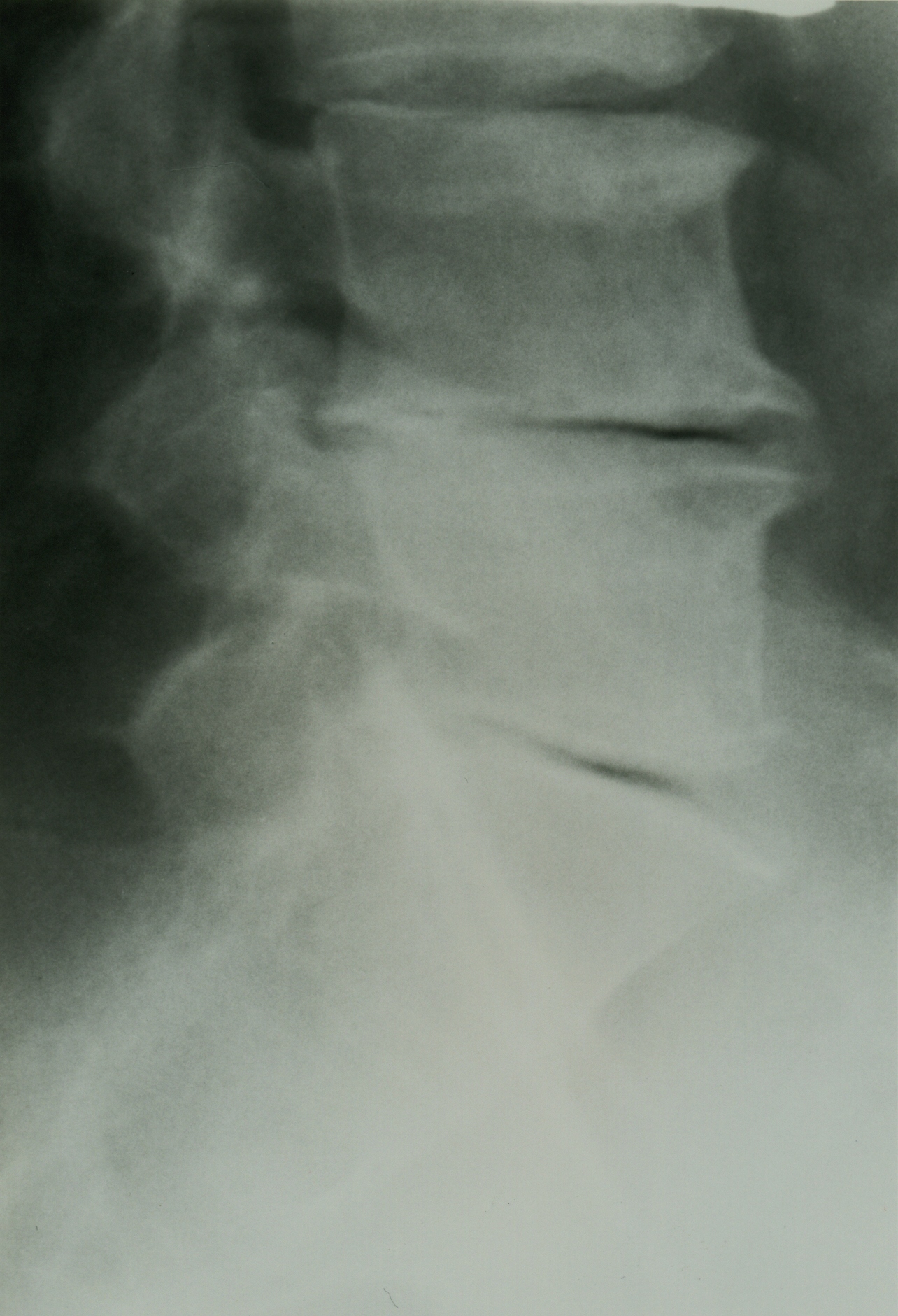
These results scream out to the profession that we, as a profession, do not understand MRI, a critical practice tool, as much as we need to. In 2009, an integral part of our diagnostic ability is predicated on MRI’s and they are all not alike, nor are MRI companies compelled to follow quality standards, in either performing the test or reporting the results, thereby often rendering blind results with potentially false negatives at the peril of the chiropractor and his/her patients. These false negatives are not a result of the radiologist missing a finding on a film but, many times, result from improper imaging protocols with slices that are “too thick.” Not improper, just clinically non-diagnostic and not regulated.

These false negatives can be eradicated if the chiropractor understands MRI protocols and mandates the correct image sequences and slices in his/her orders. This can only be accomplished with education and it is the responsibility of each individual chiropractor to get the training required to ensure an accurate MRI scan and results. Too many imaging companies, due to economic pressure, will cut corners on these blind items and compromise the results, leaving us in the dark, and that is our fault as much as theirs if we are ignorant.

Many will opine that it is a fault of our professional education that we do not have a greater understanding of MRI and they couldn’t be more wrong. The purpose of our formal education is to give us a starting point to care for our patients and to be able to attain licensure by meeting the minimum national and state standards of practice. It is the responsibility of each individual to then take it to the next level. DD and BJ had the big idea. However, if you study history, you will find that BJ Palmer did more research in concert with his medical counterparts than most, if not all, in contemporary research. He created his own team and maintained control of all chiropractic decisions, as you should.

Twenty years ago when MRI became readily available, there were no courses for chiropractors to learn how to read images and to teach them what protocols to order. I learned how to read the hard way; every time I ordered an MRI, I would go to the radiology office and ask them to show me what they were diagnosing and have them explain the anatomy and the pathology to me. After 100 to 200 patients and lots of trip to the radiologist, I became proficient in interpreting my own MRI films. Although an ineffective way to learn (formal training is much better), it opened my eyes on how to interpret MRI images, understand protocols and gave me an appreciation for the neuroradiologist.

The solution is obviously education and creating your own team. The time has long passed that the chiropractor needs to be part of the medical team. That notion is so long over, it should be dead! Each chiropractor needs to create their own team that includes the medical specialist, when needed. As a result, the clinical decision making is not abdicated to the MD, who has no training in chiropractic, and allows us to maintain full control of our patients using our team for input. Without the requisite training in understanding image protocols and the basics of reading images, we are left in the above paradigm that the survey results brought out and then are forced to be part of the medical specialist’s team, because we will not have the necessary knowledge, mandating someone else, usually the MD specialist, to make the tough decisions and dictate to us the care of our patients.

Does this mean we need to become radiologists? No, we need the knowledge and the credentials in reading MRI images and then we can utilize the radiologist as part of our team, to confirm what we are seeing. This brings us to the first member of our team, the radiologist, or more specifically, the neuroradiologist, who is a radiologist that spends an additional one to two years in training focused solely on brain and spine. I not only choose to be the best-of-the-best in clinical excellence through hard work; I also choose to only work with the best-of-the-best and the neuroradiologist is it.

As a note, once I started to interpret my own MRI’s, I had numerous disagreements with the general radiologist’s conclusions and, after bringing the neuroradiologist onto my team, I rarely had a disagreement.

The second member of my team is the neurosurgeon. If there is a space occupying lesion, such as a herniated disc on the cord or root, I confer with the neurosurgeon to ensure an accurate diagnosis, so I can make the clinical decision for my patient. There is no need for “gray areas” in disc-related diagnosis with today’s technology and your ability to assemble your team. This ensures an accurate prognosis and treatment plan.

Over time, the medical specialists will see you as their equal, based upon your clinical excellence and the feedback your patients are giving them in consultation. In the end, this will equate to increased referrals and help to eradicate any negative stigmas that some of the “medical fringe” still harbor towards chiropractic. When you are in conference with co-treating doctors, either primary care or specialists, and you know as much or more than they do, it is only a matter of time before they will be reaching out to you for the solution to their patient’s non-surgical spinal-related care.

This is how to build a referral network from the medical community. This is another example of how the only way to win for a lifetime is through clinical excellence. All it takes is a commitment to learn and the direction is clear. Once you have that level of knowledge with the understanding that the learning is ongoing, all that remains is how you communicate with your patients and community.

The message is simple; get the knowledge, the credentials, create your own team and be in control.

Dr.-Mark-StudinDr. Mark Studin is the President of CMCS Management which offers the Lawyers Marketing Program,Family/MD Marketing Program and Compliance Auditing services. He can be contacted at www.TeachChiros.com or call 1-631-786-4253
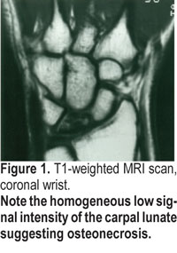
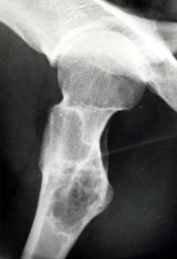