A Case of RA/DISH

Discussion:


RA/DISH is a disorder involving two conditions: rheumatoid arthritis (RA) and diffuse idiopathic skeletal hyperostosis (DISH), which is also known as Forestiers disease or ankylosing hyperostosis.1,3,5

RA is considered the most common inflammatory arthritide.1 It is a systemic connective tissue disorder that primarily affects the synovial lined joints.1 It is characterized by an inflammatory, hyperplastic synovitis (pannus formation), which causes cartilage and bone destruction ultimately leading to loss of joint function.1 It typically affects the small joints of the body, i.e., hands, wrists, elbows, shoulders, feet, knees and hips.1,5 It is usually seen bilaterally and affects the joints symmetrically.1,5 The onset of RA is generally between the ages of twenty and fifty and affects women more often than men.1,5 When RA is seen in older individuals above sixty years of age, the ratio of occurrence between men and women becomes almost equal.1 Generally speaking, rheumatoid arthritis rarely affects the axial skeleton, but when it does, the cervical spine is the most common region of involvement, resulting in anterior translation (movement) of the atlantoaxial complex 9.5 percent to 36 percent of the time.1 This forward translation is most often caused from erosion of the odontoid process or transverse ligament laxity from pannus formation.1 Laboratory tests such as the rheumatoid factor (RF) assist in confirming a diagnosis. A positive rheumatoid factor is usually seen in 70 to 80 percent of the patients with rheumatoid arthritis.1 Other laboratory tests such as a complete blood count (CBC), erythrocyte sedimentation rate (ESR) and anti-nuclearantibodies (ANA) assay may help to confirm and evaluate patients with rheumatoid arthritis.1

DISH is classified as a rheumatological abnormality characterized by marked proliferation of bone where the ligamentous and tendinous attachments to bony sites are found.1 DISH can affect the spine or extraspinal areas, but tends to develop most commonly in the anterior longitudinal ligament (ALL) of the spine.1 It tends to affect males more than females and is usually seen in patients over the age of fifty.5 The typical radiographic findings are characterized by large flowing ossifications, sometimes referred to as candle wax drippings, located along the anterolateral aspect of the vertebral bodies of at least four contiguous vertebrae, with relative preservation of the disc heights and absence of facet ankylosing and sacroiliac erosion/fusion.1,5 It should be noted, as a result of the large flowing ossification about the anterolateral aspect of the vertebral bodies, secondary complications of dysphagia can arise in approximately 20 percent of the patients, causing displacement of the pharyngeal air shadow.5 In addition, there is a higher incident of patients diagnosed with DISH having associated diabetes mellitus, approximately 13-49 percent of the time.1 Laboratory tests are generally unremarkable; however, patients with DISH tend to show hyperinsulinemia following glucose challenge tests.1,5 Evaluation of the HLA B8 antigen may be positive in approximately 40 percent of the patients with DISH.5


Differential Diagnosis:


Differential diagnoses should include other more common inflammatory arthritides like anyklosing spondylitis (AS) also known as Marie-Strumpell’s disease, as well as other inflammatory arthritides like psoriatic and Reiter’s arthritis.5 In addition, a common non-inflammatory arthritide like degenerative joint disease (DJD), also known as osteoarthritis or wear and tear disease, should not be overlooked as a possible differential diagnosis.5


Radiographic Findings:


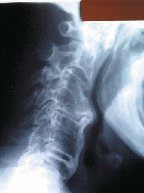
The radiographs being depicted are those of an eighty-nine-year-old male patient who consulted me because of pain, stiffness, spasm and restricted range of motion in his cervical and cervicothoracic spine.

The patient’s X-rays revealed an increase in the atlantodental interspace (ADI), which normally should measure no greater than 3mm in an adult and 5mm in a child.1,5 Also noted is the presence of forward displacement of the posterior cervical line, prominent flowing bone spurs (candle wax drippings) and fusion of several vertebral segments along the anterior longitudinal ligament. The articular facets and many of the disc spaces of this patient’s cervical spine are still preserved. Reference X-rays for these abnormal findings.


Treatment:


Treatment for RA/DISH should be directed toward obtaining meaningful symptomatic improvement, because there is no known cure for these two disorders.2 Patients afflicted with RA and/or DISH require that treatment goals be directed toward relieving pain, spasm, inflammation and trying to slow down or stop joint damage, as well as improve the patient’s sense of well-being and his ability to function.2,4 Medical physicians usually prescribe medication to help control these symptoms.2,4 Physical therapy, along with mild non-strenuous movements and exercises, may also help stabilize and strengthen joints and muscles to help to increase articular range of motion.2,4 Goals should also include emphasizing that patients obtain adequate rest and exercise.2,4 The X-rays depicted in this case revealed significant osseous changes resulting from DISH, along with anterior displacement of C1 from RA, which precluded any osseous manipulation or adjustments from being administered to this patient’s cervical spine. However, in carefully selected patients without significant upper cervical pathology, where there is no concern for injury to the spinal cord or other neurological elements, gentle and measured manipulation may be an option the chiropractic physician would want to consider.

Physiotherapy modalities, such as electro-muscle stimulation, ultrasound, hydrocollator therapy, soft tissue manipulation, trigger point therapy and light stretching, may be of help in reducing the patient’s symptomatology. It is prudent to remember, treatment should always be designed to fit the patient’s needs rather than the patient being required to fit the therapy.

All or some of the above procedures, depending upon the stage of the patient’s RA/DISH, may be of clinical benefit in ameliorating the patient’s active symptoms, which is a result of these two disorders. Stress reduction techniques can be helpful in assisting the patient in dealing with the emotional and physical challenges patients experience with these disorders.4

In some cases, where severe joint damage has occurred, surgery may be required to reduce pain and improve joint function, which may also help improve the patient’s ability to perform his normal activities of daily living. The procedures to accomplish this may include joint replacement, tendon reconstruction and synovectomy.4 In rare cases involving DISH, where ossification of the anterior longitudinal ligament (ALL) becomes so significantly calcified wherein it causes the patient difficulty with swallowing, the patient may need to undergo surgery in order to remove the bony spurs.2 In any event, the patient’s age, health status and stage of his disease would determine the most appropriate course of treatment.

Dr. Paul Sherman practiced in New Jersey for sixteen years. Presently, he is an assistant professor of clinical sciences and a post-graduate faculty instructor at the University of Bridgeport College of Chiropractic, Bridgeport, Connecticut. He is also an instructor and writer for Chirocredit.com, a chiropractic continuing education website. He can be reached at [email protected].


References

1. Marchiori, Clinical Imaging with Skeletal, Chest and Abdomen Pattern Differentials, Mosby, 1999

2. MayoClinic.com article on diffuse idiopathic skeletal hyperostosis, May 2006

3. Resnick and Niwayama, Diagnosis of Bone and Joint Disorders, 2nd Edition, WB Saunders, 1988

4. U.S. Department of Health and Human Services, National Institutes of Health (NIH) Publication No. 04-4719, May 2004

5. Yochum and Rowe, Essential of Skeletal Radiology, 2nd Edition, Williams and Wilkins, 1996


The Cervical Acceleration / Deceleration Practitioner

So, you want to treat patients that have suffered a cervical acceleration/deceleration (CAD) trauma. Then you must learn to examine and treat a CAD injured person properly and with the proper tools. First of all, if you have never treated these types of injuries before, then you should arm yourself with the knowledge of the etiology of CAD trauma. Courses like those given by Dr. Arthur Croft (CRASH) and Dr. Dan Murphy (CCST) can help you understand the mechanics behind the injury. But this, alone, won’t help you. Imagine that you have a real-life patient that is hurting in front of you and you need to evaluate that patient in such a way that you take that patient’s subjective symptoms and turn them into objective evidence—FACTS, the type of facts that a jury can understand and use in order to make an informed verdict.

Let’s take a look at a typical CAD injury evaluation. It looks like this in its simplest form:

HISTORY  EVALUATION  DIAGNOSTIC TESTING

According to the above examination flow chart, the doctor takes the history of the injury and records the appropriate responses that lead to the mechanism of injury. The history should focus on the patient’s past and current history so that the doctor can apply the latest evidence in the form of research studies to formulate the answer to questions on whether or not this injury could have happened—questions that relate to prior accidents, size of vehicles, seatbelts on/off, loss of consciousness, etc.

The doctor then performs the standard cervical evaluation by performing the needed orthopedic examination, neurological examination, ROM and muscle tests. This paints a picture as to how the patient was doing physically when the patient was initially examined. However, the examination process is very subjective and should not stop there. The doctor should take the subjective examination results and examine them in an objective manner by utilizing diagnostic tests.

All of the subjective complaints and tests that were found during the evaluation process need to be evaluated by utilizing diagnostic tests. The diagnostic tests that are chosen should be reliable and reproducible for what they are examining. For instance, the Guides to the Evaluation of Permanent Impairment, 5th edition, (AMA Guides), states that ROM testing should be evaluated utilizing dual inclinometry. Dual inclinometry is the most accurate and reliable means to perform ROM tests. The research literature also states that computerized duel inclinometry is the gold standard along with computerized muscle testing.1,2,3 Diagnostic tests then serve two purposes. Firstly, diagnostic tests are able to document the injury of the patient and, secondly, they are able to provide information that can be readily retested so that the doctor can prove whether or not the patient is improving, staying the same or getting worse. The subjective complaints and the diagnostic tests that should accompany them are listed in Table 1.

TABLE 1

Subjective Complaint
Diagnostic Tests That Should Accompany Them
Pain OA Questionaires (Oswetry, Neck Pain, Roland Morris, VAS, Headaches Disability Index), Pain Presure Treshold Testing (Algometry)

Numb/Tingling/Burning Pain

NCV testing, EMG testing, DSEP Testing
Disc Lesions
MRI
Fractures CT, Radiographs
Loss of Range of Motion

Loss of strength
cMT Testing
LMS Radiographs


Questionnaires provide a reliable means of documenting the patient’s level of pain and its impact on their activities of daily living (ADL’s).4,5 Questionnaires such as the Neck Pain Disability Index and the Rand-36 should be utilized. Even though they are considered to be subjective, they are a high form of subjective data. So high, in fact, that insurance companies such as United Healthcare with ACN require that the patient complete a health-oriented questionnaire to pre-authorize care and every time there is a request for more visits.

Nerve conduction velocity (NCV), electromyography (EMG), and dermatomal somatosensory evoked potential (DSEP) testing provide a valid and reliable means of documenting the patients neurological pain.6 The AMA Guides place objective neurological findings in a DRE Category III 15-18 percent Impairment of the Whole Person (AMA Guides Table 15-5). Foreman and Croft place the patient in a Grade III Major Injury Category (MIC) when there are neurological findings present. This is significant, because the treatment recommendations for a patient in a Grade III MIC are seventy-six visits or more. Therefore, the significance of performing neurological tests and other diagnostic tests on your patients goes beyond your simply evaluating the patient. You are able to create an objective treatment plan, a treatment plan that is not based on your opinion but based on the objective data and placed in a set of guidelines (Whiplash Injuries, 3rd edition, by Foreman and Croft).

MRI studies are very useful in evaluating the patient for disc lesions. Computed tomography (CT) is very useful in evaluating the skeletal structures for fractures, especially those that are missed by radiographic analysis. MRI studies that demonstrate a herniated disc at the level and on the side that would be expected for a subjective radiculopathy equates to a DRE Category II 5-8 percent Impairment of the Whole Person, according to Table 15-5 of the AMA Guides.

Computerized range of motion testing (cROM) is very useful in determining the degree of the loss of cervical function. ROM testing is able to reliably indicate a physical impairment in people suffering from chronic whiplash.2

Computerized muscle testing (cMT) is an excellent way of determining a loss of muscle function, because it is not based on a subjective standard. In order to reliably determine that a patient has a decrease in muscle strength, the patient must have at least a 35 percent loss in strength. This means that a doctor would not be able to reliably determine a loss in strength if it is only a 25 percent loss. Muscle strength should be compared from left and right measurements and they should not exceed more than 10 percent.

Radiographs should be taken and then digitized to examine for motion segment integrity loss (MISL) as defined by the AMA Guides. The AMA Guides state that the only way to evaluate a patient for MSIL is by utilizing the flexion/extension films and measuring for an increase in translational or rotational movement. Translation and rotation are biomechanical terms. Translation is movement of a body in straight line and should not exceed 3.5mm in the cervical spine. Rotation is defined as movement of body about a fixed point and should not exceed 11 degrees in the cervical spine (AMA Guides Table 15-5). These are significant findings according to the AMA Guides because, if the patient has MSIL, as defined by the flexion/extension radiographs, then the patient is placed in a Category IV 25-28 percent Impairment of the Whole Person. This equates to the same thing as a greater than 50 percent compression fracture of a vertebral body. What does this mean clinically? It means that you would not adjust someone at C5 if they had a 50 percent fracture of the C5 vertebrae and, therefore, you should not adjust someone that has an increased motion segment at C5 either. The only objective way to determine the MSIL is by having the radiographs digitized. This allows an outside source to determine the injury and how bad that injury is. If the defense attorney tries to refute the findings, he looks very incompetent in front of the jury because he is arguing against the facts.

As you can imagine, the jury likes to have concrete facts of the case to make an informed decision. This is why shows like CSI are very popular right now. For those of you that get frustrated at the thought of treating personal injury cases, it’s probably due to the fact that you are not collecting all of the objective evidence and providing only subjective data to the attorneys. Give the plaintiff’s attorney facts to argue with, not opinion. Believe me, there will be enough opinions flying out of the defense attorney. Get the equipment that will objectively gather the data, like computerized ROM testing and computerized muscle testing and algometry. The proper treatment protocols for treating CAD injuries can be found in Foreman and Croft’s, Whiplash Injuries, 3rd Edition, and the AMA Guides, 5th Edition. If you are not following the protocols set forth in these two texts, you are going to become increasingly frustrated. A very good friend of mine once stated, “Learn the rules, play by the rules, and win by the rules.”

Dr. Dwight C. Whynot is in fulltime practice in Johnson City, Tennessee. Dr. Whynot gives license-renewal lectures on Evidence-Based Chiropractic Practices which are promoted by the International Chiropractors Association and sponsored by Myologic and Spinal-logic Diagnostics. For questions regarding evidence-based practice procedures, email questions to [email protected].

For 12-hours CCE license renewal lecture dates and places call the ICA at 1-800-423-4690. For more information on Myologic or Spinal-logic, go to www.myologic.com or www.spinallogic.com.

References
1. Bohannon RW, Andrews AW. Standards for Judgments of Unilateral Impairments in Muscle Strength. Perceptual and Motor Skills 1999, 89, 878-883.
2. Dall’Alba P., Sterling M., Treleaven J., Edwards S., Jull G.. Cervical Range of Motion Discriminates Between Asymptomatic Persons and Those With Whiplash. Spine 2001; 26; 2090-2094 (October 1, 2001)
3. Jasiewicz J., Treleaven J., Condie P., Jull G.. Wireless Orientation Sensors: Their Suitability to Measure Head Movement for Neck Pain Assessment. Manual Therapy. September 2006.
4. Ware JJ, Sherbourne CD. The MOS 36-Item short form survey (SF-36). I. Conceptual framework and item selection. Medicare 1992; 30:473-83.
5. Hsieh JCY, Phillips RB, Adams AH, Pope MH. Functional outcomes of low back pain: comparison of four treatment groups in a randPhysical Medicine Rehabilitation 80:1273-1281, 1999omized controlled trial. Journal of Manipulative & Physiological Therapeutics 1992; 15(1):4-9
6. Haig AJ, Tzeng H-M, LeBreck DB. The value of the Electrodiagnostic Consultation for patients with upper extremity nerve complaints: A Prospective Comparison with the History and Physical Examination. Archives of Physical Medicine Rehabilitation 80:1273-1281, 1999.

Osteitis Pubis

HISTORY:

Both of these female patients are complaining of focal pain at the pubic articulation. One of them developed this pain after a difficult delivery of a child and the other patient after surgery for a retroverted uterus.


Figures 1 & 2. Characteristic changes are visible at the pubic symphysis with subchondral sclerosis, articular erosions, small osteophyte formation and a slight offset of the pubic bones. This radiographic appearance is characteristic of osteitis pubis.

Discussion:
Osteitis pubis is a painful condition of the pubic symphysis articulation characterized by bony resorption and spontaneous reossification. Although the pathogenesis is uncertain, the most common related antecedent event is surgery within close proximity to the symphysis.1

Clinical Features
Onset of signs and symptoms is usually within one to three months after surgery in the locality of the symphysis pubis articulation. The frequency of this postsurgical complication is between 1 percent and 3 percent. The most common types of surgery are for the prostate, bladder, urethra, uterus and cervix. Statistically, prostate surgery is the most commonly associated surgical procedure. Additional causes include pregnancy, trauma, and, often, unknown factors.1

Symptomology may be localized or referred and is usually described as groin burning.2 Pain is often excruciating on direct palpation. Exercise or activities involving thigh adduction, trunk flexion or even walking may refer pain to the perineal, testicular, suprapubic or inguinal area. In addition, an audible click in the area of the pubic symphysis may be heard during these activities. Postejaculatory pain referral to the scrotum and perineum has been noted in males. The symptoms are generally relieved by rest. Redness or heat is usually not present. The gait is antalgic, with trunk flexion and waddling to prevent symphyseal stress.

Pain typically subsides over an indefinite period up to one or two years, but may require arthrodesis.1,3 With persistent pain and biomechanical alterations in gait, early sacroiliac degenerative changes could ensue.2
Radiologic Features

There is a radiographic latent period after the onset of symptoms of one to three weeks; however, some patients will never manifest definitive radiologic changes. When present, the findings may simulate joint infection.1,2

The most characteristic radiographic appearance is a bilateral and usually symmetric involvement of the pubic bone and adjacent rami. Irregularity of the joint margin, subchondral sclerosis, and a moth-eaten type of osteoporosis, with widening of the joint space can be striking. 3 With resolution, there is reconstitution of normal bone density, but the joint margin frequently remains irregular and may even be ankylosed.1


References
1. Yochum TR, Rowe LJ: Essentials of Skeletal Radiology, 3rd ed., Williams & Wilkins, Baltimore, Maryland, 2005.
2. Cibert J: Post-Operative Osteitis Pubis Cause and Treatment, Br. J. Urol, 24:213, 1952.
3. Pauli S, et al., Osteomyelitis Pubis Versus Osteitis Pubis, Br. J. Sports Med. (1):71, 2002


Dr. Terry R. Yochum is a second generation chiropractor and a Cum Laude Graduate of National College of Chiropractic, where he subsequently completed his radiology residency. He is currently Director of the Rocky Mountain Chiropractic Radiological Center in Denver, Colorado, and Adjunct Professor of Radiology at the Southern California University of Health Sciences, as well as an instructor of skeletal radiology at the University of Colorado School of Medicine, Denver, CO. Dr. Yochum can be reached at 303-940-9400 or by e-mail at [email protected].

Dr. Chad J. Maola is a 1990 Magna Cum Laude Graduate of National College of Chiropractic. Dr. Maola is a Chiropractic Orthopedist and is available for post-graduate seminars. He may be reached at 303-690-8503 or e-mail [email protected].

Vacuum Phenomenon

DISCUSSION
The vacuum sign (of Knuttson) is an important early radiographic finding.1 Essentially, this represents collections of nitrogen gas in nuclear and annular fissures and presents as an area of linear radiolucency in the disc space.2  Studies have shown this to be a common sign of disc aging and degeneration, with an incidence of two to three percent in the general population.2 The collection of nitrogen in the discal fissures is thought to originate from adjacent extracellular fluid. 

In movements of the spine that produce a lowered pressure in the disc, such as in extension, nitrogen is released from the adjacent extracellular fluid and, due to the pressure gradient, accumulates in the discal fissures.  On Magnetic Resonance imaging, the disc shows diminished signal intensity due to dehydration, and a signal void at the vacuum site.  This collection of gas can be made to disappear with spinal flexion and reappear with spinal extension.1,2  Disc infections do not demonstrate this sign, due to fluid collections in the fissures.1,2 Central vacuum phenomena correspond to fissuring of the nucleus pulposus, while peripheral lesions represent rim lesions where the annulus fibrosus has been disrupted from its attachment to the vertebral body margin.2

In the peripheral joints, especially the hip, shoulder, and knee, a vacuum sign does not denote degenerative joint changes.2  This is produced as an accompanying physiologic phenomenon, usually induced by the position of the patient in a position of traction when the exposure was made.  Gas in the symphysis pubis is a normal finding during pregnancy and up to three weeks postpartum and may be seen as a vertical, thin radiolucency.

As the disease process progresses in the disc, the degenerative signs become more severe.  Subluxation phenomena are more readily recognizable, and lateral, anterior, and posterior vertebral body displacements of a measurable degree occur.  Flexion/extension films usually reveal decreased motion in these displaced segments.  Disc height is markedly diminished, with greater than twenty-five percent loss of its vertical dimension.  Loss of disc height can also be due to infection, which should be excluded by careful scrutiny for the loss of the vertebral body endplates.2

Dr. Terry R. Yochum is a second generation chiropractor and a Cum Laude Graduate of National College of Chiropractic, where he subsequently completed his radiology residency.  He is currently Director of the Rocky Mountain Chiropractic Radiological Center in Denver, Colorado, and Adjunct Professor of Radiology at the Southern California University of Health Sciences, as well as an instructor of skeletal radiology at the University of Colorado School of Medicine, Denver, CO. Dr. Yochum can be reached at 303-940-9400 or by e-mail at [email protected].

Dr. Chad J. Maola is a 1990 Magna Cum Laude Graduate of National College of Chiropractic.  Dr. Maola is a Chiropractic Orthopedist and is available for post-graduate seminars.  He may be reached at 303-690-8503 or e-mail [email protected].

Librarian Overdue for Pelvic Pain Relief

hipimbalanceHistory and Presenting Symptoms

A 37-year-old female describes a history of occasional pain in her lower back region, most noticeable on the left side.  She states that her low back pain “just seems to come and go,” with no obvious triggering activities.  Her pain is localized to the posterior hip region, and does not extend below the pelvis.  She is a recreational runner who usually enjoys hiking, snowboarding, and occasional soccer games with friends, but she denies any specific injury or trauma to her lower back.  On a 100mm Visual Analog Scale, she rates the pain in her lower back and pelvic region as varying from 30mm to 50mm.  She takes over-the-counter NSAID’s when the pain in her lower back interferes with her daily activities or her job duties as the manager of the local library.

Exam Findings

Vitals.  This active female weighs 162 lbs. which, at 5’10’’, results in a BMI of 23—she is not overweight or obese.  She reports that she works out regularly on the resistance machines at a local exercise center, and runs at least twelve miles each week.  She is a non-smoker, drinks wine moderately, and her blood pressure and pulse rate are within normal ranges (BP: 118/76 mmHg; pulse rate: 64 bpm).

Posture and gait.  Standing postural evaluation finds evidence of a lower iliac crest on the right, but the greater trochanters are level.  The left ilium is rotated forward, with prominence of the left ASIS.  Her knees and ankles are well aligned, but there is obvious medial bowing of both Achilles tendons, with pes planus and hyperpronation bilaterally.  During gait, both feet demonstrate a moderate toe-out.  Inspection of her shoes finds scuffing and wearing at the lateral aspect of both heels.

Chiropractic evaluation.  Motion palpation identifies limitations in segmental motion at the left SI joint, with localized tenderness.  The left SI joint demonstrates loss of end range mobility, and pain is reported during motion testing.  Gaenslen’s and Yeoman’s tests for SI joint dysfunction both cause increased pain when performed on the left side.  Lumbar ranges of motion are within expected norms, and neurologic testing is negative for sensory, motor, and reflexive disorders.

Imaging

A lumbopelvic series (AP and lateral lumbopelvic views) is taken in the upright position during relaxed standing.  The sacral base angle is 38° and the lumbar lordosis measures 46°.  There is no significant discrepancy in femur head or iliac crest heights, and no lateral listing or lateral curvature is noted.

Clinical Impression

Moderate lumbopelvic misalignment, with chronic mechanical dysfunction of the left sacroiliac joint.  Poor biomechanical support is noted in the lower extremities, which exacerbates her lumbopelvic dysfunction syndrome.

Treatment Plan

Adjustments.  Specific chiropractic adjustments for the lumbosacral and sacroiliac joints were provided as needed.  Side-posture adjustments were well-tolerated and resulted in good articular releases.

Support.  She was fitted with custom-made, flexible stabilizing orthotics, based on foot imaging in mid-stance.  The inserts were designed to provide support for her arches and decrease the biomechanical stress on her pelvis and sacroiliac joints during the entire gait cycle.

Rehabilitation.  She was instructed in a daily core strengthening program at home using elastic exercise tubing.  The focus was on activation of her transverse abdominis musculature, for improved spinal-pelvic stability.

Response to Care

This patient responded well to her spinal adjustments, and she adapted very quickly to her orthotics.  She performed her daily home exercise program regularly, and demonstrated good exercise performance at her weekly rehab review sessions.  After eight weeks of adjustments (twelve visits) and daily home exercises, she was released to a self-directed maintenance program.

Discussion

This patient had a chronic pelvic misalignment, which was associated with pronation and biomechanical dysfunction in the lower extremities.  Her chiropractic treatment plan included orthotics to support her strained lower extremities, and specific exercises to improve her core pelvic stability.  She responded well to her adjustments, but also needed support from the orthotics and professional guidance for her corrective exercises.

Dr. John J. Danchik is the seventh inductee to the American Chiropractic Association Sports Hall of Fame.  He is the current chairperson of the United States Olympic Committee’s Chiropractic Selection Program and lectures extensively in the United States and abroad on current trends in sports chiropractic and rehabilitation.  Dr. Danchik is an associate editor of the Journal of the Neuromusculoskeletal System.  He can be reached by e-mail at [email protected].


What Practice Structure Suits You Best? Starting a New Practice V

Solo Practitioner (No Incorporation)
This type of practice structure is when a doctor is not incorporated.  In this case, the doctor does not have the personal asset protection advantage that a corporate shield has to offer.  Being non-incorporated, if someone files a malpractice suit against the doctor, they will not only be after all his business assets (equipment, accounts receivables, checking/savings accounts, insurance coverage), they will also be able to easily attach claim to his personal assets (home, furniture, vehicles, savings accounts, checking accounts, stocks, bonds, etc.).

Obviously, practicing solo (unincorporated) is very risky.  The average doctor will have three malpractice cases filed against him during his career.  Are you going to leave all your assets exposed?  I hope not.  There is no sure thing when it comes to lawsuits, but a corporate shield is usually effective in protecting your personal assets from business related claims.  Therefore, incorporating your business is strongly recommended. 

Partnerships
What is your motive for seeking a partner?

The two main reasons people seek a partnership are:

1. Sharing the stress. Starting a new practice on your own is extremely stressful. However, if you choose a partnership, you are setting yourself up for even more stress when the partnership inevitably fails.

2. One doctor wants the use of another doctor’s money. Remember, the person who puts in most money will want—and demand—most of the control.

Do you really want a partner?  Consider the following statistics:

•  70% of partnerships break up within two years.
•  90% of partnerships end in divorce.
•  Partners never share the same passion, work ethics, work  habits, management views, etc.
•  A divorce in business can be just as traumatic and expensive as one in marriage.

The break-up of your partnership will undeniably confirm that entering into a partnership was the most expensive mistake you ever made.  
 
If You Are Determined to Start a Practice with a
Partner, Follow this Rule
Sign a partner separation settlement agreement before you start your practice. You need to be able to end the partnership in a non-threatening manner. Decide, in advance, who gets the account receivables, X-ray equipment, adjusting tables, therapy, who leaves the facility, etc.

Don’t fool yourself into thinking that you don’t need this kind of agreement because you’re going into practice with your best friend.  There is a tremendous amount of truth and warning in the phrase “best friends make the worst enemies.”  If you execute a partner separation settlement agreement in advance, your best friend will probably remain a friend. It’s like having a roll bar on your Jeep. You hope you’ll never need it but, if you do, you’ll be happy it’s there.

Five Reasons Why Partnerships Blow Up
1. One partner wants to build the practice faster, work harder, and practice more hours than the other partner. The other partner works less, but still wants to share the income fifty-fifty. It won’t work.

2. One partner hires a spouse or relative to work in the practice. How can the other partner say, “No.”  This always causes problems.

3. One partner takes cash from the cash drawer; the other partner does not appreciate being “stolen” from.

4. One partner becomes disabled. The other partner covers his practice for six weeks to ninety days. Any longer and the partnership will end in a divorce.

5. One partner wants to gouge insurance companies; the other partner is ethical. The ethical DC will end the partnership.

Advantages of a Partnership
The advantages of a partnership are sharing responsibilities, sharing expenses, and covering for each other.

Disadvantages of a Partnership
A major disadvantage of a partnership is that almost all partners are considered equally liable for the other partner’s mistakes. Depending on the type of partnership, if your partner is sued for malpractice, you will also be sued. If one partner gets a bad reputation with insurance companies, the same brush will paint the other partner. If one partner is over-friendly with patients of the opposite sex, the other partner’s reputation will also be tarnished.  There will also be arguments over money when one doctor is generating more income or contributing more toward the growth of the practice than the other. 

Should you incorporate?
Talk to your accountant—he’s the expert on your particular tax situation. As previously discussed, an advantage of having a corporation is that it usually limits your liability. If a creditor sues your corporation, they can go after the assets of your corporation, but probably will not be able to go after your personal assets. There are also a few possible tax advantages, i.e., you may be able to deduct your health insurance premiums, create a pension plan, buy your automobiles through your corporation, etc. However, it’s usually more expensive to run a corporation than it is to practice solo, and there’s definitely less flexibility of operation.

Three Types of Corporations
1. A Professional Corporation (PC) or Professional Association (PA). These corporations can only be owned by professionals.  Most states that have PC or PA corporations require professionals to use these types of corporations.

2. An incorporation (Inc.) – If it’s legal to practice under an “Inc.” in your state, you may be able to hire MD’s as employees. Non-DC’s may be able to own a portion or all of the corporation.

3. A “Limited Liability” Corporation (LLC). This is a fairly new type of corporation in which your liability is usually limited to the amount of money you have invested in the corporation.  Many attorneys prefer LLC’s because of their liability protection.  However, most accountants don’t recommend LLC’s because of increased accounting expenses.

Again, check with your accountant to determine if you should incorporate and, if so, what type of corporation would best suit your needs.

When you’re through being an associate and are ready to start your own practice, hire a consultant who specializes in starting practices to guide you.  Don’t think that you now have the experience necessary to start and build a new practice…you don’t!  Yes, you’ve gained the experience of caring for patients and learned some good office procedures, but that’s not enough knowledge to start a successful new practice.  You still have to learn how to find a great office location, acquire effective bank negotiating strategies and cost-cutting remodeling negotiations, as well as how to market a new practice, etc.  It’s the lack of this specialized knowledge that dooms new practices, not the lack of knowledge regarding patient care.

Next in this series on “How To Start A Practice,” I’ll discuss whether or not you should start a practice by entering into an independent contract arrangement.

Dr. Peter G. Fernandez is the world’s authority on starting a practice.  He has 30 years’ experience in starting new practices, has written four books and numerous articles on the subject, and has consulted in the opening of over 3,000 new practices.  Please contact Dr. Fernandez at 10733 57th Avenue North, Seminole, Florida 33772; 1-800-882-4476; [email protected] or visit www.drfernandez.com.


Heavy Metal Poisoning

DISCUSSION

The major metals involved in producing visible radiologic changes are lead, phosphorus, and bismuth. Of these, lead is the most frequent, but lead intoxication is still a rare skeletal disorder.

Lead may be ingested, inhaled, or implanted. Clinical symptoms occur abruptly, with abdominal pain, encephalopathy, and paralysis. Radiologically, the most definitive signs are the linear, transverse densities at the metaphyses (lead lines). The deposition of lead may also precipitate remodeling abnormalities. Phosphorus and bismuth exhibit similar changes.

Workers involved in the polymerization of polyvinyl chloride (PVC) may develop a peculiar form of acroosteolysis.

Dr. Terry R. Yochum is a second generation chiropractor and a Cum Laude Graduate of National College of Chiropractic, where he subsequently completed his radiology residency. He is currently Director of the Rocky Mountain Chiropractic Radiological Center in Denver, Colorado, and Adjunct Professor of Radiology at the Southern California University of Health Sciences, as well as an instructor of skeletal radiology at the University of Colorado School of Medicine, Denver, CO. Dr. Yochum can be reached at 303-940-9400 or by e-mail at [email protected].

Dr. Chad J. Maola is a 1990 Magna Cum Laude Graduate of National College of Chiropractic. Dr. Maola is a Chiropractic Orthopedist and is available for post-graduate seminars. He may be reached at 303-690-8503 or e-mail [email protected].


Vertebra Plana: What Is the Real Cause?

History

This 12-year-old male patient presents with neck pain, following heading a soccer ball.

Discussion

Eosinophilic granuloma is the least severe but most common (60-80%) of all the histiocytoses.1,2 Peak incidence comes between the ages of 5 and 10 years and 75% of cases occur in individuals less than 20 years of age. Symptoms primarily are localized pain and swelling of less than 2 months’ duration. Eosinophilic granuloma typically lacks the systemic features of Hand-Schüller-Christian or Letter-Siwe disease. Occasionally, pathologic fracture will be the presenting problem. Vertebral involvement is characterized by pain and, in some cases, complicating myelopathy may ensue secondary to cord and nerve root compression. Temporal bone involvement may produce otitis media-like symptoms, and the diagnosis of eosinophilic granuloma should be suspected when patients with suspected otitis media do not respond to traditional medical treatment.1

More than 50% of cases involve the skull, mandible (25%), spine (6%), pelvis (20%), and ribs (7%). Of the long bones, the femur (15%), tibia and humerus (8%) are involved most often. The bones of the hands and feet are affected rarely.2 Monostotic presentations are three times more common than polyostotic presentations.2 Monostotic lesions progress to lesions elsewhere in 20% of cases.1 Pain may be the primary symptom and the diagnosis often requires histopathologic analysis of the lesions, as well as the detection of S-100 CD1 antigens on immunohisto-chemical analysis of the tissues.

Spine

More than 50% of cases involve the thoracic spine; 35%, the lumbar spine; and less than15%, the cervical spine.1 Solitary vertebral involvement is more common, though multiple levels are occasionally involved. With cervical involvement, C2 is the primary target in adults, whereas the middle cervicals are normally affected in children.1,2

The vertebral body is usually involved with relative sparing of the neural arch structures. In contrast, cases have been reported of lesions affecting the posterior arch and lateral masses in the absence of vertebral body involvement; however, this is rare. An osteolytic lesion is the expected radiographic appearance.1,2

Neural arch involvement, when present, destroys the internal matrix, but preserves the cortical outline, which has been described as a ghostly appearance. The most prominent feature in the lumbar and thoracic spine is pathologic fracture, with dramatic loss of vertebral height as thin as 2 mm, involving both the anterior and posterior vertebral body surfaces (vertebra plana, silver dollar vertebra, coin-on-edge vertebra).1 This is rare in the cervical spine. Areas of destruction within the centrum may be observed before collapse. A short-segment kyphosis usually accompanies thoracic vertebral involvement. Paravertebral swelling can be prominent and is more likely to be associated with increased risk to the cord. On CT examination, osteolytic destruction can be seen, which simulates aggressive neoplasm; occasionally a sequestrum may be visible.

Bracing is the treatment of choice, and surgical intervention is rarely necessary. Restoration of height with healing is to be expected in at least 90% of cases and can be rapid over 1 year. The majority of cases reconstitute to 48-95% of normal height, especially in patients under 15 years of age at the time of onset. Residual sclerosis and trabecular accentuation always remain. Rarely, interbody fusion may occur after radiotherapy and, occasionally, there is a bone-within-bone appearance. Healed lesions do not appear to increase the risk for long-term back disability in adulthood.

Dr. Terry R. Yochum is a second generation chiropractor and a Cum Laude Graduate of National College of Chiropractic, where he subsequently completed his radiology residency. He is currently Director of the Rocky Mountain Chiropractic Radiological Center in Denver, Colorado, and Adjunct Professor of Radiology at the Southern California University of Health Sciences, as well as an instructor of skeletal radiology at the University of Colorado School of Medicine, Denver, CO. Dr. Yochum can be reached at 303-940-9400 or by e-mail at [email protected].

Dr. Chad J. Maola is a 1990 Magna Cum Laude Graduate of National College of Chiropractic. Dr. Maola is a Chiropractic Orthopedist and is available for post-graduate seminars. He may be reached at 303-690-8503 or e-mail [email protected].

The Upper Thoractic Hump Pattern

“Doctor, I’ve been worried about my hump. Will it get worse? Can you fix it?”

Unlike the better-known dowager’s hump, the upper thoracic hump (see photos and illustrations) is more common and is found in younger as well as older people.

Patients are concerned about their humps and are very motivated to get them fixed. They should be. Along with its unattractiveness, the hump affects the heart, lungs and thyroid.

Increased thoracic curve (hyperkyphosis) has even been linked to increased mortality. One research paper finds, “The hyperkyphotic posture was specifically associated with an increased rate of death due to heart disease.”1

The hump can also cause or contribute to hand, arm, shoulder, neck, lower back, sacrum and sciatica pain. Patients fear it is a harbinger to little-ol’-man/woman status, i.e. the dowager’s hump.

The classical dowager’s hump or dorsal kyphosis (forward curve in the mid and upper thoracic spine) is associated with old age and may involve wedged or triangular shaped vertebral bodies, compression fractures and osteoporosis. Undoubtedly, it is the end-stage of a chronic subluxation pattern and is probably related to the upper thoracic hump.

Subluxation patterns

Pattern: A consistent, characteristic form, style, or method, as:

  •  A composite of traits or features characteristic of an individual or a group: one’s pattern of behavior.2

In medicine, a “syndrome” denotes a group of symptoms that characterize a disease condition. Chiropractic, being a vitalistic healing profession rather than disease and symptom oriented, refers to a group of subluxations as a pattern.

There are a few common patterns we can quickly locate (analyze) and correct (yes, correct) with Koren Specific Technique (KST). In addition to the hump pattern, there are the panic pattern, various cranial patterns, the femur head pattern and the upper cervical pattern.

What is the hump pattern?

A bump is usually easily palpated at the top of the thoracic spine or at the thoracic/cervical area.  The head is often anterior to the shoulders, exposing the upper thoracic area. The body deposits fat over the exposed area, which has been  referred to as the “hump pad.”3

The hump pattern causes loss of height and diminished lung capacity. Diminished lung capacity may not be noticed until the pattern is corrected and you tell the patient to inhale. (“I didn’t realize it, but now I can take a deep breath.”).

In illustration “A” on the far left, the woman’s head sits evenly over her shoulders; this is “normal” posture. In illustration “B”, her head is anterior to her shoulders and, over time, the hump may develop fatty tissue to protect the “exposed” area.

Because of her anterior head carriage the trapezius muscles are often tight and there is greater stress on the mid-thoracic spine and her lower back and sacrum.

When the hump is corrected…

When the hump pattern is corrected or adjusted, patients often experience immediate improvement in posture, balance, breathing and loss of inflammation along the anterior ribs. A greater overall sense of relaxation and well-being is often noticed as deep subluxation stress releases.

The goal of chiropractic care is not to correct the hump per se, but to correct the subluxations associated with the hump (the hump pattern). Once the subluxations are corrected and the segments are no longer fixated, the body is better able to restore a healthy posture.

For many, the hump may not appear much different at first; however, for some people, the hump may dramatically reduce within a few days and may disappear within weeks. Others, depending upon age and lifestyle, may take longer. But, as long as the hump remains unsubluxated, it will be healing.

Fixing the hump pattern

The key to a proper correction is specificity: knowing exactly what is out of alignment and the direction of the misalignment (listing).  This permits you to use a minimal amount of force/energy/information to correct the subluxation. Corrections will also be more long lasting.

Note: With KST hump pattern adjusting, the patient is standing or sitting.

What needs to be adjusted?

This is the typical hump pattern:

1. Upper thoracic: T-1, T-2 spinous processes superior, T-3 spinous process inferior.

2. Ribs: 1st, 2nd and 3rd anterior ribs are usually inferior on the right and superior on left. This is important. The reason the hump persists, even after vertebrae adjustments, is because the ribs are locked. There may be inflammation and sensitivity over the anterior ribs when they are challenged.

3. Other thoracics: A mid thoracic vertebrae (usually T-7 to T-9 inferior) is involved.  It may be rotated left or right. The ribs are usually inferior on one side and superior on the other side.

Note: Hump pattern patients often have lumbar and/or sacral problems/subluxations. This may be compensation for the anterior head carriage.

Slight variations

Some hump patterns have complications (oh, no!) and, unless corrected, the hump will not release. Here they are:

1. T-1 and T-2 counter-rotation: In addition to T-1 and T-2, superiority, T-1 and T-2 may be counter-rotated, meaning that T-1 is rotated left, while T-2 is rotated right or vice versa (challenge the spinous processes). In some cases, counter-rotation can cause severe nerve impingement and pain, numbness and paresthesias and weakness in the shoulders, arms, wrists, hands and fingers.

2. Transverse process (TP) and rib involvement: Occasionally, you’ll find T-1 and T-2 tilt. The transverse processes (TPs) may be anterior/superior on one side and posterior/inferior on the other. The rib articulations can be involved, causing brachial plexus problems. It isn’t easy to adjust the TP and ribs from the inferior and posterior (unless you’d like to do some surgery), so contact the superior/anterior side.

3. Thoracic discs: On occasion you’ll find thoracic disc subluxations. C-7/T-1, T-1/T-2 or T-2/T-3 discs may be subluxated on the left or right side.  Adjust, using the ArthroStim™.

4. Sternum and clavicles: On rare occasions, a patient may have the sternum and/or clavicles out of alignment.  This is often as a result of trauma.

Adjustment/correction

Finally! Let’s fix that hump. Koren Specific Technique (KST) is a quick and easy method of analyzing and correcting any part of the body.  It will quickly tell you if there is a hump pattern. After you determine the listings involved, we recommend you use the ArthroStim™ adjusting instrument to correct the involved segments.

The ArthroStim™ is a “toggle in a bottle.” I set it at 12 taps per second. In a pinch, a hand-held adjusting instrument may work. You can also use a thumb toggle (á la DNFT) but, in my experience, nothing corrects subluxations as easily as an ArthroStim™.

1. Correcting the upper thoracics: T-1 and T-2 are adjusted contacting the spinous or the lamina pedicle junction superior to inferior (S to I). T-3 is adjusted inferior to superior (I to S). If there is counter-rotation of T-1 and T-2 (i.e., one goes left, one goes right), it must be corrected and contact is usually on the spinous processes.

2. Correcting the ribs: This is very important. If the ribs are not released, the hump will not release and the upper thoracics will re-subluxate. The ribs are adjusted at their anterior. Contact is just lateral to the sternocostal junction on both sides. Line of drive is usually I to S on the right and S to I on the left. Use the sleeve with the narrow fork with the ArthroStim™.  The ribs usually correct very easily but, be gentle, because they may be inflamed from years of subluxations.

3. Correcting disc subluxations: To locate a disc subluxation in KST, we use the negative finger (2nd or index).  The procedure is as follows: Touch the area where the disc is located (between the vertebrae) and the body will tell you by the occipital drop or other biofeedback mechanism (i.e., muscle goes weak, reactive leg goes short) if the disc is subluxated. Adjustment is in the direction the negative finger is pointing; the negative finger “points” to the subluxation.

4. Fixing the lower thoracic(s): Usually T-7, T-8 or T-9, etc., is adjusted I to S. Adjust L to R or R to L if there is rotation.

5. Fixing the thoracic transverse process/rib articulation: Adjust the transverse process (TP) I to S on the high side and A to P on the anterior side. If there’s counter-rotation, either side may be out.

Will I need to correct the hump pattern on every visit?

KST adjustments usually hold for a long time. However, everyone is different and patients with severe hump patterns should be checked, at least initially, on every visit. You’ll find a re-adjustment is rarely needed.

Dowager’s Hump

Can KST procedures help dowager’s hump? Since the dowager’s hump (DH) kyphosis is due to compression fractures, unlike the upper thoracic hump, it will most likely never return to normal shape. However, people with DH don’t have to live in pain or get worse. When their subluxation patterns are corrected, the nervous system will function better. This will promote decreased pain and other symptoms, while promoting overall healing.

This is in contrast to the medical approach which is exercise, osteoporosis medications and a procedure called vertebroplasty or kyphoplasty, wherein a radiologist injects a cement into the porous sections of the fractured vertebra to stabilize the fracture, strengthen and raise the vertebral body to normal height.

KST adjustments offer a safer alternative.

Dr. Tedd Koren is the developer of Koren Specific Technique, a  quick and easy way to locate and correct subluxations anywhere in the body. For information on KST seminars, go to www.teddkorenseminars.com or call 800-537-3001.

For infor­mation on the ArthroStim™ ­adjusting instrument, go to www.impacinc.net. You can email Dr. Koren at [email protected].

References

1. Kado DM, Karlamangla AS, Barrett-Connor E and Greendale G. Hyperkyphotic posture predicts mortality in older community-dwelling men and women: a prospective study. Journal of the American Geriatrics Society. 2004;52(10):1662.

2.  The American Heritage® Dictionary of the English Language, Fourth Edition. Copyright © 2000 by Houghton Mifflin Company.

3. Aldrete JA, Mushin AU, Zapata JC and Ghaly R. Skin to cervical epidural space distances as read from magnetic resonance imaging films: consideration of the “hump pad.” J Clin Anesth. 1998;10(4):309-313. http://www.ncbi.nlm.nih.gov/entrez/query.fcgi?cmd=Retrieve&db=PubMed&list_uids=9667347&dopt=Abstract )

Compression Fractures

History: This 60-year-old male patient presents with acute lower thoracic pain after a fall while skiing.  Axial flexion compression force occurred during this fall.  The radiographic examination demonstrates how many compression fractures?  Are these compression fractures benign or malignant?

DISCUSSION

Compression fractures of the lumbar spine are the result of a combination of truncal flexion and axial compression.  The extent of the vertebral compression and degree of comminution are dependent upon the severity of the force applied and the relative strength of the vertebra.  In the aging and geriatric patient with osteoporosis, compression fractures are quite common.  The most common segmental levels to develop compression fractures are T12, L1 and L2.1,2

Radiographic signs of vertebral compression fracture include a step defect, wedge deformity, linear zone of condensation, displaced endplate, paraspinal edema, and abdominal ileus.1,2,3

The Step Defect. Since the anterior aspect of the vertebral body is under the greatest stress, the first bony injury to occur is a buckling of the anterior cortex, usually near the superior vertebral endplate.  This sign is best seen on the lateral view as a sharp step off of the anterosuperior vertebral margin along the smooth concave edge of the vertebral body.  In subtle compression fractures, the “step” defect may be the only radiographic sign of fracture.  Anatomically, the actual step off deformity represents the anteriorly displaced corner of the superior vertebral cortex.  As the superior endplate is compressed in flexion, a sliding forward of the vertebral endplate occurs, creating this roentgen sign.1

Wedge Deformity.  In most compression fractures, an anterior depression of the vertebral body occurs, creating a triangular wedge shape.  The posterior vertebral height remains uncompromised, differentiating a traumatic fracture from a pathological fracture.  This wedging may create angular kyphosis in the adjacent area.  It has been estimated that a 20 percent or greater loss in anterior height is required before the deformity is readily apparent on conventional lateral radiographs of the spine.1

Linear White Band of Condensation (Zone of Impaction). Occasionally, a band of radiopacity may be seen just below the vertebral endplate that has been fractured.  The radiopaque band represents the early site of bone impaction following a forceful flexion injury where the bones are driven together.  Callus formation adds to the density of the radiopaque band later, in the healing stage of the fracture injury.  This radiographic sign is striking when present; however, it is an unreliable sign, since it is not present as often as might be expected.  Its presence, however, denotes a fracture of recent origin (less than 2 months’ duration).1,3

Disruption in the Vertebral Endplate.  A sharp disruption in the fractured vertebral endplate may be seen with spinal compression fracture.  This may be difficult to perceive on plain films and tomography provides the definitive means to identification.   The edges of the disruption are often jagged and irregular.1,3

Paraspinal Edema.  In cases of extensive trauma, unilateral or bilateral paraspinal masses may occur which represent hemorrhage.  These are best seen in the thoracic spine on the anteroposterior projection, but may occur adjacent to the lumbar spine, creating asymmetrical densities or bulges in the psoas margins.1,3

Determining an Old versus a New Compression Fracture

Since the wedge shape deformity of the vertebral body persists after the compression fracture heals, additional roentgen signs are necessary to evaluate the time status of a compression fracture.  The presence of a “step” defect and the white band of condensation are signs of an active or current fracture (less than 2 months old).  These two roentgen signs will vanish once the fracture totally heals, which may be as long as 3 months in the adult spine.

When the question of presence or recent origin of the fracture arises, a radionuclide bone scan may be helpful.  Such scans are positive (hot) with recent fractures undergoing active repair.  These scans may stay positive (hot or warm) for as long as 18 months to 2 years following injury.  This complicates the evaluation of a patient who has been injured previously (within 2 years). A bone scan may not prove helpful in this type of presentation.1,2,3 Additionally, MRI scans are the most accurate way of determining whether a fracture is old or new. Bone marrow edema will be present in recent fractures of 6 weeks or less duration and is more accurate than a bone scan to determine the chronology of a compression fracture anywhere in the spine.

References

1. Yochum TR, Rowe LJ, Essentials of Skeletal Radiology, 3rd ed.  Lippincott, Williams and Wilkins, Baltimore, 2005.

2. Roof R, A Study of Mechanics of Spinal Injuries, J Bone Joint Surg. (Br) 42:810, 1960.

3. Gehweiler JA, et al, The Radiology of Vertebral Trauma, Philadelphia, W.B. Saunders, 1980.

Dr. Terry R. Yochum is a second generation chiropractor and a Cum Laude Graduate of the National College of Chiropractic, where he subsequently completed his radiology residency.  He is currently Director of the Rocky Mountain Chiropractic Radiological Center in Denver, Colorado, and Adjunct Professor of Radiology at the Southern California University of Health Sciences, as well as an instructor of skeletal radiology at the University of Colorado School of Medicine, Denver, CO. Dr. Yochum can be reached at 303-940-9400 or by e-mail at [email protected].

Dr. Chad J. Maola is a 1990 Magna Cum Laude Graduate of the National College of Chiropractic.  Dr. Maola is a Chiropractic Orthopedist and is available for post-graduate seminars.  He may be reached at 303-690-8503 or e-mail [email protected].