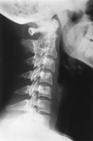
C2 to C3 Congenital Block Vertebra

Description. When two adjacent vertebrae are osseously fused from birth, this joined unit is called a congenital block vertebra. Embryologically, this is the result of failure of the normal segmentation process of the somites during the period of differentiation at 3-to-8 fetal weeks.1 The block vertebra, by itself, is clinically insignificant. As there is no motion allowed at the fused level, there is no potential for degenerative disease of the disc or posterior apophyseal joints. The foramina at the blocked level may be smaller than normal, normally sized, or enlarged, but have not been shown to cause nerve compression. However, because of the lack of a motion segment, the free articulations above and below the block segment are stressed and usually result in premature degenerative discogenic spondylosis and arthrosis at the fully articulated levels, especially below the fusion site. Fusions are partial (i.e., do not completely involve the anterior and posterior spinal units) and may result in abnormal spinal curvature, usually scoliosis, because of a unilateral bar. Block vertebrae are most commonly found at C5-C6, C2-C3, T12-L1, and L4-L5, in decreasing order of incidence.1,2 C2-C3 Congenital Block Vertebra

A recent report suggests that long-standing congenital or acquired fusion of upper cervical vertebrae may lead to stretching and laxity of the ligaments between the occiput and the atlas, resulting in excessive motion and brainstem or cord compression.2

Radiologic Features. A typical congenital block vertebra will demonstrate the following roentgen signs: a diminished AP diameter of the vertebral body; a hypoplastic or rudimentary disc space that may show faint calcification; possible fusion of the apophyseal joints (50% of cases); and possible malformation or fusion of the spinous processes.

The anterior margins of the involved vertebrae form a concave surface, because of the decreased AP diameter at the fusion that is visible on plain film and MRI. This “wasp waist”3,5 or “C” shape can serve as a mnemonic device, to indicate that this fusion is “congenital.” Another helpful sign of this congenital anomaly is osseous fusion of the neural arches, that is almost never associated with infectious, traumatic processes or other causes of block vertebrae. 4,5 TAC

Dr. Terry R. Yochum is a second-generation chiropractor and a cum laude graduate of the National College of Chiropractic, where he subsequently completed his radiology specialty. He is currently Director of the Rocky Mountain Chiropractic Radiological Center, in Denver, CO, an Adjunct Professor of Radiology at the Los Angeles College of Chiropractic, as well as an instructor of Skeletal Radiology at the University of Colorado School of Medicine, Denver, CO. Dr. Yochum is, also, a consultant to Health Care Manufacturing Company that offers a Stored Energy system. For more information, Dr. Yochum can be reached at: 303-940-9400 or by e-mail at [email protected]. Magna Cum Laude graduate of National College of Chiropractic.

Dr. Chad Maola is a 1999

Magna Cum Laude graduate of National College of Chiropractic.

An Occult Lesion of the Calcaneus

HISTORY

This active adolescent athlete sprained his foot while playing tennis.  Radiographs revealed a large radiolucent lesion in the calcaneus.  What is this?

DIAGNOSIS:  SIMPLE BONE CYST

The geographic lytic expansile lesion in the floor of the calcaneus is well circumscribed and has all the characteristic appearances of a benign lesion.  Benign lytic lesions of the calcaneus are most commonly simple bone cysts (unicameral); however, the possibility of an aneurysmal bone cyst, giant cell tumor, or other benign tumors cannot be ruled out. 

Figure 1. Observe the geographic lytic expansile lesion present in the floor of the calcaneus.GENERAL  CONSIDERATIONS

Simple bone cyst (SBC), sometimes referred to as a unicameral bone cyst (UBC), solitary bone cyst, or juvenile bone cyst, is not a true neoplasm of bone, but rather a fluid-filled cyst that is lined with a thin layer of fibrous tissue.  Nonetheless, it is frequently classified under the heading of primary bone tumors.  Jaffe and Lichtenstein1, who described the first cases in 1942, clearly delineated this cyst as a distinct disease entity, naming it unicameral bone cyst.  The term unicameral, meaning one house, has actually created some confusion, since many lesions present with a bubbly or chambered appearance; it is not single chambered at all.  All of the cases that Jaffe and Lichtenstein originally reported were of the single-chamber cystic variety, which led them to use the term unicameral initially.  However, many multichambered lesions have been reported since, which are better termed simple bone cysts rather than unicameral.  In fact, histologically, they are the same lesion.

INCIDENCE

SBC’s represent slightly more than 3% of biopsied primary bone tumors.2
An SBC most commonly occurs between the ages of 3 and 14 years (80% of cases).  They have been reported in a 2-month-old infant3, and in patients over 50.3  Males predominate, 2:1.

SIGNS AND SYMPTOMS

Most lesions are totally asymptomatic until a pathologic fracture occurs, not uncommonly in athletic activities.4,5  Approximately two-thirds of the SBC’s eventually undergo pathologic fracture.6

TREATMENT AND PROGNOSIS

The traditional treatment of choice has been surgical curettage with cauterization of the cyst.  Packing of the hollow cavity with bone chips following surgery is necessary.  However, recurrence rates with this technique have been high, approximately 30 to 40%.7   More recently, the injection of steroids has significantly reduced the recurrence rates, thus providing effective treatment for the cyst.5,8-11

Dr. Terry R. Yochum is a second-generation chiropractor and a cum laude graduate of the National College of Chiropractic, where he subsequently completed his radiology specialty.  He is currently Director of the Rocky Mountain Chiropractic Radiological Center, in Denver, CO, an Adjunct Professor of Radiology at the Los Angeles College of Chiropractic, as well as an instructor of Skeletal Radiology at the University of Colorado School of Medicine, Denver, CO.  Dr. Yochum is, also, a consultant to Health Care Manufacturing Company that offers a Stored Energy system.  For more information, Dr. Yochum can be reached  at: 303-940-9400 or by e-mail at [email protected].

Dr. Chad Maola is a 1999  Magna Cum Laude graduate of  National College of Chiropractic.

References

  1. Jaffe HL, Lichtenstein L:  Solitary unicameral bone cyst, with emphasis on the roentgen picture, the pathologic appearance and the pathogenesis. Arch Surg 44:104, 1942.
  2. Mirra JM:  Bone Tumors—Diagnosis and Treatment. Philadelphia, JB Lippincott, 1980.
  3. Jaffe HL:  Tumors and Tumorous Conditions of the Bones and Joints, Philadelphia, Lea & Febiger, 1958.
  4. Garceau GJ, Gregory CF:  Solitary unicameral bone cyst. J Bone Joint Surg (Am) 36:267, 1954.
  5. Rowe LJ, Brandt JR:  Simple bone cysts in athletes. Chiro Sports Med 2:33, 1988.
  6. Wilner D:  Radiology of Bone Tumors and Allied Disorders, Philadelphia, WB Saunders, 1982.
  7. Smith RW, Smith CF: Solitary unicameral bone cyst of the calcaneus. J Bone Joint Surg (Am) 56:49, 1974.
  8. Capanna R, Albisinni U, Campanacci M, et al: Contrast examination as a prognostic factor in the treatment of solitary bone cyst by cortisone injection. Skeletal Radiol 12:97, 1984.
  9. Capanna R, DalMonte A, Campanacci M: The natural history of unicameral bone cyst after steroid injection. Clin Orthop 166:204, 1982.
  10. Scaglietti O, Marchetti PG, Bartolozzi P: Final results obtained in the treatment of bone cysts with methylprednisolone acetate (depo-medral) and a discussion of results achieved in other bone lesions. Clin Orthop 165:33, 1982.
  11. Yochum TR, Rowe LJ:  Essentials of Skeletal Radiology, ed. 2. Baltimore, Williams & Wilkins, 1996.

Lumbar Spine Trauma

Compression fractures are the most common fractures of the lumbar spine.  They result from combined flexion and axial compression.1,2  The most common segmental levels to develop compression fractures are T12-L1.3  The extent of the vertebral compression and degree of comminution are dependent upon the severity of the force applied and the relative strength of the vertebra.4  In children, they are torus-type fractures.  In the elderly, osteoporosis can precipitate spontaneous compression fractures during everyday activities, which technically render them classifiable as insufficiency fractures (“grandma fractures”).  Up to 35% of compression fractures in female patients over the age of forty-five years may be due to early menopause and 30% to secondary osteopenia, most often from corticosteroids (15%), hyperthyroidism (8%), and malignancy (less than 2%).5  The disruption of the cortical vertebral endplate causes acute symptoms of only ten-to-fourteen days, duration, as long as no dislocations accompany the fracture.6

Below are X-rays of different patients who have sustained traumma to the lumbar spine.  The two most common fractures in the lumbar spine are demonstrated in the figures below.  Can you identify them?

Figure 1

Figure 2

Figure 1. Compression Fracture of the L1 Vertebral Body.  Observe the linear white band of condensation (zone of impaction) seen throughout the mid vertebral body.  This is a positive radiographic sign for vertebral compression fracture, along with the wedge-shaped vertebral body.

Figure 2. Transverse Process fractures of L1, L2 and L3.  Note the transverse process fractures on the left side of the L1, L2 and L3.

 Post-fracture stability is determined based on the classification by Denis.7  Three columns are recognized:  anterior column (from the anterior longitudinal ligament to the midvertebral body), middle column (from the midvertebral body to the posterior longitudinal ligament), and the posterior column (from the posterior longitudinal ligament to the supraspinous ligament).  If two or more compartments are disrupted, the fracture complex is unstable.  The likelihood of neurologic injury is high and interventional surgery is likely to be necessary.

 
Radiographic Signs of
Vertebral Compression Fracture

Radiographs of optimum quality are necessary in order to adequately demonstrate these fractures.  Lateral radiographs best demonstrate fracture features.  Radiographic signs of vertebral compression fracture include a step defect, wedge deformity, linear zone of condensation, and displaced endplate.

The Step Defect

Since the anterior aspect of the vertebral body is under the greatest stress, the first bony injury to occur is a buckling of the anterior cortex, usually near the superior vertebral endplate.  This sign is best seen on the lateral view as a sharp step-off of the anterosuperior vertebral margin along the smooth concave edge of the vertebral body.  In subtle compression fractures, the “step” defect may be the only radiographic sign of fracture.  Anatomically, the actual step-off deformity represents the anteriorly displaced corner of the superior vertebral cortex.  As the superior endplate is compressed in flexion, a sliding forward of the vertebral endplate occurs, creating the roentgen sign.

Wedge Deformity

In most compression fractures, an anterior depression of the vertebral body occurs, creating a triangular wedge shape.  The posterior vertebral height remains uncompromised, differentiating a traumatic fracture from a pathologic fracture.  This wedging may create angular kyphosis in the adjacent area.  The superior endplate is far more often involved than the inferior endplate.  Up to 30% or greater loss in anterior height may be required before the deformity is readily apparent on conventional lateral radiographs of the spine.8  Normal variant anterior wedging of 10-to-15%, or 1-3 mm, is common throughout the thoracic spine most marked at T11-L2.9,10
In all compression fractures, there should be clear differentiation from an underlying pathology that has produced the fracture. Key features of pathologic fractures may be identified by loss of the posterior body height, pedicle, and other sites of destruction. A paraspinal mass on MR imaging with abnormal marrow can be demonstrated.11

Linear White Band of Condensation
(Zone of Impaction)

Occasionally, a band of radiopacity may be seen just below the vertebral endplate that has been fractured.  The radiopaque band represents the early site of bone impaction following a forceful flexion injury where the bones are driven together.  Callus formation adds to the density of the radiopaque band later, in the healing stage of the fracture injury.  This radiographic sign is striking when present; however, it is an unreliable sign, since it is not present as often as might be expected.  Its presence, however, denotes a fracture of recent origin (less than two months’ duration).  It is nicely demonstrated in Figure 1.

Disruption in the Vertebral Endplate

A sharp disruption in the fractured vertebral endplate may be seen with spinal compression fracture.  This may be difficult to perceive on plain films and tomography; CT (computed tomography) provides the definitive means to identification.  The edges of the disruption are often jagged and irregular.  The superior endplate is more commonly fractured than the inferior endplate.

Transverse Process Fractures

Transverse process fractures are the second most common fractures of the lumbar spine, with compression fracture being the most common.  They occur, usually, secondary to a severe hyperextension and lateral flexion blow to the lumbar spine from avulsion of the paraspinal muscles.  The most common segments to suffer transverse process fractures are L2 and L3.
Radiographically, the fracture line appears as a jagged radiolucent separation, usually occurring close to its point of origin from the vertebra.  Frequently, the separated fragment is displaced inferiorly.  If the fracture line is horizontal, close inspection for a transverse or Chance fracture should be performed.  Fractures often occur at multiple levels.  Fractures of the fifth lumbar transverse process are frequently found in association with pelvic fractures, particularly fractures of the sacral ala, or disruption of the sacroiliac joint.  Occasionally, loss of the psoas shadow may occur secondary to hemorrhage.  Ossification within the hemorrhage (myositis ossificans) can result in bony bridging between transverse processes (lumbar ossified bridging syndrome, LOBS).12   Renal damage may occur which may be associated with hematuria.
A pseudofracture of the transverse process can be simulated by developmental nonunion, especially at L1, the psoas margin where it crosses the tip of the transverse process and overlying fat lines, or intestinal gas.  Oblique or tilt views may be necessary to rule out fracture.13 TAC

Dr. Terry R. Yochum is a second-generation chiropractor and a cum laude graduate of National College of Chiropractic, where he subsequently completed his radiology specialty.  He is currently Director of the Rocky Mountain Chiropractor Center, in Denver, CO, and Adjunct Professor of Radiology at the Los Angeles College of Chiropractic, as well as an instructor of Skeletal Radiology at the University of Colorado School of Medicine, Denver, CO. Dr Yochum is, also a consultant to Health Care Manufacturing Company that offers a Stored Energy system. For more information, Dr. Yocum can be reached at: (303) 940-9400 or by e-mail at [email protected].

Dr. Chad Maola is a 1999 Magna Cum Laude Graduate from National College of Chiropractic.

TAC’s MISSION:

We are dedicated to the continuing education & advancement of the chiropractic professional – YOU!

The Positive fat-pad sign: what is it?

A useful sign of an intraarticular fracture of the elbow is the clear depiction of displaced humeral capsular fat-pads.1  In the normal elbow, a layer of fat (“fat-pad”) lies between the synovial and fibrous layers of both the anterior and posterior joint capsules.  In the lateral projection of the normal elbow, the anterior fat-pad is seen as an obliquely oriented radiolucency.  When acute intracapsular swelling is present from any origin (hemorrhagic, inflammatory, or traumatic), the anterior fat-pad is elevated to be oriented horizontally, while the posterior fat-pad, when visible, is the most reliable sign of intraarticular effusion.  In children and adolescents, 90% of posterior fat-pad signs will have an associated fracture.1  In adults the sign is less frequently seen, and its absence does not exclude the presence of a fracture.2, 3 


Figure 1. Observe the displacement of the posterior fat-pad from the olecranon fossa (arrow). This usually indicates a hidden fracture Figure 1
Figure 2 Figure 3


Figure 2 and Figure 3. There is an impaction fracture of the radius (arrow). This patient fell on outsreched hand, locked her elbow and had a positive fat-pad sign.


References

  1. Norell G:  Roentgenologic visualization of the extracapsular fat.  Its importance in the diagnosis of traumatic injuries to the elbow.   Acta Radiol 42:205, 1954.
  2. Hunter RD:  Swollen elbow following trauma.  JAMA 230:1573, 1974.
  3. Yochum TR, Rowe LJ:  Essentials of Skeletal Radiology, 2nd ed., Williams & Wilkins, Baltimore, Maryland, 1996.

Dr. Terry R. Yochum is a second-generation chiropractor and a cum laude graduate of the National College of Chiropractic, where he subsequently completed his radiology specialty.  He is currently Director of the Rocky Mountain Chiropractic Radiological Center, in Denver, CO, an Adjunct Professor of Radiology at the Los Angeles College of Chiropractic, as well as an instructor of Skeletal Radiology at the University of Colorado School of Medicine, Denver, CO.  Dr. Yochum is, also, a consultant to Health Care Manufacturing Company that offers a Stored Energy system.  For more information, Dr. Yochum can be reached  at: (303) 940-9400 or by e-mail at [email protected].

Dr. Chad Maola is a 1999 Magna Cum Laude Graduate from National College of Chiropractic.

Partial Agenesis of the C1 Posterior Arch

Embrylology.  Ossification of the first cervical vertebra begins about the seventh fetal week at the lateral masses, and proceeds perichondrally in a dorsal direction, creating the posterior arch of the atlas.  In the second year of life, a secondary growth center for the posterior tubercle develops between these neural arches.  Complete fusion of the posterior arch should be noted between the third and fifth years.1

Description.  The basic defect in agenesis of the posterior arch of the atlas is the lack of a cartilage template on which the ossification process builds.  Complete or partial agenesis of the posterior arch is rare, and the posterior arch defects, by themselves, should not be the cause of neurologic or biomechanical findings unless found in association with other anomalies such as Klippel-Feil syndrome.1,2,3

Radiologic Features.  An absent posterior arch can be easily visualized on standard lateral cervical radiographs by the lack of a bony posterior neural arch.  A commonly associated finding with complete agenesis is enlargement of the superior aspect of the second cervical spinous process, which has been referred to as a mega-spinous process, representing fusion of a rudimentary posterior arch and posterior tubercle of the atlas.2  One may also observe increased size of the anterior arch of C1, which is thought to be stress related.  This is a helpful radiographic sign and suggests a long-standing and probable congenital origin to the missing posterior arch of the atlas.3 TAC

Dr. Terry R. Yochum is a second-generation chiropractor and a cum laude graduate of the National College of Chiropractic, where he subsequently completed his radiology specialty.  He is currently Director of the Rocky Mountain Chiropractic Radiological Center, in Denver, CO, an Adjunct Professor of Radiology at the Los Angeles College of Chiropractic, as well as an instructor of Skeletal Radiology at the University of Colorado School of Medicine, Denver, CO.  Dr. Yochum is, also, a consultant to Health Care Manufacturing Company that offers a Stored Energy system.  For more information, Dr. Yochum can be reached  at: (303) 940-9400 or by e-mail at [email protected].