The Misunderstood Component in Musculo-Skeletal Health Care

backadjustmentmay:dropcap_open:I:dropcap_close:s it possible to walk through a doorway when the door is closed? That strange question is a metaphor that can be used to describe the difference in research-based rehabilitation to prepare the spine for corrective procedures with little to no pain versus no preparation at all or, worse yet, substituting bilateral strengthening exercises for rehab to prepare the spine for corrections that are often both painful and ineffective.

Gym type exercises are often called rehabilitation exercises. However, the two have little in common. Gym type exercises are isotonic that work and shorten muscles, thereby opposing positional changes. Phasic muscles are enervated by a1-motor fibers and are consciously controlled and, when totally fatigued or injured, become flaccid. Postural muscles are enervated by a2-motor fibers and are autonomic nerve controlled. The strengthening of these muscles requires isometric exercises. When they are fatigued or injured, they become spastic. It is critical to understand that postural muscles dominate one’s spinal position and function as well as posture, stance and gait.

What is rehabilitation and why is it necessary for quality patient care? Dorland’s Medical Dictionary 26th Ed. defines rehabilitation as:

1. “The restoration of normal form and function after injury or illness.”

2. “The restoration of an ill or injured patient to self-sufficiency or to gainful employment at the highest attainable skill in the shortest period of time”.

The profession of chiropractic claims that the adjustment is for the restoration of normal form and function of the spine through correction of spinal displacement subluxations. We can agree that abnormal/subluxated spinal forms cause abnormal functions.

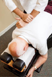
Surveys show that the clinical procedures taught in chiropractic colleges and practiced by 84% of the profession involve the use of a sudden applied force into the subluxated spine, referred to as an adjustment or treatment.

What do the adjusting forces actually do? Is it possible to physically correct spinal displacement subluxations without first preparing the spine to welcome the force through rehabilitating the soft tissues that hold the static spine upright while in a normal or abnormal (subluxated) position?

:dropcap_open:We can agree that abnormal/subluxated spinal forms cause abnormal functions.:quoteleft_close:

Research published in 1996, by JMPT, titled “Lasting Changes in Passive Range of Motion after Spinal Mobilization,” a Randomized, Blind Controlled Trial, tested spinal changes produced by diversified and toggle recoil adjusting. Their findings were that these adjustments caused about a 5% increase in mobility and the changes lasted for about 7 days. A more recent article published in the Spine Journal demonstrated with pre and post MRI studies that the same kinds of adjusting resulted in the spine becoming measurably more displaced than before it was adjusted. Chiropractic adjusting, without first performing rehabilitation to prepare the spinal soft tissues for change, typically has shown little success in making gross structural spinal and postural changes. This also was tested and published in JMPT. The authors were Harrison, et al in 1997, and Lantz et al, in 2001. In 1975, Jowett and Fidler published research in Orthop. Clin., N. Amer. that proved the body changes phasic fibers into postural muscle fibers on the convex side of spinal displacement subluxations; hypo mobility and nerve root compression were also noted.

Neuro-muscular research has proven that the dynamic stretch reflex guards all body parts including spinal position, even if it is in a subluxated position from being changed by sudden applied forces. Guyton’s Physiology 5th and 6th Edition, explains how the dynamic stretch reflex causes muscles to replace body parts after a dynamic force has displaced them. Muscles intercept sudden applied forces of up to .3 of a second in speed. The phasic muscle fibers can contract fast enough to counteract these sudden applied forces. This reflex protection is automatic, unless the force is faster than .3 seconds or so great that they tear the muscles and then the ligaments, thereby causing spinal subluxations.

The protective energy of the muscles can be greatly reduced by slowly stretching the phasic and postural muscles rather than attempting to exercise them. To be effective, stretching should last at least 40 seconds and include all muscles involved.

:quoteright_open:Hysteresis can temporarily remove up to 95% of the ligament’s holding energy and lasts for approximately six minutes.:quoteright_close:

Ligament research has proven that to instantly change a vertebrae’s position, the adjusting force would have to overcome 40% of the ligament’s resisting force. Such high forces are impossible for a doctor to produce and could crush the vertebrae before the ligament entered the plastic range necessary, before instant positional change of a vertebra can occur. However, rehabilitation procedures that cause disks and ligaments to go through full range loading and unloading cycle’s for 2 to 5 minutes produces a condition called hysteresis, which gradually lowers the resistance of spinal disks and ligaments, thereby making changes in spinal position possible, easy and pain free. Hysteresis can temporarily remove up to 95% of the ligament’s holding energy and lasts for approximately six minutes after loading and unloading cycles are discontinued. Within 15 to 20 minutes after loading and unloading cycles are discontinued, the ligaments regain all of their holding energy.

Loading and unloading cycles in the cervical spine requires slow, intermittent traction, while effective loading and unloading cycles in the lumbo-sacral spine require full range, figure 8 cycles that are slowly applied, requiring 4 seconds to complete one cycle after the ligaments are completely stretched. The figure-8 cycles also mix and re-mix the protoglycine aggregate of the disk’s nucleus necessary for making and maintenance of a perfect jell. Like fluids, the jelled nucleus is non-compressible. When perfected, it equally transfers body weight from the vertebrae above to the vertebrae below, thereby providing a foundation for maintenance of the corrected spine’s form and function. In addition, the figure-8 motion pumps the cerebral-spinal fluids necessary for the metabolic interchange of glucose to feed the brain, thereby giving the patient a feeling of mental and physical well being—an additional benefit of the rehab procedures! Home care instructions work in tandem with clinical care and will include the use of deep, diaphragmatic breathing in coordination with specific rehabilitation procedures. These procedures take into account the fact that the thoracic spine is approximately 13 times less flexible than the cervical spine and approximately 3 times less flexible than the lumbar spine; therefore, thoracic loading and unloading cycles require more time and concerted rehabilitation procedures. Patients are taught to prepare their spine both in the clinic before being seen, as well as at home before daily activities and rest.

Change is inevitable for the growth of any profession; without it, stagnation and eventual decline is the end result. Through the use of clinical research and documentation with new and more efficient methodologies, we grow in our knowledge and our abilities. The Pettibon Biomechanics Institute has published several retrospective as well as blinded studies, proving through pre- and post- X-rays that permanent spinal corrections are possible after the spinal soft tissues have been prepared through our holistic rehabilitation procedures. Research can be found at www.pettiboninstitute.org.


Burl R. Pettibon, DC, FABCS, FRCCM, PhD. (Hon) has guided The Pettibon Institute’s direction, continuing education offerings, and research since the Institute’s inception as the Pettibon Spinal Bio-Mechanics Institute in 1981. As a teacher, inventor, and researcher, Dr. Pettibon’s influence and contributions to the science of chiropractic are legendary. Dr. Pettibon has been an extension faculty member and lecturer at Palmer College of Chiropractic for more than 35 years. He has also been an extension faculty member at Life University, Logan College of Chiropractic, Parker College of Chiropractic, and Cleveland Chiropractic College—where he received his degree in 1956. Dr. Pettibon has written more than 65 papers and books on chiropractic care and research. Over the course of his career, he has developed 25 clinics. At the present time, the profession is using the more than 50 rehabilitative products that he has invented to make the detection and correction of vertebral displacements both easier and more accurate. He currently holds four patents. Papers and books are also available through www.pettibonsystem.com.

References

1.Morningstar MW, Strauchman MN, Weeks DA, Spinal Manipulation and Anterior Headweighting for the Correction of Forward Head Posture and Cervical Hypolordosis: A Pilot Study, Spring 2003, Number 2, Volume 2, Journal of Chiropractic Medicine.

2.Horseman I, Morningstar MW, Radiographic disk height increase after a trial of multimodal spine rehabilitation and vibration traction: a retrospective case series, 2008 7, 140-145, Journal of Chiropractic Medicine, 2008.

3.Schwab MJ, Chiropractic management of a 47-year-old firefighter with lumbar disk extrusion, 7, 146-154, Journal of Chiropractic Medicine, 2008.

4.Saunders ES, Woggon E, Cohen C, Robinson DH, Improvement of Cervical Lordosis and Reduction of Forward Head Posture with Anterior Head Weighting and Proprioceptive Balancing Protocols, J. Vertebral Subluxation Res., April 27, 2003.

5.Morningstar MW, Cervical curve restoration and forward head posture reduction for the treatment of Mechanical thoracic pain using the Pettibon corrective and rehabilitative procedures, Summer 2002, Number 3, Volume 1, Journal of Chiropractic Medicine.

6.Morningstar MW, Cervical hyperlordosis, forward head posture, and lumbar Kyphosis correction: a novel treatment for Mid-thoracic pain, Spring 2003, Number 3, Volume 2, Journal of Chiropractic Medicine.

Don’t Let a Lifetime Be Defined by Scoliosis

scoliosisblue2
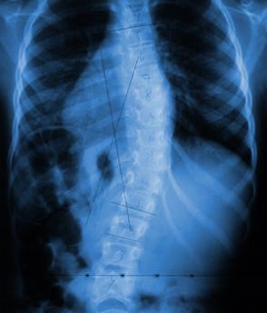
Now that genetic pre-disposition testing for scoliosis progression risk is available, An Early Stage Intervention Program has also been developed to provide scoliosis patients a non-bracing, non-surgical treatment option that allows them to take immediate action in the prevention of the next stage of the standard treatment process.

The three medically-sanctioned methods of scoliosis treatment—observation, bracing, and surgery—have been around for decades. A great deal of research has been done on the risks and benefits of each option. However, the general conclusion of this research suggests that a new paradigm is desperately needed, as there are many conflicts and inadequacies present in the current model.

Observation Only or the “Watch & Wait” Stage

Once an individual has been diagnosed with scoliosis, no treatment is initially prescribed, and no action is immediately taken, until the cobb angle has progressed to 25 degrees.  At this point, bracing is typically prescribed. This period, which is termed “watch & wait,” consists only of regular visits to an orthopedic.

Spinal brace treatment (generally recommended for curvatures 25 degrees and larger)

If there ever was a time when a patient could benefit most greatly from chiropractic, therapeutic exercise, or non-surgical intervention, it would undoubtedly be during the mild stages of the disease—before the muscles and tissues of the body have been deformed by months or even years of compensating for the abnormal twisting and bending of the spine.

Bracing dates back to approximately 650 AD, when Paul of Aegina suggested bandaging scoliosis patients with wooden strips. The first metal brace was developed by Ambroise Pare in the 16th century. Today, there is a bewildering assortment of braces in use, ranging from the venerable and bulky Milwaukee brace, to the traditional TLSO braces such as the Boston and the Wilmington brace. There are “part-time” braces, designed to be worn at night: the Providence brace, and the Charleston brace. There are also “dynamic corrective braces” (SpinCor), which may use soft, elastic materials and claim to be able to do more than simply stabilize the progression of scoliosis.

This dizzying variety is further complicated by the fact that not every doctor prescribes the braces to be used in the same manner, and not every patient may follow their doctor’s recommendations to the same extent. As a result, research is often conflicting in regard to the true effectiveness of bracing in scoliosis treatment. Some studies have shown very little difference between patients who wore the brace for the prescribed time.  In every case, all corrective benefit is lost very quickly once the patient stops wearing the brace.

Spinal fusion surgery (Generally recommended of curvatures 40 degrees and larger)

Those patients for whom bracing fails to prevent the progression of their scoliosis are left with only one option: surgery. Those who are confronted with this choice may be told that having a metal rod fused to their spine will not impair their daily activities, and will reduce the rib arch and improve their cosmetic appearance. However, research has consistently shown that surgery—which primarily focuses upon the sideways bending, and does little to address the rotation of the spine (and hence the rib protrusion)—will actually cause the rib arch to worsen (Chen 2002, Goldberg 2003, Hill 2002, Pratt 2001, Weatherly 1980, Wood 1991, Wood 1997).

The rate of hardware failure is virtually 100% over the course of a normal lifetime. It may occur immediately after the surgery or several years later, but one or more components of the hardware placed inside the body is highly likely to fail or break. The author of one study stated, “One would expect that, if the patient lives long enough, rod breakage will be a virtual certainty”. Another study found that, amongst seventy-four patients who underwent the surgery, failed fusion occurred in 27% of patients within a few years after the procedure.

The truth of the matter is that scoliosis is an abnormality of the spine which involves much more than merely a sideways curve. Yet the “effectiveness” of surgery is measured only by the degree to which it can reduce the lateral deviation through the application of brute force, and a fused spine is every bit as abnormal and dysfunctional as a scoliotic spine.

We can alter the natural course of this disease by identifying which patients are at the highest risk for severe progression via genetic testing (Scoliscore) and by implementing an aggressive, non-invasive Early Stage Scoliosis Intervention program that re-trains the brain’s involuntary postural controls centers before the spinal curvature reaches the 30 degree “buckling” point.


Dr Stitzel graduated from Palmer College of Chiropractic in 2002. Dr Stitzel practices in Lititz,PA and specializes in scoliosis rehabilitation. Dr Stitzel is a former director of the CLEAR-Institute and lectures both nationally and internationally on the topic of scoliosis rehabilitation.

Dr. A. Joshua Woggon, a 2010 Graduate of Parker College, is the Clinic Director  of the CLEAR Scoliosis Treatment and Research Clinic on the campus of Parker College of Chiropractic in Dallas, Texas (www.clearscoliosisclinic.com), and the Director of Research for CLEAR Institute, a Non-Profit Organization dedicated to advancing chiropractic scoliosis correction (www.clear-institute.org).  He can be contacted at [email protected].


Oh, Baby, My Back Hurts

As if the prospect of morning sickness, constipation, rashes and changes in hormone and wardrobe weren’t daunting enough, 80% of pregnant women will develop back pain and almost 100% will develop poor posture. The pain can last up to three months past the delivery date. Typically pain is experienced in the following areas:pregnantbelly

  • Hip Joints
  • Low Back
  • Mid-back
  • Neck
  • Shoulders
  • TMJ or Jaw joint

Unfortunately, the poor posture can become permanent.

It’s easy to see why. First there are the hormone changes that help relax ligaments and joints throughout the body. Next is the extra weight. Here is a breakdown of weight gain for a woman of normal weight.pregnancychart

The Body’s Balance System

The human body has a magnificent system of adapting to keep us upright under many different conditions. This “Balance Control System” is comprised of two parts; Sensory (Where am I) and Motor (What am I going to do). But this adaptation comes at a price. As the extra 25 or so pounds are added to the woman’s general mid-line area, the initial reaction is for the Center of Gravity and Center of Mass to shift forward. However, the body’s Balance Control System kicks in and begins a slow but steady realignment of the pelvis, spine, head and shoulders to act as a counter-balance for the extra weight of pregnancy. Eventually the Center of Gravity shifts backward. This short term solution can have long lasting consequences.

The spinal changes are very predictable.

  1. Pelvis tilts forward
  2. Mid back drifts backward
  3. Head/Neck assume a position in front of the shoulders (know as Forward Head Posture)

How This Affects the Spinepregnancyskeleton

The natural “S” shape curve of the spine is created from the attachment of muscles and ligaments. Any changes in the curves of the spine will cause some muscles to stretch
and some muscles to shorten, both situations contributing to instability in the spine. This instability leads to chronic tension in certain muscle groups and increased strain on joints.

The study of Physiology tells us that a muscle kept in a stretched position will eventually elongate its fibers and become weak (Stretch Weakness). Conversely, a muscle held in an over-relaxed position will shorten its fibers and also become weak (Short Weakness). Because the postural changes are so predictable, it is clearly understood which muscle groups are involved and whether they have become stretched or shortened. Unfortunately  after delivery and the loss of the extra weight normally gained during pregnancy, the mother’s posture does not “snap” back into place. The stretched and shortened muscles stay that way until there is an intervention. That Intervention is known as Posture Reprogramming™.

The Shear Truth

pregnancyspine
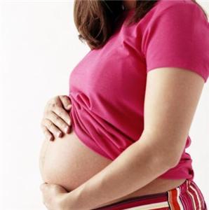
As if all this wasn’t enough, other forces come into play to create a low back that is less stable and more prone to create chronic pain and discomfort. Shear forces to be exact. In a person with normal posture, downward Gravitational and directional shear forces work with the body to “lock” joints into position under load, thereby providing strength and stability to the spine. In a pregnancy posture, the Gravitational forces remain essentially the same, but the shear forces change direction in such a way as to open the low back joints, providing less stability in the spine. The body’s exquisite feedback system senses this and the muscles in the low back become tighter in an effort to take up the slack.

First the bad news: Poor posture is NOT self-correcting.

Now the good news: Poor posture can be corrected in as little as four weeks!

Identifying Poor Posture Through the Use of the Posture Numberpregnancytop

Correcting posture begins with a simple non-invasive digital analysis to establish the patient’s Posture Numbertm. This number is used to identify and quantify the extent of the poor posture, and provide guidelines for the cost of posture reprogramming.

Next a series of gentle spinal mobilization sessions will be started and a set of custom stretches and exercises will be assigned. A special therapeutic cushion may be prescribed. These protocols are designed to reverse spinal muscle changes and reprogram muscles back to their original positions. This process takes about four weeks. 

About the Posture Reprogramming Stretches and Exercises

Because the pelvic and spinal changes are so predictable, it is possible to map which muscles will undergo “Stretch Weakness” and pregnancymiddlewhich will exhibit “Short Weakness”. With that knowledge, a Posture Reprogramming professional can create an exercise program to strengthen the muscles that have become stretched, and a stretching program for the muscles that have become shotened.

In addition to these maneuvers, a new therapeutic cushion that helps reduce  the effects of Forward Head Posture may be recommended. It’s called Posture Blocks™ (Patent Pending).

Gravity Works Against You All Day Long. Now, Make It Work For You!

The special contours and foam density of the Posture Blocks cushion causes the person’s body weight, along with the pull of Gravity, to stretch muscles that need to be stretched and relax muscles that need to be relaxed. Imagine an exercise program that works simply by doing no more than lying on the floor!

Here are some of the innovative features:pregnancybottom

  • Adjustable Headrest allows the head to assume a neutral or slightly backward position simultaneously stretching the muscles in the front of the neck and relaxing the muscles in back.
  • The soft neck bridge encourages a normal curve in the neck.
  • Special cut-out encourages external rotation of the shoulders.
  • The upper back bridge exerts a gentle forward force that relaxes the back and expands the chest area.
  • The upper back bridge has a gentle decline to the low back area.

The special features of the Posture Blocks cushion are so unique the company was awarded a Patent Pending designation by the USPTO. 

Reimbursement for Posture Reprogramming

Although there is an ICD-10 code for Abnormal Posture  (R29.3), third-party reimbursement for abnormal posture is rare and is generally considered an out of network service, Most insurance companies will allow its use as a compound or complicating code. This essentially means that Posture Reprogramming is a cash based service. The fee is based on the results of the posture exam. The Posture Pro software will generate a Posture Number™, which is the accumulation of deviations from normal. The general fee guideline is $50 for every Posture Number unit of deviation. For the average patient that would be in the $500-$1,000 range for the Posture Reprogramming.

Using the Posture Reprogramming System a doctor of chiropractic can market this service directly to pregnant patients or to local OB/GYN clinics.

Conclusion

Back pain and postural changes from pregnancy are both inevitable and manageable. If started early, a prescribed exercise and stretching program can help reverse muscle changes and improve posture during and after the pregnancy. A new therapeutic cushion, the Posture Blocks, shows great promise in helping with this universal problem.


Joseph Ventura D.C. is owner of VenturaDesigns, a 32 year company that develops software and marketing strategies for the chiropractic profession. Dr. Ventura can be reached at (888) 713-2093 or at [email protected]


Posture Reprogramming and Athletic Performance

:dropcap_open:T:dropcap_close:o coax the most out of the human body during training or actual performance, there are certain principles, or building blocks that must be present. Good breathing, proper nutrition, hydration and intensity are just a few of these principles. But, at the top of the list is good posture.

Proper posture extends performance, reduces injury, speeds healing, builds more muscle and increases efficiency. Good posture also releases more energy to the primary muscles of the task at hand by not having to engage “secondary” postural muscles.

posturereprogramming

What is Good Posture?

For the purpose of this article, the focus will be on postural alignment from the side. Normal, neutral posture is present when a plumb line passes through five anatomical landmarks: Center of the ear, center of the shoulder, greater trochanter, center of the knee and just in front of the ankle.

This is illustrated in the picture on the left. Notice how straight the black plumb line is.

posturereprogramminghamstrings
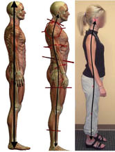
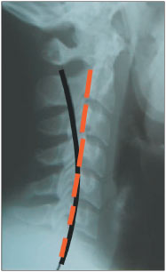
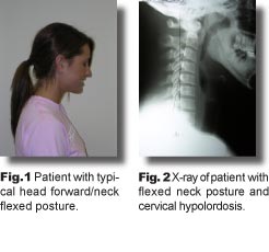
The most common abnormal posture profile is illustrated on the right. The head sits forward of the shoulders, the upper back has drifted backward and the pelvis has tipped forward. This is commonly known as Forward Head Posture (FHP). Notice the straight plumb line we expect to see in good posture now has a substantial curve in FHP. It’s been estimated that 80% of the general population has varying degrees of FHP.

Why Forward Head

Posture Is Detrimental to Athletic Performance

First and foremost, FHP places an abnormal stress on every core muscle. For example, in FHP the pelvis tips forward, causing the hamstrings in back to stretch and pre-load. This tilting also causes the Quads in front to shorten and become weak. Here’s an illustration of that.

The hamstring muscles attach to the bottom of the pelvis, the ischial tuberosity. The Quads attach to the front of the pelvis.

When the pelvis tilts forward in Forward Head Posture, it causes the hamstrings to stretch and the quads to shorten. Physiology of the body tells us that a muscle that is stretched and held in that position for a long period of time becomes weak (Stretch Weakness). Conversely, a muscle that is shortened and held in that position also becomes weak (Short Weakness). Having these two things happen to antagonistic muscle groups is quite detrimental to performance.

If one only focused on the effects of FHP on the hamstrings and quads, the need to identify FHP in the athlete becomes apparent.  However, due to compensatory changes in the spine and other areas, these kinds of muscle changes occur up the entire kinetic chain, causing compromises in the integrity of the low back, changes in breathing, changes in shoulder positioning, range of motion deficiencies, and instability of the neck motor unit.  As far as the professional athlete and weekend warrior is concerned, there is nothing good about bad posture.posturereprogrammingrehab

The Posture Reprogramming SystemTM

Forward Head Posture can be corrected. Recognizing FHP is the first step in correction.  While a quick visual check to see if the head is resting over or in front of the shoulder can provide a visual clue of the presence of FHP, it cannot quantify the full extent of the problem and it can’t be used to track progress. To do that, you need a method of capturing and measuring posture. The Posture Reprogramming SystemTM developed by the author utilizes a software program called Posture ProTM to analyze static posture and to track progress over time.  By capturing digital images of static posture and using the Posture Pro software to plot screen coordinates that represent anatomical landmarks known to be either level or plumb in neutral posture, the operator can establish baseline posture. Future exams can then track progress by comparing to the baseline values. Posture Pro has several methods of tracking progress.  One of the most effective methods is to create a plot graph of all the exams.

Yes, You Can Change Posture in as Little as Four Weeks

The author has found the profession’s biggest hesitation to focus on posture is the lack of posture correction education, either in or out of school.  For the past ten years, thousands of doctors of chiropractic around the world have been changing posture using a three-fold approach.  First, is spinal mobilization. This is a general spinal manipulation of the spine and pelvis to ensure joint mobility in advance of the changes about to happen in the muscles.  Second is the patient performing a specific set of exercises and stretches to target the muscles involved in FHP. These maneuvers were developed by John Christman, Ph.D., and refined by the author. The third protocol is the prescription of a set of Posture BlocksTM (Patent Pending). These foam cushion shapes are designed to use the weight of the body, the pull of Gravity and the resistance of the foam to stretch and relax different areas of the FHP target area.  Using spinal mobilization techniques, specific muscle stretches and exercises and utilizing a special therapeutic cushion at home, the muscles attached to the pelvis, shoulders, spine and head can be reprogrammed back to their original neutral positions. In a healthy, motivated person, this can mean a return to neutral posture in about four weeks.

Reimbursement for Posture Reprogramming

Although there is an ICD-10 code for Abnormal Posture (R29.3), third-party reimbursement for abnormal posture is rare and is generally considered an out of network service. Most insurance companies will allow its use as a compound or complicating code. This essentially means that Posture Reprogramming is a cash-based service. The fee is based on the results of the posture exam. The Posture Pro software will generate a Posture NumberTM, which is the accumulation of deviations from normal. The general fee guideline is $50 for every Posture Number unit of deviation. For the average patient, that would be in the $500-$1,000 range for the Posture Reprogramming.

Using the Posture Reprogramming System, a doctor of chiropractic can market this service to health clubs and high school, college or professional sports departments, as well as private athletes, as a method of performance screening and enhancement.


Joseph Ventura D.C. is the owner of VenturaDesigns a private company specializing in Chiropractic Consulting services and software development, He is the developer of the Posture Reprogramming System, His full bio can be found at www.posturepro.com/bio2.htm. He can be reached at [email protected]


High School Athletes – The new injury prevention plan

athletesstructuralfingerprint32 years ago, when just starting in practice, I contacted our local high school football coach and offered my services to his team.  A couple times a week I would work on any players who asked for help.  As great as the job seemed, it was short lived.  As soon as the school physician got wind of my involvement, I was introduced to the politics of sports.  The athletic director informed me I was not to come onto the campus again.

So, my simple goal was to get back onto campus, and through the front door this time, not some side door where no one knew I was there.  Along the way, I’ve learned a lot.   We live with a broken sporstmedicine system, and this age group is all but ignored.


Here’s the deal:

Just about all healthcare decisions are based upon health insurance guidelines. Most high school athlete’s care will depend upon what their insurance covers.  So, they have to be injured before they can do anything.  Anyone in the healthcare delivery industry knows that health insurance coverage is shrinking at a severe rate (increased co-pays and deductibles with decreased coverage) and, unfortunately, the care of high school athletes is jammed underneath this broken healthcare system umbrella.  Athletes, especially middle and high school athletes, have needs that are much different and far greater than the needs of the general population.  These needs are ignored.

All middle and high school athletes receive a physical prior to the season beginning, but the majority of this examination is a medical exam, i.e., eyes, ears, nose and throat.  Yes, these tests are needed, but the biomechanical exam, which checks the muscular, neurological and skeletal systems, is absent.  The examiner will perform a scoliosis screening.  (This is like saying the absence of terminal cancer means you’re healthy).  As in, there’s a lot more to biomechanics than a scoliosis screening.

We live in a reactive healthcare system dictated by economics.  These kids are never looked at until they’re hurt.  Secondly, our front line docs (primaries) are not trained in biomechanics, therefore, are not qualified to accurately diagnose or treat these injuries.  The “System” then kicks into a costly referral system, going from the primary to the orthopedist to the physical therapist or chiropractor, and the treatment goal is to get rid of the pain/injury.  No biomechanics involved.

The New York Giants and Chicago Bulls used a conditioning pyramid, with the base of that pyramid containing 6 categories; aerobic capacity, body composition, joint mobility, strength endurance, core strength and aerobic capacity.  These professional athletes needed to pass tests in all 6 categories before they could enter the weight room.  In almost every high school in this country, kids begin aggressively working out with weights with questionable supervision and absolutely no biomechanical evaluations.  This egregious omission WILL produce devastating long term detrimental effects.

injuredathlete
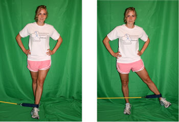
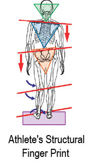
When an athlete becomes injured, the goal is to reduce the symptoms, but never correct the underlying problem.  Fig. 1 shows an example of the biomechanical imbalances that exist in all of us, and it is these imbalances that lead to the majority of injuries in athletes.  These imbalances originate in the feet (our foundation) and if this imbalance is not addressed, we have a limited potential in balancing the rest of the structure.

We’re sitting on a ticking time bomb.  Many of these kids suffer with low level injuries that don’t meet the criteria for taking action.  But, all of these kids have mild to extreme biomechanical faults, and our current sportsmedicine system prefers to perform joint replacements and prescribe a lifetime of pharmaceuticals later rather than address the issues now.


Possible solutions

Chiropractors and Physical Therapists—You’re the biomechanical providers out there, so you need to step up.  The first step is to get to the schools and educate the families and coaches.  Then offer your services for a complete biomechanical exam for all athletes, not just those injured.  This should take place before the season begins.  A biomechanical exam can be found at www.StructuralManagement.com.  Prescribe flexible custom orthotics as a first step in balancing their biomechanics.

Family Drs.—Admit this isn’t your specialty, and work with the families to find a chiropractor or physical therapist who will help with the biomechanical needs of the athlete.

Athletic Directors—Realize you are the lynchpin to all parents and athletes in your school district.  Your role is critical.  Work with those who are capable of providing more biomechanical information to this group.  Don’t settle for that age old response, “We’ve done fine without this so why do we need it now”.

Coaches—You’re the ones who have the most contact with these athletes.  Teach them about prevention and the importance of being disciplined in taking care of themselves.  Help to build the bridge between families and biomechanical providers in your communities.  Small injuries are warning signals.  Please don’t promote the “No Pain, No Gain” mantra.  Realize that pain is a warning signal for underlying biomechanical imbalances.

Parents—Don’t stop until you find someone who will perform a biomechanical exam on your child.  It will help detect the predictable sights of injuries, and will uncover the reasons why chronic injuries persist.  And, encourage corrective recommendations.

Other than that, have a great month.


Dr. Maggs currently practices full time, while also lecturing for Foot Levelers. He is the developer of The Structural Management Program, as well as the 10 Week Webinar Series, “How to Build Your High School Athlete Practice”.  He can be reached at 1-518-393-6566 or [email protected]. His website is www.StructuralManagement.com

Back Injuries in the Young Athlete

hurtathlete:dropcap_open:B:dropcap_close:ack injuries in adolescents are not very common, but add participation in athletics and you increase the opportunity of experiencing some form of low back pain. According to Micheli, back injuries of young athletes are a significant phenomenon, estimated to occur in 10 to 15 percent of participants1. The prevalence will vary, based on certain sports. In sports like gymnastics back injuries are 11 percent whereas, in football linemen, it has been recorded as high as 50 percent2.

Back injuries can occur from single episodes of blunt trauma -—like being pushed or hit in the back-— to repetitive microtrauma (overuse) from activities of repetitive lumbar extension from practicing sports like gymnastics and diving. Commonly seen back-related injuries in adolescents can result in fractures, which usually are not associated with the severity of a cervical spine fracture that can lead to catastrophic spinal cord injuries. Acute disc herniation, contusions, sprains, strains, spondylolysis and spondylolisthesis, facet syndromes and lordotic low back pain are common. Injuries associated with flexion can be both Atypical and Typical Scheuermann’s Kyphosis, compression or end plate fractures. All of these mechanisms will result in and contribute to vertebral subluxation complexes.

:dropcap_open:All of these mechanisms will result in and contribute to vertebral subluxation complexes.:quoteleft_close:

I would like to focus on just one of the above-mentioned conditions due to it being commonly missed diagnostically. This is “lordotic low back pain.” As doctors of chiropractic, we commonly see adolescents in our office with low back pain that ranges from many causes, and often the patient or parent cannot give a mechanism of injury. One of the most important consideration factors when treating an adolescent is to first determine if they are going through a growth spurt.

Let us look at Elyse, a 12-year-old female gymnast who enters our office with low back pain. Elyse complains of lower back pain which she describes as being on and off for several weeks in duration. The severity ranges from a 0 to a 10 on a pain scale and can change for no related reason. Today, the pain is at a 10, which is why the appointment was made. History indicates Elyse is an active gymnast who practices four days a week, averaging about two hours per practice. Some days the pain restricts her practice time. She describes that location of the pain varies from along the tops of both hip bones (crest of the ilium) to the middle of her low back. Both mother and Elyse deny any recent fall or trauma in the last six months.

Examination shows a normal 12-year-old in height and weight. She can heel-toe walk normally. Balance and coordination appear normal. Ranges of motion demonstrates full ranges of all extremities, cervical spine, and dorsolumbar spine with exception of pain on dorsolumbar extension and restricted flexion that was less painful. Palpation notes symmetrical tightness of the lumbar musculature with no spasms. Palpation of the iliac crest and especially the lumbar spinous process bring about a response of soreness.

Based on our examination findings, we determine the need for lumbar radiographs. The AP view shows a nice straight spine with no rotation. It also shows normal height and formation of the vertebrae with no wedging. Iliac crest and femoral heads all are equal in comparison. Open growth places are noted along the iliac crest and pelvis. The lateral view also shows normal vertebral heights and disc heights. No evidence of degeneration, fracture or other pathology. With additional viewing of the lateral film, we note a slight hyperlordosis of the lumbar spine with no interruption of George’s line.

I am sure I am not the only DC who has looked at an X-ray and found very little of anything structurally wrong on the films. You then question yourself as to how you are going to explain to the parents and child why they are experiencing such pain. It’s easy when we can reference a scoliosis, or point out disc wedging caused by misaligned vertebrae, rotated spinous processes, and phases of disc degeneration. But what are we going to tell the parents and child now as to why Elyse is having low back pain and why she needs chiropractic care? What are we going to adjust? Yes, chiropractic care should be part of everyone’s health program, but we have a child in pain that is looking for answers to why she hurts.

This is why this condition is commonly misdiagnosed. The key diagnostic signs here are the areas of pain, restricted flexion and extension ranges of motion, and the only radiographic finding is the hyperlordosis of the lumbar spine. Above all, the key sign is her age, because she is going through a growth spurt!

:quoteright_open:This is a time that the spine and its supporting tissues are usually very elastic and pliable.:quoteright_close:

In this case, as the lumbar vertebrae are growing, the thoracolumbar fascia is not stretching at the same rate. This phenomenon is what causes the hyperlordosis of the lumbar spine. The fascia is so strong and isn’t expanding, so the growing lumbar vertebrae have no place to go but forward, causing the increased hyperflexion. Considering the anatomy of the fascia and where it originates from across the iliac crest, the body is actually causing a tractioning on the apophysis. The low back pain can be caused from the lumbar spinous processes being jammed upon each other.

Commonly, when we adjust this patient, the lumbar spine is very tight and you will usually not get any movement of the spine. The patient may tell you it was very painful when the lower back was adjusted. I am sure as we described this episode, many of you were able to recall patients of this age that you encountered with the same scenario, and often we lose this patient because he or she gets discouraged because the episodes of pain are so sporadic. More commonly than not, on the days the pain is worse, they are going through a growth spurt.

This patient is truly a chiropractic patient. We must be able to educate the patient and parents on what is actually happening during the child’s development. This is a time that the spine and its supporting tissues are usually very elastic and pliable. Therefore, monitoring structural alignment during these episodes of growth is essential to maintain good spinal development and prevent possible structural problems from developing in the future. Additional considerations you could recommend to help with pain are stretching and use of an anti-lordotic brace.

A 1980 graduate of Palmer College of Chiropractic, Dr. Kirk Lee is a member of the Palmer College of Chiropractic Post Graduate Faculty and Parker College of Chiropractic Post Graduate Faculty. He has lectured nationwide on sports injuries and the adolescent athlete, and currently practices in Albion, Michigan.

References:

1. Micheli, LJ. “Back Pain in Young Athletes.” Arch Pediatr Adolesc Med 149:15-18, 1995.

2. Mundy, DJ. “Epidemiologic study of sports and weight lifting as possible risk factors for herniated lumbar and cervical disc.” Am J Sports Med 21:854-860, 1993.


Building Your Practice on a Strong Foundation

Building Your Practice on a Strong Foundation

by Dr. Mark R. Payne D.C.


In keeping with this month’s focus on practice management and coaching, I wanted to take a slightly different twist with this article.

While I make no pretense at being a practice manager or coach, I have started and operated two successful practices in my career plus a couple of other successful businesses, and there is one principle which should be carved in stone. You can never have any measure of long term success unless you actually deliver a real and valuable service/product to your patient or client. Oh, sure, there are plenty of folks in every line of business who talk a great game, but never actually deliver very much of what they promise. Such businesses generally start out great but fade in the stretch. Practices which are built on little more than hype and patient motivation generally start to implode after a few years when the community realizes the doctor “just keeps you coming back for the money.” There’s just no staying power to any business that doesn’t honestly serve the best interests of its customers.

On the other hand, thousands of doctors all over the country are quietly doing their job, delivering what they promise, and serving the needs of their patients; and patients intuitively know who has their best interest at heart. With a bit of patient education, patients can understand the difference between short term relief and true correction. And, sometimes, with a little patience, they even come to understand the concept of prevention. But there’s one thing that patients always understand and that is objective results they can clearly see with their own eyes. There is nothing more powerful than when a patient sees and understands their problem at the beginning of care and then can clearly visualize their own improvement as care progresses.

It probably comes as no surprise that I believe a postural chiropractic paradigm has numerous advantages for both doctor and patient. At the same time, I realize and respect that many of you have a fundamentally different approach to practice. Where I might choose to concentrate on improving the biomechanical (structural) status of the patient, another doctor might focus on various parameters of patient function like range or quality of joint motion or enhanced ability to perform various activities of daily living.

Whenever we treat a patient, it is incumbent upon us to objectively document the results of our care. Obviously, some methods of documentation are apparent only to the doctor, such as improvement in palpated joint motion. Others, like X-Ray, or visual postural analysis, may be easily seen and understood by the patient as well. The degree to which patients can see and comprehend their problem and the results of your treatment will determine, in large part, the patient’s perceived value of your care and, consequently, the referral value of your services. It is here that a strong focus on posture rehabilitation can be a significant tool in building your practice.


Figure 1: The PRE treatment film here clearly shows the patient’s loss of the normal cervical lordosis (the red dotted line) as compared to a more normal or optimum lordosis (the black solid line). Patients understand such simple, concrete demonstrations of their problems.


Doctors who focus on biomechanical correction have no problem educating patients as to the nature of their postural problems. These are problems which don’t require a sophisticated or trained eye to see. Once patients understand what their spine should look like (shown here with the black solid line), they can easily understand why they need to perform various rehab activities like exercise, extension traction, or lifestyle modifications to help their posture return to normal. Likewise, the clinical advantages of a structural practice paradigm are numerous as well. Unlike the small, single segment subluxations you learned about in chiropractic college, large scale, multi segmental postural misalignments can be easily and objectively identified, accurately measured, and effectively treated in the majority of cases.


Figure 2: The POST treatment film here clearly shows the patient’s lordosis returning to a more normal or optimum position. Patients can clearly see and understand results like these.


Including postural care brings patient benefits that simply can’t be achieved in any other way, as well. Simple, low tech methods of postural rehab, such as extension traction and reverse posture exercise, as were used on the patient above, have been shown to produce long term structural improvements, far exceeding those of adjusting or functional based rehab alone. Such postural improvements are key to reducing long term pain and spinal degeneration. Obviously, that’s in the best interest of your patients in terms of their long term health. Hopefully, the above example will stir your thoughts as to how the simple, “common sense” paradigm of postural based chiropractic can be an effective tool for building the health of your practice as well.

Special thanks to our client Dr. Mark Berry of Vestavia Hills, AL, for sharing his pre and post care results. Congratulations, Dr. Berry, on a job well done.


Dr. Mark Payne is the president of Matlin Mfg., a manufacturer and distributor of postural rehab products since 1988. To learn more about implementing postural rehab into your practice, call 1-334-448-1210 or link to www.MatlinMfg.com to request our FREE REPORT, “The Best Corrections of Your Career.”

Preventing Injuries Requires More Than a Strong Core

Preventing Injuries Requires More Than a Strong Core

by Dr. Kirk A. Lee, D.C.


Whether we are confronted with developing an exercise rehab program for a male patient (who is sedentary, overweight, and experiencing low back pain) or developing a strengthening program to fine tune a professional athlete, some emphasis must be placed on the core musculature. With today’s marketing of the “infomercial,” we have seen everything from machines that promise six-pack abs to cures for low back pain.

Before we start any patient on a rehabilitation program, we must consider several things. It must be designed to improve flexibility and increase strength but not create overload or stresses on the musculoskeletal system. Too much stress can prevent the body from continuing to heal and repair and, ultimately, lead to an exacerbation of the patient’s present injury or lead to a new injury.


One of the easiest means of addressing core strength is through the use of exercise balls. A patient who has balance problems can achieve help from just sitting on the ball. Sitting on the pliable-moveable ball causes the stimulation and recruitment of the core muscles to maintain stability. Most balls come with a booklet of choice exercises. It is your job, as the doctor, to determine what activities best suit your patients’ needs.

Keeping in mind the importance of a good core muscular strength, another area of consideration must be focused around the hip or pelvic musculature. When we consider the biomechanics of walking and running we know that, during the stance phase or weight-bearing phase of gait, we have a sub phase called “mid-stance.” During this sub phase of weight bearing, all the body’s weight is placed on one leg at a time. It takes strong abductor muscles (gluteus minimus and medius) to maintain a level pelvis, while weakness in the muscles can allow a drop of the unsupported side. Weakness of these muscles could lead to repetitive stress and a familiar gait pattern called a “trendelenburg.”


The importance of maintaining a strong hip and/or pelvic musculature is clearly pointed out in a research study completed at the University of Calgary in 2007 by Dr. Reed Ferber, a professor in Kinesiology. During a seven-month period, they assessed 284 patients who entered his running injury clinic at the University of Calgary. The study showed that 92 percent of the patients demonstrated weakness within the hip musculature. Of those 284 patients, 89 percent showed marked improvement in four to six weeks from performing specific exercises to strengthen the hip/pelvic musculature. The exercises included hip abduction, hip extension and flexion, and external rotation. These were performed in a sequence of:

1. Day one—1 set of 10

2. Day two—2 sets of 10

3. Day three—2 sets of 10

4. Day four and after—3 sets of 10


He recommended performing these exercises for four to six weeks and then continuing two times a week thereafter to maintain the strength that was gained.


HIP ABDUCTOR—STANDING

1. Place foot not attached to the exercise band behind the leg that is attached to the band.

2. Move leg attached to the band outward, keeping the knee straight.

3. Count two seconds going out and four seconds coming in, controlling the
motion throughout.

HIP FLEXOR—STANDING

1. Place foot not attached to the exercise band beside the leg that is attached to the band.

2. Move leg attached to the band forward, keeping knee straight.

3. Count two seconds going out and four seconds coming in, controlling the
motion throughout.


HIP EXTERNAL ROTATION—SEATED

1. Move leg attached to the exercise band outward, then return to starting position.

2. Keep knees together throughout the motion.

3. Count two seconds going out and four seconds coming in, controlling the
motion throughout.


In his study, Dr. Ferber recommended using a two-second count while taking the leg out as well as bringing it in. I like adding the four-second count to allow an eccentric unloading of the muscle as you return the leg to the starting point.

As you assess your patients’ rehabilitation needs, remember to emphasize not only the core musculature but the hip and pelvic musculature as well for additional stabilization.

A 1980 graduate of Palmer College of Chiropractic, Dr. Kirk Lee is a member of the Palmer College of Chiropractic Post Graduate Faculty and Parker College of Chiropractic Post Graduate Faculty. He has lectured nationwide on sports injuries and the adolescent athlete, and currently practices in Albion, Michigan. Dr. Lee can be reached at [email protected].

The Crossroads of Rehab and Technique

The Crossroads of Rehab and Technique

by Mark R. Payne D.C.


Adjusting technique and spinal rehabilitation have always been viewed as two completely separate subjects. Chiropractic’s rich history is replete with innovative ways of adjusting misaligned or dysfunctional spinal segments. Most such chiropractic techniques would be classed as “passive care” in that the patient is a non active recipient of care administered by the doctor.

Continue reading “The Crossroads of Rehab and Technique”

Importance of Foot Function in the Gait Cycle

Importance of Foot Function in the Gait Cycle

by Dr. Kirk A. Lee, D.C.


An essential part of our chiropractic examination should include evaluation of the gaitcycle, with special consideration toward the patientwho has chronic musculoskeletal-related pain.

Continue reading “Importance of Foot Function in the Gait Cycle”