 CASE HISTORY:
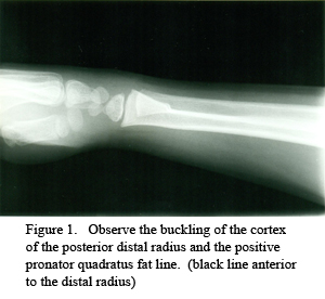
This 8-year-old fell and hyperextended his wrist.
Diagnosis: Torus fracture–distal radius.
FRACTURES OF THE WRIST
Distal Radius Fractures.
The distal radius is one of the most common sites of fracture in the wrist. Care must be taken to search all projections for these fractures, many of which are often subtle and obscure. In the absence of an obvious fracture, close observation on the lateral projection for displacement of the pronator quadratus fat line is a useful indicator for the presence or absence of fracture. Normally, this fat line appears as a well-defined linear lucency, oriented parallel with the plane of the radius, 2 to 5 mm from its anterior surface. In almost all cases of distal radius fractures, the pronator quadratus fat line will be altered.1 These alterations include anterior displacement, blurring, irregularity and obliteration of the line. The most common fracture of the wrist in the elderly is Colles’ fracture and in a child is a torus fracture of the distal radius.
Colles’ Fractures.
In 1814, Abraham Colles wrote the definitive description of this fracture which, consequently, bears his name. The injury is defined as a fracture of the distal radius, approximately 20 to 35 mm proximal to the articular surface, with posterior angulation of the distal fragment. More than 60% will have an accompanying fracture of the ulnar styloid process. The usual mechanism is a fall on an outstretched, extended hand. The physical appearance of the fractured distal forearm and wrist has led to its being called the dinner fork deformity. The incidence of the fracture increases with age, and this increase is so rapid in women that, by age 65, the lesion is six times more common in women than in men. Osteoporosis appears to be the underlying influencing factor. Complications are common and may be severe.
Torus Fracture.
This is the most common fracture of the wrist between 6 and 10 years of age. Typically, the fracture is located 2 to 4 cm from the distal growth plate. A torus fracture can occur in any long bone and is the term applied to a buckled cortex following trauma. As such, the key radiologic sign is a localized cortical bulge, bump or offset.1
Dr. Terry R. Yochum is a second generation chiropractor and a Cum Laude Graduate of National College of Chiropractic, where he subsequently completed his radiology residency. He is currently Director of the Rocky Mountain Chiropractic Radiological Center in Denver, CO, and Adjunct Professor of Radiology at the Southern California University of Health Sciences, as well as an instructor of skeletal radiology at the University of Colorado School of Medicine, Denver, CO. Dr. Yochum can be reached at 1-303-940-9400 or by e-mail at [email protected].
Dr. Chad Maola is a 1990 Magna Cum Laude Graduate of National College of Chiropractic. Dr. Maola is available for post-graduate seminars. He may be reached at 1-727-433-0153 or by email at [email protected].
References
1.Yochum TR, Rowe LJ, Essentials of Skeletal Radiology, 3rd ed. Lippincott, Williams and Wilkins, Baltimore, 2005.