 History and Presenting Symptoms
A 62-year-old female presents with a recent history of moderate pain in her lower back. Her back pain responds well to chiropractic adjustments, but recurs within a couple of days. There are no specific triggering activities, although being up and active seems to bring on the pain more rapidly. She describes her current level of low back pain as usually around 30mm to occasionally 40mm on a Visual Analog Scale.
Exam Findings
Vitals: This aging, but physically active woman (she plays golf at least twice each week, and walks about a mile every day) weighs 148 lbs., which, at 5’ 7”, results in a BMI of 23; she is not overweight. She reports that she has been a non-smoker since she quit 22 years ago, and she is a social drinker of alcohol, with an average of one glass of wine each day. Her blood pressure is 124/84 mmHg and her pulse rate is 76 bpm. These findings are within the normal range.
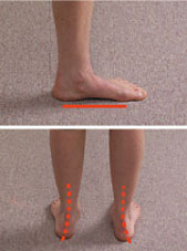
Posture and gait: Standing postural evaluation finds a lower iliac crest on the right, and a low right greater trochanter. The left shoulder is slightly lower than the right, with no history of fracture or surgery. She has a mild bilateral knee valgus and moderate calcaneal eversion and hyperpronation on the right side, with a noticeable outward flare of her right foot. Palpation of the right arch, when standing, finds it significantly lower than the left, but it is not tender to direct pressure. The Navicular Drop test demonstrates greater excursion of the right navicular bone when moving from sitting to standing (non-weight bearing to weight bearing).
Chiropractic evaluation: Motion palpation identifies limitations in segmental motion at L4/L5 and L5/S1, with some local tenderness. These segmental dysfunctions demonstrate loss of end range mobility in all directions. Additional subluxations are noted at T9/T10, C5/6, and C2/3. Lumbar ranges of motion are full and pain-free, and neurological testing is negative.
Imaging
Upright, weight-bearing X-rays of the lumbar spine demonstrate moderate loss of intervertebral disc height at L4/L5 and L5/S1, with small osteophyte formation at those levels. A discrepancy in femur head heights is seen, with a measured difference of 6mm (right side lower). A moderate lumbar curvature (6°) is noted, convex to the right side, and both the sacral base and the iliac crest are lower on the right side. The sacral base angle and measured lumbar lordosis are within normal limits.
Clinical Impression
Moderate lumbosacral osteoarthrosis and disc degeneration, with mechanical dysfunction associated with poor biomechanical support from the lower extremities. There is a functional short leg on the right side. The asymmetry in the lower extremities is clearly demonstrated by the loss of right arch stability seen on the Navicular Drop test. There is noticeable hyperpronation, arch collapse, and foot flare consistent with right arch collapse, with the expected effects in the pelvis and spine.
Treatment Plan
Adjustments: Specific chiropractic adjustments for the lower extremities and the involved spinal regions were provided as needed.
Support: Custom-made, stabilizing orthotics were provided to support the right arch and calcaneus (pronation correction) and decrease the asymmetrical stress on the knees and back.
Rehabilitation: This patient was instructed to perform an at-home series of back exercises using elastic tubing to develop and maintain coordinated strength in her spinal stabilizers (paraspinal musculature) and core (trunk and pelvic) musculature.
Response to Care
She responded well to the adjustments and exercise, and reported a rapid decrease in symptoms. Within two weeks of receiving her orthotics, she related that she had more energy and no longer had the previous nagging low back pain. She was released to a self-directed maintenance program after a total of 10 treatment sessions over two months.
Discussion
This patient had no foot or arch pain, but was undergoing plastic deformation of her arches. For unknown reasons, the deformation was accelerated in the right foot, producing a chronic asymmetrical strain on her pelvis and spine. Her condition was documented with a test for stability of the arches—the Navicular Drop test. This highlighted the asymmetry in her lower extremities and provided for an easy discussion of the benefits of long-term orthotic support.
Dr. John J. Danchik is the seventh inductee to the American Chiropractic Association Sports Hall of Fame. He is the current chairperson of the United States Olympic Committee’s Chiropractic Selection Program. He lectures extensively in the United States and abroad on current trends in sports chiropractic and rehabilitation. Dr. Danchik is an associate editor of the Journal of the Neuromusculoskeletal System. He has been in private practice in Massachusetts for 29 years. He can be reached by e-mail at [email protected].