Motion Limitations Hamper Runner

History and Presenting Symptoms
The patient, a 33-year-old female, is a marketing director who took up running for stress reduction and weight loss. She has been running for six months and has progressively increased her mileage. She is experiencing recurring and worsening pain in her left foot, as well as increased low-back tightness. She recalls no specific injury to her foot or back, but thinks she may favor her left foot while running.
 
Exam Findings
runner3Vitals. This active woman weighs 154 lbs, which at 5′6″ results in a BMI of 24.9—she is right on the normal/overweight borderline. She mentions that she has lost about 14 lbs since beginning regular running, and hopes to lose about 10 more (which would be appropriate for her height). She reports that she has never smoked, and that her blood pressure and pulse rate are both at the lower end of normal range. She drinks one to two glasses of white wine each week, usually with meals.

Posture and gait. Standing postural evaluation finds basically good alignment with intact spinal curves and no lateral listing of her pelvis or spine. She demonstrates bilateral calcaneal eversion, worse on the left with a lower left arch. Treadmill gait evaluation finds obvious hyperpronation of the left foot and ankle when walking, which is noticeably worse when running.

Chiropractic evaluation. Motion palpation identifies a limitation in right sacroiliac motion with moderate tenderness and loss of end-range mobility. Several compensatory fixations are identified throughout the thoracolumbar region. Yeoman’s provocative test elicits moderate pain upon prone extension of the right leg. All other spinal and neurological tests are negative, including sensory and reflex testing of the lower extremities.

Primary complaint. Palpation of the left foot finds the fourth metatarsal bone to be quite tender to digital pressure just proximal to the metatarsal head. Manual testing finds no specific muscle weakness, nor is there significant asymmetry in muscle mass or leg diameter. All ankle joint ranges of motion are full and pain free, bilaterally.
 
Imaging
An x-ray series of the left foot finds an area of slightly increased density in the distal third of the fourth metatarsal bone. Based on the clinical and plain radiographic findings, she was referred for a bone scan of the lower extremities and feet. This study identified an area of increased uptake in the distal third of the fourth metatarsal bone, consistent with a stress response.
 
Clinical Impression
There is early stress fracture of the fourth metatarsal bone. While no actual fracture line is present, the plain film and bone scan findings support the clinical indication of a “stress reaction” of bone. This is in response to the increased biomechanical strain of her running program, which is accompanied by sacroiliac joint motion restriction and compensatory thoracolumbar fixations associated with altered gait.
 
Treatment Plan
Adjustments. Specific, corrective adjustments for the right sacroiliac joint and the thoracolumbar region were provided as needed. The left cuboid and navicular were adjusted while carefully avoiding placing pressure on the fourth metatarsal bone.

Support. Individually designed stabilizing orthotics were supplied to help provide support through the entire gait cycle, maintain the arches, limit calcaneal eversion, and decrease heel-strike impact. Two pairs of stabilizing orthotics were ordered, one designed specifically for her running shoes and the other for job-related dress shoes.

Rehabilitation. All weight-bearing exercise was restricted for two weeks. Then, towel scrunching exercises were initiated to strengthen the intrinsic foot muscles. After four weeks, she was permitted to gradually return to her distance running program.
 
Response to Care
She responded well to the spinal and foot adjustments, and reported a rapid decrease in her foot symptoms with rest. After four weeks away, she built back up to her previous running program. She reported no return of the left foot pain, and also noted a subjective feeling of smoother and more efficient gait with the orthotics. She has now been running regularly and without difficulty for the past four months.
 
Discussion
Metatarsal stress fractures often occur when moderate biomechanical asymmetries are stressed by rapid increases in weight-bearing exercise. Flexible, shock-absorbing orthotics will incorporate arch support while reducing pronation and decreasing the stress of repetitive heel strikes on the foot and spine. 

Lower Extremity Aging Concerns

History and Presenting Symptoms
A 60-year-old female presents with recurring episodes of moderate pain in her lower back and right knee. As a full-time waitress/cashier, she spends almost half her workday on her feet. She recalls no specific back or knee injuries, and states that these problems have developed over the past couple of years and have been more noticeable in the past four months. On a 100 mm visual analog scale, she rates her low back pain as usually 45 mm, while her right knee varies from 25 to 60 mm. The knee feels worse after walking, and she takes over-the-counter NSAIDs for relief.
 
waitressExam Findings
Vitals.  This 5’ 5’’ female weighs 166 lbs, which results in a BMI of 27.6. She knows that she is overweight and is following a sensible diet, but is having difficulty doing the needed walking because of pain. She quit smoking 15 years ago. Her blood pressure is 118/79 mmHg and her pulse rate is 73 bpm. These findings are within the normal range.

Posture and gait.  Standing postural evaluation finds generally good alignment with intact spinal curves and no significant lateral listing of her pelvis or spine. She demonstrates bilateral calcaneal eversion that is worse on the right with a lower right arch. During gait, both feet pronate substantially, and both feet flare outward (toe-out).

Chiropractic evaluation. Motion palpation identifies numerous limitations in spinal motion: the right SI joint, the lumbosacral junction on the right, L2/L3 on the left, T11/12 generally, and at the cervicothoracic junction. Palpation finds no significant local tenderness or muscle spasm in these regions, and all active thoracolumbar spinal ranges of motion are limited slightly by aging, but are pain-free. Provocative orthopedic and neurological tests for nerve root impingement and/or disc involvement are negative.

Lower extremities.  Both knees demonstrate full and pain-free movements, and no provocative orthopedic tests are positive. Closer examination finds a low medial arch on the left foot, and no arch remaining on the right when standing. Her right calcaneus also demonstrates greater eversion when bearing weight. Manual testing finds no significant muscle weakness in the fibular (peroneal) or anterior tibial muscles on either side.
 
Imaging
Lumbopelvic and knee x-rays in the upright, standing position are taken while weight bearing. There is some loss of lumbar disc heights, most obvious at the lumbosacral joint, and decreasing cephalad. A slight discrepancy in femur head heights is noted, with a measured difference of 5 mm (right side lower). A moderate lumbar curvature (4°) is also seen, convex to the right side, and both the sacral base and the iliac crest are slightly lower on the right. The sacral base angle and measured lumbar lordosis are increased, but still within normal limits. No significant loss of joint spacing or osteophyte formation is seen in the knee joints.
 
Clinical Impression
Moderate lumbopelvic imbalance and spinal dysfunction associated with generalized loss of arch height (worse on the right) and aging of the knee joints and feet.
 
Treatment Plan
Adjustments. Specific, corrective adjustments for the SI joints and the lumbar and cervicothoracic regions were provided as needed with good response. Manipulation of the right foot and knee was also performed.

Support. Individually designed stabilizing orthotics were supplied, which included bilateral pronation correction (varus wedges). She described no problems in adapting to the stabilizing orthotics, although close inspection found that her shoes were too small for her feet and she did need to purchase better-fitting shoes.

Rehabilitation.  She received instruction in a comprehensive spinal wellness exercise program using elastic resistance tubing. She brought her exercise log to each visit so her adherence to the program could be encouraged.

Response to Care
The adjustments were well tolerated and the stabilizing orthotics made a noticeable improvement in her postural alignment at the feet and the lumbopelvic region. After eight treatment sessions over two months and daily home exercises, including wearing the orthotics, she was released to a self-directed home stretching program.
 
Discussion
This patient’s history and physical examination are consistent with the commonly seen spinal effects of aging on the feet. The combination of lower spinal symptoms with knee pain made worse by walking prompted an evaluation of the lower extremities during weight bearing. The problem most in need of correction was the excessive pronation that caused functional imbalance.

Athlete Back on the Ice Thanks to Chiropractic Care

History and Presenting Symptoms
The patient, a 24-year-old male semi-professional hockey player, reports a recent right hamstring “grab” that just does not seem to be improving. His history includes several injuries to the same hamstring muscle during his collegiate playing career with mixed attempts to stretch and rehab. He also describes varying levels of pain in his lower back and left hip region, which has led him to ask for a complete musculoskeletal evaluation.
 
Exam Findings
hockeyplayerVitals.  This athletic young male weighs 175 pounds, which at 5’11’’ results in a BMI of 24.4: He is not overweight. He is a non-smoker, does not drink alcohol, and his blood pressure and pulse rate are at the lower end of the normal range.

Posture and gait. Standing postural evaluation finds relatively good alignment with intact spinal curves but a slightly lower iliac crest on the right along with a lower-right greater trochanter. His spine appears to be well balanced above with no evidence of lateral curve or list. He also demonstrates right calcaneal eversion and a low medial arch (hyperpronation). A tendency to toe out on the right is seen during a standard gait screening, and he confirms his need to be conscious of the positioning of his right foot when skating.

Chiropractic evaluation.  Motion palpation identifies a mild limitation in left sacroiliac motion with moderate tenderness and loss of endrange mobility. Compensatory fixations are identified at L4/5 and L2/3. Otherwise, all orthopedic and neurological testing is negative.

Primary complaint.  Examination of his right lower extremity finds tenderness to palpation at the ischial insertion and in the medial belly of the hamstring muscle group. Straight leg raise is limited to 58° by hamstring tightness and pain, and active knee extension is restricted when the hip is placed in 90° of flexion. Manual muscle testing finds painful weakness in the right hamstring when only moderate resistance is provided.
 
Imaging
Because of his history of recurrent strains and the evidence for postural asymmetry, an upright lumbopelvic series is obtained. The standing AP lumbopelvic view shows a lateral pelvic tilt, a low sacral base on the right, and the right femur head is five millimeters lower.
 
Clinical Impression
Hamstring muscle injury associated with chronic biomechanical strain. A functional short leg on the right is accompanied by sacroiliac joint motion restriction and compensatory lumbar fixations.
 
Treatment Plan
Adjustments.  Specific, corrective adjustments for the SI joints and lumbar region were provided as needed. Mobilization and manipulation were performed on his right arch, calcaneus, and navicular bone.

Support.  Individually designed stabilizing orthotics were ordered for his street shoes, and a second pair was fitted into his hockey skates. They were designed to support the arches, decrease calcaneal eversion, and reduce the asymmetrical biomechanical forces being transmitted to the spine and pelvis.

Rehabilitation.  He was initially instructed to perform sustained functional hamstring stretches four times a day. After one week, daily strengthening exercises for the right hamstring were progressed from light to strenuous resistance using elastic exercise tubing.

 
Response to Care
The spinal and pelvic adjustments were well tolerated, and once he began wearing the stabilizing orthotics, this active hockey player required very few re-adjustments. His compliance with the recommended exercises was excellent because he was quite motivated to improve his performance and to prevent future hamstring injuries. He was immediately able to wear his orthotics full time and reported an improved sense of edging and control in his skates. The right hamstring muscle regained full flexibility and balanced strength, and he was released from acute care to a self-directed home stretching and sports performance program after a total of eight visits over two months.
 
Discussion
While a hamstring muscle “grab” or “pull” is often just a simple strain injury, in some cases there is an underlying biomechanical fault that makes it recurrent. Careful evaluation of the patient’s spine and lower extremities identified the asymmetry in the feet and ankles as a major contributor to his symptoms. Fitting stabilizing orthotics into his skates helped to provide a better boot fit, improved his edge control, and enhanced long-term support for his lower extremities. As with this young man, athletes at all levels appreciate a thorough evaluation and advice that can help to improve sports performance.
 

Joint Pains Bother Fitness Seeker

History and Presenting Symptoms
jointpainsfitnessThis female patient is a 51-year-old small business owner and exercise enthusiast.  She reports pain in the front of her left knee over the past several months, especially when exercising at the gym.  She has no obvious swelling or discoloration and denies any specific injury.  She reports that she takes over-the-counter medication for relief, but is worried about taking drugs in order to complete her exercise regimen.
 
Exam Findings
Vitals.  This active, middle-aged woman weighs 138 lbs, which at 5’4’’ results in a BMI of 23.7 – not overweight, but at the higher end of normal. She is a non-smoker, and her blood pressure and pulse rate are both at the lower end of the normal range.

Posture and gait.  Standing postural evaluation finds generally good alignment throughout her pelvis and spine.  She has a mild bilateral knee valgus and moderate calcaneal eversion and hyperpronation on the left side.  Measurement finds a standing Q-angle of 27° on the left and 23° on the right (20° is normal for women).

Chiropractic evaluation.  Motion palpation identifies a limitation in lumbosacral motion, with moderate tenderness and loss of endrange mobility to the left at L5/S1.  A compensatory fixation is also noted at T10/T11.  Neurologic testing is negative.

Primary complaint.  Examination of the left knee indicates no ligament instability, but there is a positive patellar grinding test.  All knee ranges of motion are full and pain free, bilaterally.  Manual muscle testing finds no evidence of muscle weakness.
 
Imaging
No x-rays or other forms of musculoskeletal imaging were requested.
 
Clinical Impression
Patello-femoral arthralgia on the left, associated with an elevated Q-angle and foot pronation.  This is accompanied by lumbosacral joint motion restriction and compensatory lower thoracic fixation.
 
Treatment Plan
Adjustments.  Specific, corrective adjustments for the lumbosacral joint and the lower thoracic region were provided as needed.  Manipulation of the left knee into external rotation was performed to decrease the internal rotation associated with hyperpronation and her elevated Q-angle.

Support.  Individually designed stabilizing orthotics were provided to support her arches and decrease her Q-angles. Two pairs of stabilizing orthotics were ordered – one designed specifically for her gym shoes and the other for her job-related dress shoes.

Rehabilitation.  Due to her active lifestyle, no specific rehabilitation exercises were provided. She continued with her frequent activities at the local gym.
 
Response to Care
The adjustments for the spine and knee were well-tolerated, and she reported what appeared to her to be a rapid decrease in symptoms.  After this patient began wearing her orthotics regularly, she indicated noticing a substantial decrease in knee irritation with use, and a firmer foot plant during aerobics.  Within three weeks of receiving her orthotics, she related that she was performing all of her favorite workout routines with no knee pain or limitation.  She was released to a self-directed stretching program after a total of eight treatment sessions over two months.
 
Discussion
Several factors in this case make it quite interesting.  This high-powered business woman used her gym time as both a form of recreational relaxation and a place for business networking.  As her knee began to bother her more, she was driven to begin using anti-inflammatory drugs.  She did start wearing more supportive footwear, but her underlying biomechanical problem had not been sufficiently addressed.
 
When women are physically active, their naturally higher Q-angles are frequently a source of lower extremity pain. Patello-femoral arthralgia (previously known as chondromalacia patellae) has been recognized as a biomechanical tracking disorder of the kneecap in the femoral groove.  The best treatment is a conservative approach, with a combination of chiropractic adjustments, stabilizing orthotic support, and – when indicated – rehabilitative strengthening sessions using exercise tubing.
 

A Better Life by Avoiding the Knife

History and Presenting Symptoms
kneepain14The patient is a 44-year-old male who describes persistent pain and occasional stiffness in his right knee that is not associated with any specific activity.  He reports at least two years of recurring episodes of medial knee pain that just comes and goes with no swelling or inflammation.  Previous evaluations, including a knee MRI, have resulted in a diagnosis of a torn meniscus.  Surgery has been offered; but with no guarantee of significant pain reduction, he has declined to undergo arthroscopic repair.
 
Exam Findings
Vitals. This male patient weighs 170 lbs., which at 5’ 9.5’’ results in a BMI of 24.7. He is not overweight but getting close.  He was a moderate cigarette smoker (eight per day) from his late teens into his mid-twenties, but he has been nicotine free for almost 20 years.  His blood pressure and pulse rate are at the upper end of the normal range.

Posture and gait. Standing postural evaluation finds generally good alignment with a slight forward head carriage but otherwise intact spinal curves and a balanced pelvis. He has a mild bilateral knee valgus with moderate calcaneal eversion and hyperpronation on the right side. A tendency for the right foot to flare out is noted during gait screening.

Chiropractic evaluation.  Motion palpation identifies a limitation in right sacroiliac (SI) movement with definite tenderness and loss of end-range mobility.  Several compensatory subluxations are identified throughout the lumbar region.  Otherwise, all spinal orthopedic and neurological testing is negative.

Primary complaint. Examination of his right knee finds no ligament instability and no limitation, pain, or “click” on McMurray testing.  Mild pain and crepitus are found during Apley’s grinding test.  All knee ranges of motion are full and pain free, bilaterally.  Manual muscle testing finds mild weakness in the right sartorius muscle when compared to the left side.
 
Imaging
No X-rays or other forms of musculoskeletal imaging were requested since multiple X-rays and an MRI of the knee had been performed during the initial surgical discussion period.
 
Clinical Impression
Biomechanical asymmetry of the lower extremities with probable meniscal irritation at the right knee.  This is accompanied by SI joint motion restriction and compensatory lumbar subluxations.
 

Treatment Plan
Adjustments. Specific, corrective adjustments for the SI joints and lumbar region were provided as needed.  The right proximal tibia was adjusted into internal rotation with additional medial pressure for the valgus malposition.

Stabilization.  Individually designed stabilizing orthotics were provided to support the arches, to decrease the medial pressure on his right knee, and to reduce the asymmetrical biomechanical forces being transmitted up the lower extremity to the pelvis and spine.

Rehabilitation. This patient performed daily exercises with elastic tubing to retrain and strengthen his right sartorius muscle for improved medial knee support.  He also worked to strengthen internal rotation action of his right hip to decrease the tendency to toe out.  In addition, he was supplied with a dietary supplement for joint health that contained glucosamine and chondroitin sulfates.
 
Response to Care
The spinal and extremity adjustments were well tolerated, but knee pain recurred until he had been regularly performing his rehab exercises for almost two weeks.  He reported no difficulty in wearing the orthotics in all of his shoes.  Over the next month, he described a noticeable improvement in his knee function with an eventual complete cessation of the previous pain and stiffness.  He was released from active problem care to a self-directed home stretching program after a total of eight visits over six weeks.
 
Discussion
This patient had received the medical opinion that, at some point, surgery would be the only answer for his chronic knee pain.  Fortunately, he persisted in searching for alternatives.

The combination of spinal and extremity adjustments, nutritional supplementation, and better support for the medial knee from improved muscle function and stabilizing orthotics resulted in an excellent response.  In fact, he is now embarking on a wellness plan that includes addressing his lifestyle choices to improve his diet and overall health.

Growth Asymmetry in an Outdoorsman

History and Presenting Symptoms
:dropcap_open:A:dropcap_close: 41-year-old male presents with recurring episodes of pain in his low back and left hip. He recalls no injury to the region, and cannot identify any precipitating activities or events. On a Visual Analog Scale, he rates his low back pain as varying from 30mm to 65mm, while the left hip pain is usually around 35mm.  He takes over-the-counter NSAIDs when the pain interferes with his daily activities, and that usually provides marginal relief.  He works as an RV (recreational vehicle) salesman and is also a volunteer nature trail guide. He is seeking non-drug treatment.
 
rvExam Findings
Vitals.This male patient weighs 168 lbs, which at 5’10’’ results in a BMI of 24.1; he is not overweight. He is a non-smoker, and his blood pressure and pulse rate are both within the normal range.
Posture and gait. Standing postural evaluation finds a lower iliac crest on the right, and a low right greater trochanter. The left shoulder is noticeably lower than the right, with no history of fracture or surgery. His lower extremities are symmetrical, with no significant calcaneal eversion, foot flare, or low medial arch. 

Chiropractic evaluation. Motion palpation identifies functional limitations in right lateral flexion and ipsilateral rotation at the L3/L4 and L4/L5 levels,with moderate tenderness and loss of endrange mobility.  Hip ranges of motion are full and pain-free. All provocative orthopedic and neurological tests are negative for nerve root impingement and/or disc involvement.
 
Imaging
AP and lateral lumbopelvic x-rays in the upright, standing position are taken while weight bearing. The heels are aligned directly under the femur heads, and both knees are extended.  A discrepancy in femur head heights is seen, with a measured difference of 7mm (right side lower).  A moderate lumbar curvature (6°) is noted, convex to the right side, and both the sacral base and the iliac crest are lower on the right side.  The sacral base angle and measured lumbar lordosis are within normal limits.
 
Clinical Impression
Moderate anatomical leg length discrepancy (right short leg), with associated pelvic tilt and lumbar curvature. There is an accompanying history of recurrent mechanical low back pain and left hip pain.

Treatment Plan
Adjustments. Specific, corrective adjustments for the lower lumbar region were provided as needed, with good response.

Support. Individually designed stabilizing orthotics were supplied, and a permanent 5mm heel lift was added to the right side. These were introduced after the first week of regular adjustments.
 
Rehabilitation.  He was instructed in a daily core strengthening program (the “easy eight” exercises), to be done at home using elastic exercise tubing. His exercise log was reviewed at each visit to ensure adherence to the exercise recommendations.
 
Response to Care
This patient responded rapidly to his spinal and pelvic adjustments.  He reported no difficulty in wearing the orthotics, and no problems with the right heel lift.  He brought with him to every visit his exercise log, which documented his regular performance of the home exercises.  After eight visits over six weeks and daily home exercises, including wearing the orthotics with a heel lift, he successfully completed his re-examination and was released to a self-directed home stretching program.  He has been seen occasionally for wellness adjustments, and he reports that he now feels “unbalanced” when he is not wearing his orthotics.
 
Discussion
With no history of injury to his leg, hip, or pelvis, this patient apparently has an anatomical short leg due to growth asymmetry.  This condition, while not rare, is an often-overlooked cause of “mechanical” low back pain.  Spinal adjustments and core strengthening exercises provided relief and improved function, but the underlying structural leg length inequality had to be addressed.  Over time, this amount of discrepancy was bound to cause low back discomfort, and eventually degenerative changes in the spine and the hip joint of the longer leg.  In most cases, a permanent heel lift is best supplied with individually designed stabilizing orthotics, in order to ensure good foot biomechanics.

Consider Hobbies to Determine What’s Causing Pain

:dropcap_open:A:dropcap_close:s chiropractors, when we conduct our case histories and examinations we typically determine mechanisms of injury to be related to specific instances of trauma, repetitive macrotrauma or microtrauma. However, we should also consider posture, activities of daily living and hobbies as these can sometimes be the root cause of a patient’s pain. Today we are going to take into consideration a patient whose hobby provided the underlying cause.

golfswing
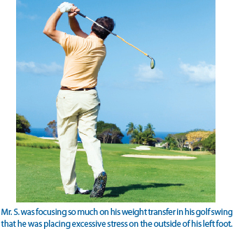
Mr. S. is a well-known golfer in our area. He is 60 years of age, has won several county senior titles and has played at a very competitive level for many years. He presently plays at a “scratch” handicap. Mr. S. has been a patient for a number of years and has always understood the importance of regular chiropractic adjustments and exercise. Early this spring, he came to us with a new complaint of pain in his left low back area radiating into his left hip. Pain was also noted in the area along the lateral tibia of the left leg and continued pain over the lateral aspect of the foot. Mr. S. also feels that when he walks he is placing more pressure down on the left foot.
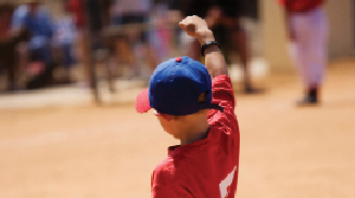
He denies any falls or accidents. When we asked him if he had changed any activity in his exercise program, he stated he was doing the same thing he had in the past. Examination of his gait cycle demonstrated a longer stance phase on the left side in comparison to the right. Video analysis also noted a slight limp with his left leg when he proceeded through the stance phase of gait. Muscle testing noted a weakened abductors bilaterally and gluteus maximus on the left. The left foot showed reduced dorsiflexion while walking and through active and passive ranges of motion. A digital foot scanner demonstrated that Mr. S. had symmetry of all three arches on the right foot, but on his left foot the lateral arch had dropped significantly. Showing Mr. S. the results of his scan, he remembered that he started doing some golf drills that helped him keep more pressure on his left side when finishing his swing, and that they put a lot of stress on the outside of his foot. He stated, “The pain did start a few days after I started those drills!”

Our treatment included stabilizing orthotics to provide his feet with symmetrical stability. Also, we adjusted the left foot after we determined his reduced dorsiflexion was due to a misaligned calcaneus and talus. Abduction exercises and glute-strengthening exercises were all recommended to provide additional stability to further enhance the chiropractic full spine adjusting. We hypothesized that Mr. S. was focusing so much on his weight transfer in his golf swing that he was placing excessive stress on the outside of his left foot. Coupled with rotation stress during his swing finish he developed a neuro-musculoskeletal condition through his changes in his normal biomechanical movement patterns.

Although he could not initially recall a mechanism of injury for the complaints that brought him back into our office, after examination and further questions about his lifestyle and hobbies we were able to determine the root cause of his pain. Since we had no history of any additional trauma, some activity in his daily living had to be a contributing factor. This is an important step to apply with any patient who complains of pain without injury. Consider a patient’s hobbies and see if what they do for fun could be the source of their pain.

A 1980 graduate of Palmer College of Chiropractic, Dr. Kirk Lee is a member of the Palmer College of Chiropractic Post Graduate Faculty and Parker College of Chiropractic Post Graduate Faculty. He has lectured nationwide on sports injuries and the adolescent athlete, and currently practices in Albion, Michigan.

Back Surgery Averted for a Teenaged Boy

History and Presenting Symptoms

The patient is a 13-year-old boy who was identified at a school screening to have a discrepancy in his shoulder heights. His parents were advised to contact an orthopedic surgeon for further evaluation. They reported that the orthopedist found evidence of a scoliosis, but recommended a “watch and wait” approach. No treatment was offered, but if the boy’s spinal curve increased, he (the orthopedist) would be available to perform spinal corrective surgery. The boy’s parents are requesting a second opinion, and any recommendations for non-invasive, conservative care. The patient has no back symptoms, and neither he nor his parents recalls any back injury. He is regularly active in several physical activities, including softball and swimming.

baseballboyExam Findings

Vitals. This 5’4’’ tall, athletic 13-year-old boy weighs 112 lbs, which results in a BMI of 19.2 – he is within the healthy range.

Postural examination. Standing postural evaluation identifies a left low pelvis, and a right low shoulder. His knees are well-aligned, but he has an obvious medial bowing of the left Achilles tendon, with a lower medial arch on the left foot.

Chiropractic evaluation. Motion palpation identifies several mild limitations in spinal motion: the left SI joint, the lumbosacral junction, T11/12, and at the cervicothoracic junction. Palpation finds no local tenderness in these regions, and he has full and pain-free active spinal ranges of motion. Thoraco-lumbar lateral bending is equal for both sides, and the Adams forward bending test finds no evidence of rib hump or persisting curve.

Lower extremities. Closer examination finds that the left medial arch of the foot is lower than the right when standing. When he is seated and non-weightbearing, the left arch appears equal to the right. And when he performs a toe-raise while standing, the left arch returns. Manual muscle testing finds no evidence of muscle weakness in the peroneal or anterior tibial muscles.

Imaging

A P-A full-spine film demonstrates a C-curve scoliosis, which encompasses the lumbar and thoracic regions. The sacral base is lower on the left by 3 mm, and the Cobb angle is 12°. A collimated pelvic view with the femur heads centered finds a difference of 6 mm in the heights of the femur heads, with the left side lower.

Clinical Impression

Here we have a classic case of a functional scoliosis associated with a unilateral flexible flat foot. By definition, this eliminates the concern of a progressive idiopathic scoliosis, which had given the parents cause for worry. The condition is accompanied by multiple areas of mild joint motion restriction and compensatory spinal fixations.

Treatment Plan

Adjustments. Specific adjustments for the lumbopelvic and thoracolumbar spinal regions were provided as needed. Manipulation of the left foot, including the navicular and cuboid bones, was performed.

Support. Individually designed stabilizing orthotics were provided to ensure balanced support for both arches and to reduce weight-bearing asymmetry. Particular emphasis was placed on wearing the supports in his athletic shoes.

Rehabilitation. Because of his age and athletic pursuits, no specific rehabilitation exercises were provided. He was able to continue in his sports activities without difficulty.

Response to Care

All spinal and foot adjustments were well tolerated, since he was young and symptom free. The orthotics improved his postural misalignment and eliminated the shoulder discrepancy. After two months of care, repeat full-spine x-rays with his orthotics in place found only a minimal (3 mm) leg length discrepancy, a level sacral base, and a 6° Cobb angle (which is considered non-scoliotic). He was released to a self-directed home stretching program after a total of eight treatment sessions over two months.

Discussion

This active 13-year-old boy responded well to a combination of spinal adjustments and stabilizing orthotics. Although he was asymptomatic, his parents worried about him being a potential candidate for spinal surgery, based on the specialist’s opinion. Chiropractic evaluation found his scoliosis to be functional, and his flat foot was found to be flexible. Appropriate conservative care was initiated, and was ultimately very successful. In most cases, a functional scoliosis responds well to chiropractic care, and is unlikely to require surgery.

Understanding the Mechanism of Plantar Fascitis

:dropcap_open:A:dropcap_close:s chiropractors, our primary focus of care is the chiropractic adjustment. Based on our education in science, art and philosophy, we decide how and where to apply the chiropractic adjustment. Our treatment of a patient’s condition is based on the reduction or stabilization of the resulting subluxation complexes. We listen to our patients’ concerns and answer their questions, but we know that the expression of pain is not the finite reason for our care. We do not let symptoms dictate what we feel is the underlying cause of a patient’s complaint. For example, we know biomechanically and physiologically that subluxations in the lumbar spine can be a contributing factor in neck pain and headaches. However, when a patient’s complaint is not spine related, we have a tendency to use modalities, hot/cold application, give a recommendation to change shoes, ergonomic correction at our work stations, and often we treat the symptom instead of the underlying factor.

footbones2
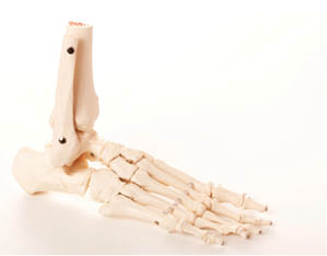
I get numerous emails from doctors who have questions regarding plantar fascia conditions. It is a common ailment for runners, but anyone can develop plantar fascitis. You probably have more patients with plantar fascia problems than you realize because the first signs and symptoms are usually foot pain with difficulty walking when arising out of bed in the morning. Once they have walked a little the pain usually resolves. When they come into our offices later in the day for their other complaints, they might not mention the condition. 
 
Let’s review the anatomy of the foot. Structurally the foot is made up of three arches: lateral longitudinal, anterior transverse and medial longitudinal. These arches form the rigid foundation of the foot, thus the importance of maintaining anatomical height. On radiographic views we can use the landmarks of the cyma line, which demonstrates an S-shaped curve between the talonavicular and calcaneocuboid joints. As the foot transfers the lateral weightbearing forces medial, the navicular drops (rolls inward or pronates), but the talus also slides anteriorly. During supination the talus posteriorly glides. The normal heel-to-toe transition that is important in having a symmetrical gait must have symmetry between pronation and supination. If the talus is constantly being translated forward excessively, the calcaneous will start to shift posterior and superior, causing more tension on the plantar fascia by further lengthening.

We know that the plantar fascia pain is the result of excessive traction of the fascia. Wolff’s law tells us the mechanical stresses will influence and cause hard and soft tissue to distort in direct correlation to the amount of stress imposed on them. 1 Thus, there is a possibility of a heel spur developing. Since the spur is last in forming, the pain is more a result of the excessive tension being placed on the plantar fascia.
 
If a patient demonstrates excessive foot pronation or hyperpronation, then the foot is more flexible, with fallen arches. Effective treatment of the condition must include stabilization of the asymmetrical patterning that the foot is going through. Commonly observed in this patient is reduced dorsiflexion of the foot and ankle. This is usually the result of a tight Achilles tendon, for which the foot must compensate throughout the stance phase of the gait cycle. This creates even more stress on the plantar fascia. For additional anatomical and biomechanical understanding, review the windlass effect that John Hicks first described in 1954.2

When we consider the biomechanical phenomenon that takes place in the foot during the weightbearing portion of the gait cycle, we can see why Leonardo Da Vinci stated, “The human foot is a masterpiece of engineering and a work of art.” So in our treatment plan we must consider adjusting the foot to make sure all joints are moving normally, as well as evaluate for weakened musculature or tight tissue structures like the anterior and posterior tibialis and the Achilles tendon. Scan the patient to identify their pronation index (which helps determine if stabilizing orthotics should be recommended). Possible shoe-type recommendations may be necessary based on foot structure and activity levels. It is extremely important to evaluate the gait cycle once you have made your corrections and recommendations. If compensatory patterns were being developed from favoring the pain and/or restricted movements resulting from subluxations, then some neuromuscular re-education may be necessary to further bring about a symmetrical gait cycle for a patient.
 
References:
  1. Frost HM. Wolff’s Law and bone’s structural adaptations to mechanical usage: an overview for clinicians Angle Orthod. 1994;64(3):175-88. 
  2. Malone TR, Bolgla LA. Plantar Fasciitis and the Windlass Mechanism: A Biomechanical Link to Clinical Practice. J Athl Train. 2004 Jan-Mar;39(1):77–82.
 
A 1980 graduate of Palmer College of Chiropractic, Dr. Kirk Lee is a member of the Palmer College of Chiropractic Post Graduate Faculty and Parker College of Chiropractic Post Graduate Faculty. He has lectured nationwide on sports injuries and the adolescent athlete, and currently practices in Albion, Michigan.

Providing the “Racer’s Edge” to a Young Runner

History and Presenting Symptoms

:dropcap_open:T:dropcap_close:he patient, a 27-year-old female, is a media designer who has been running regularly for the past eight years.  She reports the recent onset and gradual worsening of pain in the front of her left lower leg, which is now limiting her regular physical activities.  The leg pain is described as an “aching soreness” that has been getting progressively worse.  She recalls no specific injury, and has no obvious swelling or discoloration.  Her left leg pain becomes particularly noticeable when she runs downhill or tries to increase her mileage.  There is also now a mild persistent aching in her left buttock region.  She is planning on running her first 10k race in four months.

runnercrossingfinishlineExam Findings

Vitals. This active young woman weighs 122 lbs, which at 5’4’’ results in a BMI of 20.9 – she is at normal weight.  She doesn’t drink alcohol or smoke, and her blood pressure and pulse rate are both at the lower end of normal range.

Posture and gait. Standing postural evaluation finds generally good alignment throughout her spine, although she shows evidence of a left posterior ileum.  She has mild calcaneal eversion, with a lower left arch.  Treadmill gait evaluation indicates obvious hyperpronation of the left foot and ankle when running.  Standing Q-angle is measured at 27° on the left and 22° on the right (20° is normal for women).

Chiropractic evaluation. Motion palpation identifies a limitation in her left sacroiliac motion, with mild tenderness and loss of endrange mobility.  Yeoman’s provocative test elicits moderate pain upon prone extension of the left leg.  Neurologic testing is negative.

Primary complaint. Palpation of the left lower leg finds tenderness and tightness of the muscle insertions in the lower third of the tibia, along the anterolateral aspect.  Manual testing identifies mild weakness of the tibialis anterior, extensor hallucis longus, and extensor digitorum longus muscles, and the isometric testing elicits increased pain in these muscles.  There are no sensory or reflex changes, and no significant asymmetry in muscle mass or leg diameter.  All ankle joint ranges of motion are full and pain-free, bilaterally.

Imaging

No x-rays or other forms of musculoskeletal imaging were requested.

Clinical Impression

“Shin splints” in the deceleration muscles of the left ankle, along with an elevated Q-angle and foot pronation.  This is accompanied by left sacroiliac joint motion restriction and dysfunction.

Treatment Plan

orthoticblueAdjustments. Specific side-posture adjustments for the left sacroiliac joint were provided.  Manipulation of the left navicular and calcaneal bones was performed to reduce the biomechanical stress on the medial arch and sub-talar joint.

Support. Individually designed stabilizing orthotics were provided to support the arches and decrease impact at heel strike.  Two pairs of orthotics were ordered: one for her job-related dress shoes and the other for her running shoes.

Rehabilitation. Full-range resistance exercises (using surgical tubing) for the anterior tibialis muscles were performed daily; her efforts were recorded in a log.  This program progressed to focus on strengthening the eccentric (deceleration) phase in particular.  She was able to continue her distance-running training program.

Response to Care

She responded well to the sacroiliac and foot adjustments, and reported a rapid decrease in her leg symptoms.  Within two weeks (after introducing the orthotics), she was able to return to her previous distance-running training program.  She reported that she felt that her gait was smoother, and that she felt her heel strikes were less stressful.  After a total of eight treatment sessions she successfully completed her first 10k race.  She described moderate, bilateral post-run leg soreness, which resolved within two days.  She then returned to regular running with no persistent or recurrent discomfort.

Discussion

Moderate biomechanical asymmetries can become more prominent (and symptomatic) when levels of physical stress and training volume increase.  This seems to be especially true in the lower extremities.  Shock-absorbing stabilizing orthotics incorporate support for the arches while they reduce pronation and decrease the stress of repetitive heel strikes on the foot and spine.  Anterolateral shin splints indicate a problem with deceleration of the foot at heel strike, which requires improvement of eccentric strength of the anterior tibialis muscle and its co-contractors.