The “Rights” and “Wrongs” of Orthotic Selection

Why is there so much disagreement in the field of professional foot orthotics?  When I read articles and research regarding support of the feet and lower extremities, I am constantly reminded that there are two major camps.  Each side seems to make a strong argument.  If I tell a podiatrist about the types of orthotics I find to be helpful to my patients, I am told that they are no good, and can’t possibly help any patient.  Yet my own clinical experience finds the chiropractic types of orthotics to be very useful.  In fact, I have had many successful results in patients who have previously attempted—unsuccessfully—to use the rigid type of orthotic with forefoot posting.


I am still amazed that some people can walk—much less be told they should run—in the traditional, rigid-control orthotics.  And I still hear stories from my patients of a podiatrist (or even occasionally another chiropractor) who scoffs at the more flexible orthotics I have provided for them.  The two ends of the orthotic spectrum are so different that it seems that someone must be right, thereby making the other doctor completely wrong.  So the question is:  Who or what is right, and who or what is wrong?


Here’s my assessment of the situation:  As is true in many similar controversies concerning the human body, the answer depends on the patient’s needs and the training and focus of the practitioner.  The two major approaches to providing orthotics are completely different systems which are not interchangeable, and cannot be compared within the other system.  Both have their uses and value for patients.


Differing Viewpoints

Each approach has its own construct, or pattern, which is inherently valid.  If you start within the conceptual framework of one orthotic philosophy, you are OK, as long as you stay within its confines.  Each procedure begins with a different viewpoint, and continues in a rational, reasonable manner to an end result which is a very different-looking product.  In other words, each approach makes sense within itself, but the end product (the orthotic) can’t be reasonably judged using the conceptual basis of the other construct.  And, really, it is the final result—the patient outcome—which should be the final determinant.  If the orthotic achieves the purpose for which it was designed, and is, itself, the cause of no subsequent problems, then the intervention can be considered successful, and the patient is the beneficiary!


Apples Aren’t Oranges

In Table 1, I have attempted to differentiate the two major orthotic constructs.  It is evident that both approaches are inherently valid; each one makes sense when viewed from within its own pattern.  To use the concepts of one to evaluate the products of another just won’t work.  This viewpoint has allowed me to appreciate the differences and to understand better the rationales for the two very different procedures.  To jump across columns or mix concepts just doesn’t work; that’s why you can’t send a weight-bearing image of a foot to a podiatric orthotic lab and expect to receive anything useful (and vice versa).  Apples are apples and oranges are oranges; just don’t expect an apple to taste like an orange and you won’t be disappointed!

Table 1. Podiatric and chiropractic constructs for orthotic use


PODIATRIC


CHIROPRACTIC


Patient’s problem


Foot pain


Spinal problems


Biomechanical lesion


Poor foot function


Inefficient support of spine during stance and gait


Apparent cause


Abnormal foot biomechanics


Transmission of abnormal forces to pelvis and spine


Proposed fix


Control excessive pronation


Provide extremity support


Method


Maintain subtalar neutral


Improve arch/ankle function


Biomechanical concept


Forefoot/rearfoot imbalance


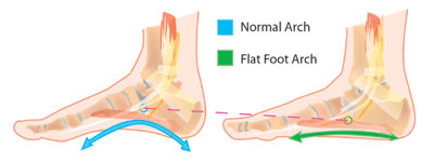
3 arches and heel support


Orthotic solution


Rigid control w/ forefoot & rearfoot posting


Flexible support for arches and shock absorption


(occasional rearfoot posting)


LABORATORY


Imaging needed


Foot in subtalar neutral (“ideal”) position


Foot in functional posture (when it’s providing support for the spine)


Provider input needed


Exact posting measurements


Special instructions (lift, etc.)


Lab procedure


Make negative image


Take measurements from weightbearing casting


PROBLEMS


Athletes


Uncomfortable—often need shock absorption


May need more torsional support  (i.e,. StanceGuard)


Orthopedics


Problems with compensatory hypermobility


Adaptation process may require period of short-term exercise of feet


Neurological


Altered proprioceptive input inhibits postural control


Learning period/muscle retraining soreness


Provider time/expertise


Lengthy/high


Moderate/moderate


Patient cost


Expensive


Moderate


Dr. John J. Danchik is the seventh inductee to the American Chiropractic Association Sports Hall of Fame.  He is the current chairperson of the United States Olympic Committee’s Chiropractic Selection Program, and lectures extensively in the United States and abroad on current trends in sports chiropractic and rehabilitation.  Dr. Danchik is an associate editor of the Journal of the Neuromusculoskeletal System, and has been in private practice in Massachusetts for 27 years. 


Adult Flatfeet and Orthotic Support

Flatfoot (the absence of a medial longitudinal foot arch) is normally found during early human growth and development.  During childhood, we all start out with little or no arch.  Some people, however, never develop normal feet arches.  This situation differs from the collapse of normal arches due to excessive loading and eventual plastic deformation.  Are “normally” flattened feet a clinical problem?  Are arch supports, special shoes, or orthotics necessary?  Chiropractors need to understand the natural history of flatfeet and the clinical presentations associated with this condition. 


In childhood, almost every foot has a large medial fat pad which slowly decreases during maturity, resulting in a more prominent medial longitudinal arch.1  A study of the developing arch in children2 has confirmed that 28% to 35% of school children have a flatfoot deformity, 80% of which are classified as “mild.”  Without treatment, over 90% of these children will have normal arches by age ten.1,3  The remaining 10% will grow into adulthood without developing normal longitudinal arches.


Adults with flatfeet demonstrate several biomechanical inefficiencies in the foot and ankle, as well as a variety of gait abnormalities.  The development of clinical problems is dependent on the levels of activity and the amount of repetitive stress which the feet must endure.  Symptoms and associated pathology arise secondary to excessive pronation of the foot and ankle, and an inability to achieve a rigid foot at toe-off.


A 1988 study assessed the effects of arch supports on oxygen consumption in 20 subjects with flatfeet who complained of fatigue and “weariness.” 4  These subjects, between 18 and 38 years old, had no specific foot symptoms.  Their heart rates, blood pressures, and walking oxygen consumption values were measured on a treadmill—first without, and then with arch-supporting orthotics.  The results demonstrated that use of the orthotics significantly improved their gait efficiency, and decreased their oxygen consumption during normal walking. 


Flexible or Rigid Arches? 


It is important to differentiate a congenital, rigid flatfoot, from the normal, flexible flatfoot, since specialist referral is often necessary for the rigid condition.  Here is a simple test to determine the existence of a rigid flatfoot (usually due to an osseous deformity, such as a tarsal coalition):  If an arch is present when the patient is sitting with the foot dangling, or when standing up on the toes, then the flatfoot is “supple and is correctable with an arch support.”5   If the foot remains flat and rigid during this test, any attempt to lift up or support the arch will be painful and unsuccessful.  In these rare cases, specialized treatment is required.  Evaluation by a foot specialist is usually necessary to determine the underlying cause of a rigid flatfoot and to develop the proper care.


Adult flatfeet and Orthotic Support


Recommendations for Adults with Flexible Flatfoot


  1. Strengthen the lower leg muscles with home exercises (exercise tubing, scrunching up a towel with the toes).
  2. Stretch the Achilles tendon regularly, which is found to be secondarily tight in many patients with flatfeet.
  3. Insist on supportive, lacing shoes with a strong, stable heel counter.
  4. If excessive pronation is present, support from flexible, custom-made orthotics is indicated.


Weight


Body weight is a major factor which can increase the levels of biomechanical stress that the feet must bear.  Adults with flatfeet can decrease their potential for developing clinical problems by keeping their weight within recommended norms.


Orthotics


Orthotics provide support for the arches and decrease the tendency for excessive pronation.  Fit patients who have flatfeet with comfortable orthotics at an early stage, before deformity develops and symptoms become intractable.  Support from flexible, custom-made orthotics will encourage normal function of the foot and ankle.  Further foot deformity can be prevented, along with reduction of abnormal kinetic chain stresses on the pelvis and spine.


Conclusion


Adults with flatfeet should be identified early in the examination process.  The likelihood of foot biomechanics being involved in their musculoskeletal complaints is high.  Corrective support for the arches is often necessary to obtain good clinical results, and custom-made orthotics should be considered from the start.  Prevention of future foot, leg, and back problems requires the chiropractor to discuss the natural history of flatfeet with the patient.  A review of the importance of maintaining normal body weight, good strength and flexibility, and the availability of orthotics will provide needed information. TAC


Dr. John J. Danchik is the seventh inductee to the American Chiropractic Association Sports Hall of Fame.  He is the current chairperson of the United States Olympic Committee’s Chiropractic Selection Program.  He lectures extensively in the United States and abroad on current trends in sports chiropractic and rehabilitation.  Dr. Danchik is an associate editor to the Journal of the Neuromusculoskeletal System.  He has been in private practice in Massachusetts for 26 years.  He can be reached by e-mail at [email protected]. 


References

  1. 1.Kemp HC.  Current Pediatric Diagnosis and Treatment.  Norwalk:  Appleton and Lange, 1984:614.
  2. Notari MA.  A study of the incidence of pedal pathology in children.  Journ Am Podiatric Med Assn 1988; 78:518-521.
  3. Wetton EA.  The Harris and Beath footprint: interpretation and clinical value. Foot & Ankle 1992; 13:462-468.
  4. Otman S, et al. Energy cost of walking with flatfeet.  Prosthets and Orthots Int 1988; 12:73-76.
  5. Hoppenfield S.  Physical Examination of the Spine and Extremities.  New York: Appleton-Century-Crofts, 1976:232.

Orthotic Support for Knee Pain

The knee has an impressive level of stability, despite the incongruent joint surfaces of the tibia and femur.  These two contrasting joint surfaces articulate only with the help of the crescent-shaped menisci.  Through their weight-distribution and shock-protection properties, the menisci help guide movements between the convex surfaces of the distal femur and the almost flat superior surfaces of the tibial plateau. 

A second knee joint involves articulation between the femur and patella.  The integrity of this joint is dependent on the coordinated contraction of the quadriceps muscles and the proper positioning of the patella on the saddle-shaped femoral surface.  Normal knee-joint movement includes pivot flexion and extension, with external tibial rotation on the femur during the last 10-to-20 degrees of extension.1
Unfortunately, conditions that produce excessive rotation of the tibia relative to the femur make the knee susceptible to the development of several degenerative conditions.    

The anterior cruciate ligament (ACL) prevents excessive anterior glide displacement of the tibia on the femur, hyperextension, and internal tibial rotation.  A review of normal and abnormal biomechanics of the gait cycle reveals why ACL injuries are among the most common ligamentous injuries of the knee.2

The Foot/Knee Connection
Pronation of the foot in the contact period helps to absorb shock, and it is normally accompanied by internal rotation of the tibia relative to the femur.  Excessive pronation during gait will transmit damaging forces up the kinetic chain.  It is the excess of these rotational forces which result in repetitive microtraumas.  If this conversion of torque, in response to pronation, occurs beyond normal limits, the tibia can subluxate in internal rotation.

The most common cause of degenerative joint disease is the presence of abnormal biomechanical forces on a normal or healthy joint.  The next most common situation is the application of normal forces on abnormal cartilage.  Excessive pronation, which causes internal tibial fixation and stretches the ACL, creates the necessary ingredient for both of these processes to occur simultaneously. 

In their study comparing measurable drop of the navicular due to pronation with the incidence of ACL injury, Beckett, et al., found the injured subjects had higher navicular drop scores.3  The results are summarized in Table 1.

The Anterior Cruciate Ligament Based.on.image.from:
http://www.willmot.com/knees/acl.html

Table 1. Navicular Drop Scores

Group

Number of Participans

Right Foot

Left Foot

The.anterior.cruciate.ligament (ACL)prevents.excessive anterior.glide.displacement of.the.tibia.on.the.femur, hyperextension,.and internal.tibial.rotation.

1. ACL uninjured 50 6.9+/
-3.2
6.9+/
-2.8
2. ACL injured 50 13.0+/
-4.4
12.7+/
-4.0

In another study, Loudon, et al., measured postural faults as predictors for the occurrence of non-contact ACL injury.  Seven postural positions were measured: pelvis, hip, sagittal knee, frontal knee, hamstring length, ankle pronation, and navicular drop test.  Postural distortions—including knee hyperextension, excessive navicular drop, and excessive pronation—were significant predictors for ACL injury.4

Remember that knee pain can often precede visual radiographic evidence of degeneration.  The navicular drop test can be a good preventative screening tool.
 
Adjust, Support, Rehabilitate
Conservative care for non-traumatic knee pain and degeneration should include:
· adjustments—to provide proper alignment and remove restricted motion (especially internal tibial rotation fixation)
· orthotic support—to control excessive pronation and internal tibial rotation
· rehabilitative exercise—to build the muscles and improve stability

Custom-made, flexible orthotics support the bones and soft tissues of the feet in their proper position of function.  By correcting pedal imbalances (which can cause excessive pronation and tibial torsion), orthotics help prevent overuse injuries and knee-joint degeneration.  Research published in the Journal of Manipulative and Physiological Therapeutics proves that custom-made orthotics improve the structural alignment of the foot, thereby creating a more symmetrical foundation throughout the entire kinetic chain.5  Further research has shown that custom-made, flexible orthotics decrease (normalize) the Q-angle and improve patellar tracking.  This indicates an improved functional alignment of the knee and leg.6

Developing muscles helps stabilize the joint and lower the incidence of serious knee injury.7  Rehabilitative exercises—including strengthening and coordinating contraction of muscles involved in flexion, extension, and rotation—will help enable the patient to perform a range of movements to build strength in muscle groups interacting with the knee. TAC

Dr. John J. Danchik is the seventh inductee to the American Chiropractic Association Sports Hall of Fame.  He is the current chairperson of the United States Olympic Committee’s Chiropractic Selection Program.  He lectures extensively in the United States and abroad on current trends in sports chiropractic and rehabilitation.  Dr. Danchik is an associate editor of the Journal of the Neuromusculoskeletal System.  He has been in private practice in Massachusetts for 26 years. You can contact him at: [email protected]

References

  1. Logan AL. The Knee: Clinical Applications. Gaithersburg, MD: Aspen Publishers, Inc.; 1994.
  2. Bergfeld J, et al. Injury to the anterior cruciate ligament. Phys Sportsmed 1982; 10:47-59.
  3. Beckett ME, et al: Incidence of hyperpronation in the ACL injured knee: a clinical perspective. J of Athletic Training 1992; 27(1):58-62.
  4. Loudon JK, Jenkins W, Loudon KL. The relationship between static posture and ACL injury in female athletes. J Orthop Sports Phys Ther 1996; 24(2):91-97.
  5. Kuhn DR, Shibley NJ, Austin WM, Yochum TR. Radiographic evaluation of weight-bearing orthotics and their effect on flexible pes planus. J Manip Physiol Ther 1999; 22(4):221-226.
  6. Kuhn DR, Yochum TR, Cherry AR, Rodgers SS. Immediate changes in the quadriceps femoris angle after insertion of an orthotic device. J Manip Physiol Ther 2002; 25(7):465-470.
  7. Roy S, Irvin R. Sports Medicine Prevention, Evaluation, Management and Rehabilitation. Englewood Cliffs, NJ:Prentice-Hall; 1983.

Orthitics for Work Boots

Many people wear special footwear at work.  Occupational health guidelines and employers often require the use of protective work boots.  When fitting patients for orthotics, ask what type of footwear is worn throughout the workday, as well as during recreation and relaxation periods.  An orthotic designed for work boots can be very helpful.  While generic insoles and off-the-shelf foot supports provide some comfort, workers appreciate a custom-made orthotic designed specifically for work boots.

Protection from Injury

Work boots protect the feet and ankles from work-related injuries.  This category of footwear contains primarily ankle-high boots made from thick, sturdy materials.  Most have a special sole, which varies depending on the work conditions (slip-resistant rubber, large lugs for traction, non-conductive to electricity, etc.).  Many also have steel, non-crushable protection for the toes.  These boots are often quite heavy.  Because weight on the feet leads to fatigue, a lightweight orthotic is appreciated.  Check the weight of the orthotics you recommend for work boots.

Support for Ankles and Arches

Better work boots provide excellent external support and help hold the foot and ankle in alignment during strenuous activities.  However, the insole must fit the many different shapes of feet that wear that same size.  Therefore, the internal support is generic, with a strong shank under the arches, but no specific support.  Foot and ankle fatigue increases as the workday progresses, especially for workers with non-standard feet, whether flat or high-arched.  A custom-made orthotic improves foot and ankle function during strenuous work activities.  It must cause little or no interference in the proprioceptive feedback system, and the foot and ankle must be kept in good balance.  This means controlling the amount of pronation during gait, preventing excessive ankle and calcaneal eversion, and supporting the three-arch complex.

Support for the Pelvis and Spine

The best work boot orthotic also ensures proper alignment of the pelvis and spine.  Biomechanical asymmetries transmit abnormal forces into these areas, resulting in persistent subluxations and susceptibility for back injuries.  The integration of a pronation wedge or heel lift, when needed, can greatly improve the function of the pelvis, sacroiliac joints, and spine.1  Many chronic back conditions associated with work postures resolve when custom-made orthotics are worn in work boots.

Shock Absorption

Long-term standing on concrete and other rigid materials can lead to back problems.  The best modern orthotics are designed to reduce shock waves generated up the leg into the knee, hip, and spine.  The best materials for work boot orthotics are the viscoelastic polymers (such as Zorbacel® and Poron®), a group of materials created to enhance the body’s ability to dissipate shock stress to sensitive and easily damaged tissues.2
Viscoelastic shoe inserts can significantly reduce reported pain levels in patients with low back pain.3  A report on soccer referees participating in a five-day tournament found, “The incidence of soreness in Achilles tendon, calf, and back were significantly reduced by the use of shock absorbing heel inserts.”4  Orthotics made for work boots should take full advantage of the newer viscoelastic materials.

Moisture Control

The sturdy construction of work boots often causes substantial heat accumulation, especially when working indoors, or during the summer.  Sweat often builds up, which can result in foot odors and fungal infections.  The feet can excrete as much as half a pint of moisture daily.5  Orthotics for work boots should incorporate special materials that pull sweat away from the foot surface.  Such an orthotic is able to control most cases of hyperhidrosis, and even normal foot moisture is handled efficiently.

Additional Orthotic Concerns

Comfort.  A good orthotic must be easy to fit into the work boot, with a minimum of modification.  There should be no sensation of “something in my shoe,” which would distract from work performance.  The newer materials tend to be more comfortable and less intrusive.  The break-in period is usually much shorter, and workers quickly become accustomed to custom-made orthotics designed with viscoelastic polymers.  An orthotic used during work must be supportive, yet flexible, so that it doesn’t hinder natural biomechanical performance.  Longitudinal flexibility (especially at the forefoot) is a major indicator of a comfortable orthotic for work boots.
Durability.  Since many work activities produce high forces and stresses on an orthotic, it must be capable of sustaining repetitive physical insults, with no perceptible decrease in support, performance, or shock absorption.  Moisture and heat must not cause significant degradation of any of the above factors.  Once again, the newer materials are usually the most durable, and their physical characteristics can be maintained the longest.

Conclusion

Orthotics need to be specifically selected for use in work boots.  They should support the arches and spine, be lightweight, and offer shock absorption, moisture control, comfort, and durability.  Patients with properly fitted and selected orthotics for their employment have noticeably less work-related fatigue. TAC

Dr. John J. Danchik is the seventh inductee to the American Chiropractic Association Sports Hall of Fame.  He is the current chairperson of the United States Olympic Committee’s Chiropractic Selection Program.  He lectures extensively in the United States and abroad on current trends in sports chiropractic and rehabilitation.  Dr. Danchik is an associate editor of the Journal of the Neuromusculoskeletal System and the Journal of Chiropractic Sports Injuries and Rehabilitation.  He has been in private practice in Massachusetts for twenty-six years.

Tarsal Tunnel Syndrome & Orthotic Support

Tarsal tunnel syndrome (TTS), while not common in the general population, is occasionally seen among athletes.  In addition, many TTS symptoms can be confused with conditions commonly treated in the chiropractic office.1  Because its etiology is often related to hyperpronation and, therefore, spinal complaints, TTS patients may be concentrated in the chiropractic office.  When a patient complains of burning pain or numbness of the foot or ankle, keep TTS on your list of differential diagnoses, including plantar fascitis, Achilles tendinitis, and lumbar radiculopathy.

Similar in function to the carpal tunnel, the tarsal tunnel is formed by the following borders:  medial calcaneus, medial malleolus, posterior talus, and flexor retinaculum.  Structures passing through these confines include tendons, blood vessels, and the posterior tibial nerve, including its branches, the medial calcaneal, medial plantar, and lateral plantar nerves.

These sensitive structures are susceptible to any direct trauma or lesion that decreases the available space.  The athlete runner with a recent history of increased activity is particularly vulnerable, but also consider any auto accident victims who have jammed their lower extremities.  The energy at impact, sent through the pedals or floorboard and into the feet and ankles, supplies the force necessary for traumatic TTS.  On the other hand, consider the simple act of walking on an excessively pronated foot, which is far more common among patients. 

The review of eighty-seven TTS cases revealed that biomechanical deformities, including tarsal jamming and hyperpronation, were to blame and could be documented radiographically. Furthermore, it has been proposed that even minimal trauma during weightbearing activities in persons with pes planus is the most likely mechanism for TTS.  This same study postulates that when pes planus is functional and associated with malposition of the tarsals, the posterior tibial nerve is stretched with each step taken.3  Tarsal malposition is evident with toe out greater than fifteen degrees and with excessive bowing of the Achilles tendon, when viewed from behind.

The symptoms of TTS can be easily confused with plantar fascitis and, in extreme cases, with lumbar radiculopathy.  In the case of biomechanical overuse, the patient will report poorly localized numbness and tingling of the medial ankle and on the plantar surface, which may extend into the lateral two toes (the lateral plantar nerve being more commonly involved). 

Unlike plantar fascitis, which is generally worse in the morning, TTS is worse at night after activity and may include pain radiating up the medial calf.  The physical exam may reveal loss of two-point discrimination and muscle strength in the distribution of the lateral plantar branch and a positive Tinel’s sign, found when tapping directly over the site of the tarsal tunnel.  A normal Achilles reflex should help rule out lumbosacral radiculopathy.

Initial treatment of TTS includes inflammation reduction of the involved tissues.  This means no weightbearing without the foot and ankle taped or without orthotics in place.  Next, adjustments should be used to restore normal biomechanics throughout the entire kinetic chain (foot, ankle, knee, hip, and spine).  Special attention must be given to the valgus misalignments of the talus and calcaneus.  Additionally, deep friction massage over the flexor retinaculum may release adhesions responsible for compression symptoms.
Most importantly, correct the underlying foot dysfunction (hyperpronation or otherwise).  Pes planus causes tightening of the flexor retinaculum, which can then compress structures within the tunnel.  Although the inflammation may go away, the nature of ligament stretch means that any plastic deformation is permanent (barring surgical intervention).  Therefore, effective and lasting treatment necessitates the use of custom-made, flexible orthotics, which have been demonstrated to control the degree of pronation, as well as the percent of time spent in pronation.

Although relatively uncommon, tarsal tunnel syndrome can present a diagnostic and treatment challenge.  Understanding the etiology of this entrapment syndrome is important for providers concerned with the treatment of athletes and auto-accident victims.  Also, because TTS is often associated with hyperpronation, patients will likely experience other conditions also associated with a faulty foundation, including knee, hip, and low back pain.  Many of these patients will seek care from chiropractors, where treatment consisting of adjustment, soft-tissue technique, and the use of flexible orthotics can offer excellent symptom relief.

Dr. John J. Danchik is the seventh inductee to the American Chiropractic Association Sports Hall of Fame.  He is the current chairperson of the United States Olympic Committee’s Chiropractic Selection Program.  He lectures extensively in the United States and abroad on current trends in sports chiropractic and rehabilitation.  Dr. Danchik is an associate editor to the Journal of the Neuromusculoskeletal System and the Journal of Chiropractic Sports Injuries and Rehabilitation.  He has been in private practice in Massachusetts for twenty-six years.

Going the “Extra Mile” with the Orthotics Process

The main values of providing custom orthotics are the postural support, protection, and comfort they offer to your patients.  Another important aspect of the orthotics process, however, is the help you can give your patients in getting reimbursed for these products. 
Many patients hope that their orthotics will be considered a covered expense by their insurance companies.  For some policies that is true; but there are exceptions.  It is a good idea to suggest that patients read through the various policy documents they were given.  If there is no specific exclusion, it is very possible that some coverage is available.  Either way, it is often helpful if patients have something in writing which details the reasons you are recommending custom orthotics.  This makes them more excited and more willing to pay the cost, even if insurance denies the expense.  Sending a copy of this written explanation to the insurance company does seem to help with payment in many cases.
Figure 1 is an explanatory letter for custom-fitted orthotics.  I recommend you re-type this onto your letterhead, and use it whenever a patient, a family member, or an insurance adjustor needs to be educated regarding the need for orthotics. Of course, any specific information you can provide to make the explanation more customized for each individual patient will be very helpful.  Your patients will appreciate the “extra mile” you have gone to help them in their dealings with insurance companies. TAC

Dr. John Danchik is the seventh inductee to the American Chiropractic Association Sports Hall of Fame.  He is the current chairperson of the United States Olympic Committee’s Chiropractic Selection Program.  He lectures extensively in the United States and abroad on current trends in sports chiropractic and rehabilitation.  Dr. Danchik is associate editor to the Journal of the Neuromusculoskeletal System and the Journal of Chiropractic Sports Injuries and Rehabilitation.  He has been in private practice in Massachusetts for 24 years. You may reaach Dr. Danchik at (617) 489-1220 or e-mail [email protected].