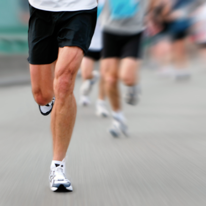
Make Me Run FASTER!

:dropcap_open:H:dropcap_close:ow many times have you heard from an athlete, “Make me run faster!” or “Improve my performance!”? Many athletes would gladly take a little pain for better performance and faster speed. However, ignoring the warning signs of pain can be a no-no! We know that the long-term outcome of living for today and not for the future can lead to chronic and disabling injuries. We have all experienced patients who do not follow our recommendations to reduce their workload, decrease training intensity, reduce the amount of miles they are running, etc. It becomes a mental block for many athletes who tell you they will, but really they won’t. First and foremost, it is not your fault. Unfortunately, many novice athletes think taking a few days off or even reducing their training program will affect their performance, whether they are runners or even football players.
 
If they don’t take your recommendation and refuse to take some time off for their bodies to heal and recover, then eventually they could develop a serious or chronic injury that begins to affect their runningfasteractivities and eventually activities of daily living. The following is a commonly used grading scale for measuring the level of overuse-type injuries in runners. This is only a guideline for you to use during consultation, similar to an Oswestry pain scale.
  1. Pain comes on after the run, but does not affect time and/or distance
  2. Pain occurs during the run, but does not affect time and/or distance
  3. Pain occurs during the run and is affecting time and/or distance
  4. Pain is severe enough that patient cannot run
  5. Pain is severe enough that it affects all activities of daily living
As chiropractors, when we think about increasing the speed of a runner or even a walker, our number-one focus must be on stabilizing any existing vertebral subluxation complexes. We should work on furthering our assessment and treatment protocols to the rest of the musculoskeletal chain. Over the years I have spent a lot of time around the track and listened to numerous high school athletes and track coaches. A popular topic of conversation is about getting the runner to “turn over” his/her stride as fast as possible. 
 
Many drills are done to work on that one phase of running, which means we are talking about power and how quickly we can generate that power. To get a better understanding we must know which muscles come into play to perform this outcome. We know that we increase our power as we go from walking to running to sprinting.
 
The primary power is generated from the hip extensors during the first half of stance phase, the hip flexors at the time of toe off, the knee extensors during the second half of stance, the ankle plantar flexors just before toe off, along with the hip abductors. (1)
 
The hamstring and gluteus maximus generate the power to pull the body forward by actively extending the hip after initial contact, when our foot is placed ahead of the body. Then, during the second half of stance phase, the quadriceps and gastrocnemius contract to push us forward by extending the knee and plantar flexing the foot. The hip abductors then contract to provide lift. Finally, the psoas propels the leg into swing phase by flexing the hip.
 
Thus the total amount of power generated increases as speed increases. The relative contribution from each of these muscle groups changes such that relatively more power is generated proximally (at the hip) as speed increases. (1) Based on this information alone, we must put our focus on a subluxation-free and strong musculature of the hip area.
 
Making Runners Faster
:dropcap_open:We also know that another leading cause of stride asymmetry is abnormal foot function caused by muscle imbalances.:quoteleft_close:
Ultimately, to make a runner faster two important keys are necessary. The first is the runner must have a consistent stride rate, or symmetrical stride. The second is a symmetrical stride length. The average stride rate ranges between 180-200 strides per minute. 
 
Have the runner count his number of steps for 15 seconds and multiply by four. He should do this over several portions of his run to come up with a reasonable average. Then you can determine the speed at which a runner runs by multiplying the steps he takes per second (known as cadence) by the stride length. 
 
To get an accurate measure of stride length I recommend doing what has worked for me for over 25 years – gait analysis using video and computer programs. Regardless of which program you use, it is important to do a follow-up analysis of your patient after a recommended course of care. You should check to see if your recommended treatment plan is improving stride length and improving symmetry in the three phases of the gait cycle. To learn more about what programs I use in my practice, visit my website at www.albionrundoctor.com or you can send a query to [email protected].
 
Research has shown that the main difference between long and short distance runners is the length of stride and not the rate of stride. (2) The average stride length is 2.5 feet long. So to walk one mile it takes approximately 2,000 steps. A common cause of overuse injuries comes from an abnormal symmetry in a patient’s stride length. Let’s do some math: Our patient is walking a one-mile race. Her right leg stride is 2 feet 3 inches and her stride with the left is 2 feet 5 inches, then, over the period of that mile, the right leg takes 100 steps that are 2 inches shorter. This totals to 200 inches or 16 feet 6 inches of distance variable. Substantial? Absolutely!
 
As doctors of chiropractic we understand the meaning and importance of symmetry. All joints from the spinal joints to the hip joints must move to their fullest innate potential. If you want to sing the bone-and-joint song, starting with “the foot bone is connected to the ankle bone,” then be my guest! We also know that another leading cause of stride asymmetry is abnormal foot function caused by muscle imbalances. 
 
These imbalances can cause inversion or eversion of the foot and the functional movement patterns of hyperpronation, resulting from an anatomical breakdown of the three arches of the foot. Stabilizing the three arches of the foot with stabilizing orthotics to allow a normal heel-to-toe transfer in walking or a midfoot-to-toe transfer in running is very important. Remember, when recommending stabilizing orthotics, it is important to also recommend appropriate footwear.
 
A 1980 graduate of Palmer College of Chiropractic, Dr. Kirk Lee is a member of the Palmer College of Chiropractic Post Graduate Faculty and Parker College of Chiropractic Post Graduate Faculty. He has lectured nationwide on sports injuries and the adolescent athlete, and currently practices in Albion, Michigan.
 
References
  1. Guten, G. Running Injuries: “Kinetics Overview”, pg 16. 1997 W.B.  Saunders Company
  2. Rompottie, K.  “A study of stride length in running” International Track and Field: 249-56.  1972

Lower Extremity Conditions and Postural Support

History and Presenting Symptoms

A 37 year-old male presents with a report of pain in his lower back and right hip. Further discussion elicits a history of previous right hamstring strain during high school soccer, along with several episodes of ankle sprains. He has had arthroscopic surgery to evaluate recurrent left knee pain and stiffness, which found no specific problem. He performs stretches daily to maintain flexibility in his iliotibial connective tissues, since they are frequently identified as tight and short. Because of his prior lower extremity symptoms, he does not currently participate in any competitive or recreational sports.


Exam Findings

Vitals. This athletic male weighs 151 lbs, which, at 5’9’’, results in a BMI of 22; he is not overweight. He is a non-smoker, and his blood pressure and pulse rate are at the lower end of the normal range.

Posture and gait. Standing postural evaluation finds generally good alignment throughout the spine, although he shows evidence of a right posterior ileum. He also has mild bilateral calcaneal eversion, with a lower right arch. There is moderate medial bowing of the Achilles tendons when standing, especially on the right, with a tendency to toe out (foot flare) that is more prominent on the right side.

Chiropractic evaluation. Motion palpation identifies a limitation in right sacroiliac motion, with moderate tenderness and loss of endrange mobility. Also identified are moderate limitations in segmental motion at L4/L5 and L5/S1, with local tenderness. Additional fixations are noted at T12/L1, T9/T10, and C5/6. Lumbar ranges of motion are generally full and pain-free. Neurologic and provocative orthopedic testing is negative. Examination of the knees and ankles finds no ligament instability, and all knee and ankle ranges of motion are full and pain-free. Manual testing finds weakness of the right psoas muscle, in comparison to other lower extremity muscles.


Imaging

Upright, weight-bearing X-rays of the lumbar spine demonstrate moderate loss of intervertebral disc height at L4/L5 and L5/S1, but no osteophyte formation is seen at those levels. There is a slight discrepancy in femur head heights (4 mm), and iliac crest heights (3 mm), but no significant lateral curvature of the lumbar spine.


Clinical Impression

Chronic mechanical dysfunction of the pelvis and lumbar spine associated with poor biomechanical support from the lower extremities. By history, there is a pattern of several lower extremity conditions, which are consistent with his identified asymmetry.


Treatment Plan

Adjustments. Specific diversified chiropractic adjustments for the sacroiliac, lower lumbar, thoracic, and cervicothoracic spinal regions were provided as indicated.

Support. Flexible, custom-made stabilizing orthotics were fitted to support the arches and decrease calcaneal eversion.

Rehabilitation. This patient was shown dynamic resistance exercises using elastic tubing to begin strengthening his spinal stabilizers and core pelvic musculature. He was also instructed to gradually initiate a daily brisk walking program while wearing his orthotics, in order to re-balance his hip and pelvic muscles.

Response to Care

He responded well to his spinal adjustments, and adapted quickly to his orthotics. His compliance with the walking and stabilization exercise recommendations was very good, once the correlation between his previous lower extremity conditions and his current back problem was explained. The home-based spinal stabilization program was also quite easy and enjoyable, so he made continued progress. After 6 weeks of adjustments (10 visits) and daily home exercises, he was symptom-free and released to a self-directed home stretching program.


Discussion

Whenever I see the combination of back complaints and a history of lower extremity problems, I look for asymmetry of the feet and legs. If this is present, I know that effective chiropractic care must address the lower extremity imbalances and also retrain the core stabilizing musculature of the lower spine and pelvis. In this case, the patient’s chiropractic care included shock-absorbing orthotics to support his strained lower extremities, and specific exercises to improve his core stability.


Dr. John J. Danchik, the seventh inductee to the ACA Sports Hall of Fame, is a clinical professor at Tufts University Medical School and formerly chaired the U.S. Olympic Committee’s Chiropractic Selection Program. Dr. Danchik lectures on current trends in sports chiropractic and rehabilitation.

Triathlete Troubled by Tarsal Tunnel Syndrome

by John Danchik, D.C., F.I.C.C., F.A.C.C


History and Presenting Symptoms
A 31-year-old male athlete presents with burning pain and paresthesias affecting the ball of his right foot and toes, and extending along the sole of his foot.  He is a competitive triathlete, but doesn’t recall any specific injuries to his right foot.  These sensations are worse at night, and are aggravated by extended standing and long runs.  The onset was insidious and intermittent, but has gradually become more intense and constant—prompting his chiropractic consultation.  On a Visual Analog Scale, he rates his right foot pain at 40mm.

Exam Findings
Vitals.  This active and fit athlete stands 5’ 10” and weighs 178 lbs; although his BMI is 26 (borderline for overweight), his muscle definition indicates that he is not truly at risk for weight-related health problems.  He does not smoke, and his blood pressure is 112/74 mmHg with a resting pulse rate of 60 bpm.
Posture and gait:  Standing evaluation finds good postural alignment with intact spinal curves, but a slightly lower iliac crest on the right, which is confirmed by a lower right greater trochanter.  He also demonstrates right calcaneal eversion with a low medial arch (hyperpronation).  A tendency to toe out (foot flare) on the right is also noted during gait screening.
Chiropractic evaluation:  Motion palpation identifies a mild limitation in right sacroiliac motion, with moderate tenderness and loss of endrange mobility.  Several compensatory fixations are identified throughout the lumbar region.  Otherwise, all orthopedic and neurological testing is negative.
Lower extremities:  Examination of the right foot finds sensory changes to pinprick and light touch testing, and Tinel’s sign (re-creation of paresthesiae when tapping over the area of entrapment) is positive.  Motor nerve involvement is checked by manual muscle testing of the intrinsic foot muscles, and finds weakness of the right digitorum brevis muscle, when compared with the flexor hallucis and flexor digitorum longus muscles.  As anticipated, the right toes demonstrate a “hammer toe” position, since the longus muscles receive their innervation from the nerve before it passes through the tarsal tunnel.


iStock_000010624539SmallA tendency to toe out (foot flare)
on the right is also noted
during gait screening.


Imaging
No X-rays or other forms of musculoskeletal imaging were requested.

Clinical Impression
Tarsal tunnel syndrome, with right foot/ankle dysfunction and subsequent entrapment of the posterior tibial nerve.  This condition is more noticeable due to his high-level sports performance, and his upcoming competitions.  He also has evidence of recurring fixations in the lumbopelvic region.

Treatment Plan
Adjustments:  Specific, corrective adjustments for the SI and lumbosacral joints were provided, with good response.  Manipulation of the right foot—including the navicular, cuboid and calcaneal bones—was also performed, in order to relieve any existing pressure on the tibial nerve.
Support:  Flexible, custom-made stabilizing orthotics were supplied, with a pronation correction added to the right side.  He had no problems in wearing the orthotics, and was able to incorporate them into his active exercising program with little difficulty.
Rehabilitation:  An exercise to stimulate the intrinsic muscles of the foot was initiated (the patient “scrunched up” a dish towel, then straightened it out again using his foot and toes).  This was done for five minutes, at least once, but preferably twice daily.  When that was progressing well, strengthening of the extrinsic muscles of the ankle was added to rehabilitate the weakened support muscles and help prepare him for a return to full athletic capability.
Response to Care:
The lumbopelvic and foot adjustments were well-tolerated, and the stabilizing orthotics made a noticeable improvement in his postural alignment, both at the feet and the lumbopelvic region.  After six weeks of adjustments (12 visits) and daily home exercises, including wearing the orthotics, he was released to a self-directed home stretching program.

Discussion
Excessive pronation and biomechanical asymmetries in the foot and ankle are often locally asymptomatic, although they can cause a variety of nerve entrapment problems in these regions.  In this active young man, his right foot symptoms and the recurring spinal symptoms were closely correlated with his foot imbalance and the resulting gait asymmetries.


Dr. John J. Danchik, the seventh inductee to the ACA Sports Hall of Fame, is a clinical professor at Tufts University Medical School and formerly chaired the U.S. Olympic Committee’s Chiropractic Selection Program.  Dr. Danchik lectures on current trends in sports chiropractic and rehabilitation.

Tight Band Sidelines Fitness Walker

Tight Band Sidelines Fitness Walker

by Dr. John Danchik, D.C., C.C.S.P., F.I.C.C.


History and Presenting Symptoms

A 34-year-old female reports persistent pain at her right knee with walking. She localizes this pain to the lateral aspect, and says that it becomes worse as she continues her regular fitness walking program. She has been trying to walk three miles daily for the past six months, in order to control her weight. On a 100mm Visual Analog Scale, the woman rates her right knee pain at about 65mm. She describes the pain as a sharp soreness in her knee upon each step, with tightness in the adjacent muscles. The tightness builds up after she has gone about a mile, and it then continues for several hours afterward. Acetaminophen has not helped, and ibuprofen provides only partial relief. She doesn’t recall any specific injury.

Continue reading “Tight Band Sidelines Fitness Walker”

Mature Knee Pain

Mature Knee Pain

by Dr. John Danchik, D.C., C.C.S.P., F.I.C.C.


History and Presenting Symptoms

The patient is an active 58-year-old male, with a history of participation in many team sports (baseball and football) in his younger years. He describes recurring pain and stiffness in his right knee, which is especially noticeable in the mornings and also when he plays racquetball or golf. He also has chronic mild-to-moderate pain in his lower back. He says he had numerous injuries to his knees many years ago, but no recent traumatic incidents. He hopes that chiropractic care can help him in both symptomatic areas.


Exam Findings


Vitals. This athletic, middle-aged man weighs 175 lbs, which, at 5’10’’, results in a BMI of 25—he is right on the borderline of overweight. He works out regularly and is in very good condition, which indicates that much of his excess weight is likely to be lean body mass. He is a non-smoker, and his blood pressure and pulse rate are both within the normal range.

Posture and gait. Standing postural evaluation finds a lower iliac crest on the right, and a low right greater trochanter. There is a lumbar list to the right, with compensatory balancing in the lower thoracic spine. His right knee demonstrates an obvious valgus alignment, and there is calcaneal eversion and hyperpronation on the right side. Gait testing finds a persistent flexion of the right knee, with an asymmetrical gait cadence.


Chiropractic evaluation. Motion palpation identifies a limitation in segmental motion at L4/L5, with moderate tenderness and painful loss of end range mobility to the right. A compensatory fixation is noted at T9/T10. Neurologic testing is negative.

Examination of the right knee finds no ligament instability, or evidence of patellar tracking problem. All knee ranges of motion are full and pain free. Manual muscle testing finds no specific muscle weakness around the knees or ankles.


Imaging

Weight-bearing X-rays of the right knee show mild loss of medial joint space, with small medial osteophytes. No advanced imaging is requested.


Clinical Impression

Moderate medial osteoarthrosis (previously osteoarthritis or degenerative joint disease) of the right knee, associated with alignment asymmetry of the right foot and ankle and hyperpronation. This is accompanied by pelvic and lumbar imbalance, with chronic lumbar spinal joint motion restriction and compensatory lower thoracic fixation.


Treatment Plan


Adjustments. Specific, corrective adjustments for the lumbar and lower thoracic spinal regions were provided as needed. Manipulation of the right knee into internal rotation was performed to decrease the external rotation associated with the chronic hyperpronation and valgus stresses.


Support. Custom-made, flexible orthotics were provided to support the arches and decrease the medial stress on the knees.


Rehabilitation. This patient was given elastic tubing exercises to strengthen his foot and ankle intrinsic musculature. He also used this device to strengthen the internal rotator muscles of his right hip.


Response to Care

He easily tolerated the spinal and right knee adjustments. He reported a rapid decrease in lumbar spine symptoms, and a substantial decrease in knee irritation during and after athletic use. Within one week of receiving his orthotics, he reported that he was making it through the whole day with no right knee aching, and that his knee was much less stiff upon arising. He returned to his racquetball and golf with no problems, and was released to a self-directed maintenance program after a total of ten treatment sessions over two months.


Discussion

This case teaches several lessons. The most important is that pronation and biomechanical asymmetry in the foot and ankle are seldom locally symptomatic. And chronic knee and back pain and degenerative processes must be evaluated fully, in order to identify the underlying problems and propose effective treatment. Anti-inflammatory drugs (which he had been using off and on for many years) did not address the primary biomechanical problem.

This condition was previously called osteoarthritis, but there is little evidence for an “itis” condition. Degenerative joint disease is also a misnomer, as this is not a “disease,” and the degeneration process is directly associated with physical activity that the abnormally functioning joint can’t tolerate. Optimal treatment of this condition is a comprehensive, conservative approach that combines chiropractic adjustments with flexible orthotic support.


Dr. John J. Danchik is the seventh inductee to the American Chiropractic Association Sports Hall of Fame. He is a clinical professor at Tufts University Medical School and for 25 years was the chairperson of the United States Olympic Committee’s Chiropractic Selection Program. Dr. Danchik lectures extensively in the United States and abroad on current trends in sports chiropractic and rehabilitation. He can be reached by e-mail at [email protected].

Popliteus Tendinitis in a Softball Player

Popliteus Tendinitis in a Softball Player

by Dr. John Danchik, D.C., C.C.S.P., F.I.C.C.


History and Presenting SymptomsThe patient is a 27-year-old male who participates in an adult softball league on weekends. He describes recurring episodes of pain and swelling along the back and outside of his right knee, which has limited his knee extension for the past several years. Upon questioning, he also reports episodes of lower back pain,with occasional limitations of activity. He presents for treatment of his lower extremity biomechanical faults, and hopes to improve his athletic performance with chiropractic care. Exam FindingsVitals. This athletic male weighs 185 lbs., which, at 6’2’’, results in a BMI of 23; he is large, but not overweight. He is a non-smoker, and his blood pressure and pulse rate are at the lower end of the normal range. Posture and gait. Standing postural evaluation finds generally good alignment, with good muscular development and intact spinal curves. There is a slightly lower iliac crest on the right, and also a lower right greater trochanter. Weightbearing evaluation of his knees and lower extremities identifies right calcaneal eversion and a low medial arch on the right (hyperpronation), amplified with dynamic examination of thegait cycle. Chiropractic evaluation. Motion palpation identifies moderate limitation in right sacroiliac motion, with slight tenderness and loss of endrange mobility. Several compensatory fixations are identified throughout the lumbar region. Otherwise, all orthopedic and neurological testing of the spine is negative. Primary complaint. Examination of his right knee finds all ranges of motion to be full and pain-free, and all ligament tests are solid and non-painful. Manual testing of the knee support muscles finds moderate weakness in the right popliteus muscle, when compared to the left side. In addition, moderate pain (4/10) is elicited when stress testing the right popliteus muscle. ImagingBecause of his prior chronic low back symptoms, a radiographic evaluation of the lumbosacral region was ordered. The basic (AP, lateral, right and left lateral obliques) lumbosacral series found approximately 20% anterior slippage of L5 on the sacrum, (Grade I spondylolisthesis) with bilateral defects in theL5 pars interarticularis.  Clinical ImpressionThis healthy athlete presents with occasional episodes of right knee pain and previous low back pain. Examination found evidence of right popliteus tendinitis and right sacroiliac fixation, with right foot/ankle instability. This is accompanied by compensatory lumbar fixations and evidence of an inactive spondylolytic spondylolisthesis. Treatment PlanAdjustments. Specific, corrective adjustments for the SI joints and lumbar region were provided as needed. The right knee was adjusted into internal rotation.Support. Custom-made, flexible orthotics were provided to support the arches and decrease calcaneal eversion, and to reduce the asymmetrical biomechanical forces being transmittedto the spine and pelvis.Rehabilitation. This patient received specific exercise recommendations to strengthen his popliteus muscle and also for lumbar spinal stabilization. He was initially instructed to perform daily internal rotation exercises of his lower leg against the resistance of elastic tubing. After two weeks, daily strengthening of the transverse abdominis muscles in the upright weightbearing position was initiated, using resistance from anat-home rehabilitative device. Response to CareThe spinal and pelvic adjustments were well-tolerated, and this active athlete required very few re-adjustments. He wore the stabilizing orthotics in his athletic shoes anddaily footwear without difficulty. He reported an immediate subjective improvement in his athletic performance, saying that his knee and leg felt “more stable.” He was released from acute care to a self-directed maintenance program after a total of eight visits over two months. DiscussionThe popliteus muscle assists in flexing the lower leg upon the thigh; when the leg is flexed, it also rotates the tibia medially. It is called into action at the beginning of knee flexion, inasmuch as it produces the slight medial rotation of the tibia that is essential in the early stage of this movement. This athlete was frustrated by the lack of answers and recommendations from various providers regarding his recurrent knee problems. He hadn’t recognized the pre-existing spondylolisthesis condition or the impact of his lower extremity mal-alignment. The asymmetry in his ankles (calcaneal eversion and poor medial support) exacerbatedand contributed to his recurring knee problems. The combination of specific adjustments, custom-madeorthotic support, and strengthening of the knee and pelvis support musculature quickly brought about an excellent response. Dr. John J. Danchik, the seventh inductee to the ACA Sports Hall of Fame, is a clinical professor at Tufts University Medical School and formerly chaired the U.S. Olympic Committee’s Chiropractic Selection Program.


Dr. Danchik lectures on current trends in sports chiropractic and rehabilitation. He can be reached at [email protected]

The Value of Observing Gait

The Value of Observing Gait

by Dr. John Danchik, D.C., C.C.S.P., F.I.C.C.


The patient is a 58-year-old female, who has recently started a personal health and wellness program. In addition to eating more sensibly, she has initiated a daily walking program. After four weeks, she has progressed to 30 minutes of brisk walking everyday. She reports that she has been noticing lower back and right hip pain as she has increased the amount of her walking. She doesn’t recall any injury or specific strain.

Continue reading “The Value of Observing Gait”

President Obama, the Insurance Industry & Chiropractic

President Obama, the Insurance Industry & Chiropractic

by Dr. Mark Studin DC, FASBE, DAAPM, DAAMLP


On May 11, 2009, in New York Newsday, The Associated Press reported a coalition between insurance companies, drug makers, hospitals and doctors to contain and control health care costs to lower the premiums, so that the 50 million Americans who are currently uninsured can get insurance. This is reported to be as a result of President Obama’s health care reform push.

Health care industry leaders have pledged a two trillion dollar voluntary reduction in spending over the next ten years in order to pay for the program and will report to the government in concert with President Obama’s health care reform plan. Although this doesn’t resolve the health care problem, it gives enough room to insure many of those uninsured, and will allow those special interest groups to maintain their influence over Congress in writing the legislative bill.

Continue reading “President Obama, the Insurance Industry & Chiropractic”

Forum [ Your Letters & Emails ]

Dear Sirs,

ForumI read with interest the two letters that appeared in your May 2009 issue regarding Personal Injury. Dr. Anonymous raised a few issues but he painted with a very broad brush. More interesting was Dr. Studin’s reply. I quite enjoyed Dr. Studin’s thoughtful, articulate and imaginative article in response to the anonymous and clearly disgruntled DC regarding the PI issue.

As a long term PI practitioner here in Boston, I have quite a few thoughts and opinions on the matter myself. My office, Boston Spine Clinics, Inc. was, once upon a time, the biggest office system in the Northeast with our six clinics, 11 doctors and support staff. We did mostly PI…..

As for the suggestion that chiropractic be legislated out of the PI arena, I must disagree. Patients do get injured in ballistic impaction accident and no amount of complaining will ever change that. These patients need care and who better to deliver the most efficacious care than chiropractors? The truth is, there has been fraud and abuse in the PI arena sadly propagated by a small group of DCs. I’ve seen it here in MA. Most of the offenders have been caught and licenses have been pulled. But it is a small group that behaved that way and the entire profession should not be smeared by these few….

… So no, don’t deny patients an opportunity to see a chiropractor for whatever reason. Better to have strict but fair Boards and stiff punishment for all abusive doctors on either side of the PI issue.

Sincerely,

John Haberstroh, DC, DABCN, DACAN, FACFE

Boston Spine Clinics

(This is a shortened version of the letter received by The American Chiropractor. To read the full body of the text, and read other letters received by TAC, go to www.amchiropractor.com/forum )

Ankle Pain Limits Runner’s Health Goals


History and Presenting Symptoms
The patient is a 32 year-old female who has been building up a running program for weight loss and wellness.  She reports a recent onset of pain around her right ankle and heel, which is becoming worse as she gradually increases the length of her longest runs.  She plans to participate in a 10-mile run in two months, but is concerned that her heel pain is worsening.  She has been taking over-the-counter anti-inflammatory drugs after each run, but has persisting pain that she rates around 4.0 on a 10cm. VAS scale.

Continue reading “Ankle Pain Limits Runner’s Health Goals”