Contrary to what you may have learned in Chiropractic College, inequalities of leg length (LLI) are extremely common and their effect on posture can be quite profound. I’m not speaking here of the simple “functional” or “apparent” short leg due to asymmetrical muscle tension in the spine. I’m talking about true discrepancies in the physical length of the lower extremity. There are a plethora of possible causes…congenital anatomical deficiencies, degenerative joint disease, surgeries, foot pronation and/or collapse of the arch of the foot, fractures, etc. Regardless of cause, such discrepancies represent an anatomical asymmetry which must often be dealt with if structural correction is the goal.
Many chiropractors perform some variation of the common supine or Derifield leg checks in their daily practice. Unfortunately, such tests serve only to give the doctor the vaguest of clues that something may be wrong. The common chiropractic “leg checks”, to the extent they are reliable at all, give absolutely no quantitative information as to the amount of deficiency. There’s a better way and it won’t require anything more difficult than a single film. The following discussion will give the busy practitioner a quick and accurate way to assess deficiencies of leg length.
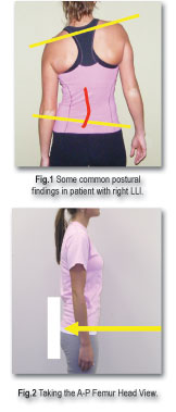
Patients with LLI may present with some or all of the following physical signs. Postural examination will generally reveal a low iliac crest ipsilaterally. The contralateral shoulder is commonly low as the posture compensates. There may be a lumbar convexity to the side of deficiency. Pronation of the ipsilateral foot is common as well. And, yes, you may well pick up the existence of a deficit on your supine or Derifield leg checks as well. Additionally, you may also get a clue of possible deficiency on the A-P lumbo-pelvic film…. Just don’t rely on it for accurate measurement.
In order to accurately diagnose and measure any suspected deficiency, it’s necessary to take a radiograph specifically for that purpose. Your traditional A-P Lumbo-Pelvic view has too much projection distortion to allow accurate measurement. Here are two easy views which will allow you to make an accurate assessment.
1. The patient stands in relaxed neutral posture (See Fig. 2) with the buttocks touching the bucky. The feet are bare, heels even, and six inches apart. Any tendency to pronation of the foot is noted.
2. The bucky is placed so as to center the tops of the femur heads on the film. (The top of the femur head may be easily located by simply having the patient flex the hip and noting the position of the anterior skin crease in flexed position).
3. Use your collimator cross hairs to project the central ray straight horizontal across the top of the femur heads.
4. Collimate as needed to avoid unnecessary exposure to unwanted areas.

By projecting the central ray directly across the tops of the femur heads, we are eliminating much of the projection distortion inherent in the traditional A-P Lumbo-Pelvic. Once we have an accurate projection, it’s simply a matter of measuring for any discrepancy between the relative heights of the femur heads.

Here’s a quick, easy way to precisely measure how much leg deficiency your patient has.
1. A true horizontal line (parallel to bottom of film) is drawn across the top of the tallest femur head and extended across until it crosses the top of the shorter femur head.
2. A dot is placed at the top of the shorter femur head. The distance from the top of the shorter femur head to the horizontal line is measured in mm.
3.The measured distance represents the amount of leg length inequality and provides an accurate measurement for the prescription of heel and/or sole lifts, as needed, to bring the femur heads back to a level position.
Author’s Note: Troyanovich has recommended an alternate view which provides for the additional possibility of measuring sacral obliquity as well as LLI. The details exceed the space available here, but I have prepared a more detailed version of this article to explain the procedure in more detail. Interested doctors may call our offices to request a copy of the unabridged article.
Once you know the amount of discrepancy, exactly when and how you might choose to intervene and try to correct the leg length discrepancy is a subject of some debate. I generally ignore discrepancies of three millimeters or less as insignificant. In cases of severe foot pronation and/or collapse of the longitudinal arch of the foot, I will place the patient in orthotics, allow six to eight weeks for the body to adapt, then take another femur head view to determine if additional correction is necessary.
If foot pronation isn’t the problem, generally proceed as follows. Short legs of four to ten mm generally get a heel lift only. Larger deficiencies are sent to a local shoe shop for a buildup of the entire shoe sole. Please don’t take these to be prescriptive of what you should do for any particular patient. They’re just generalized descriptions of what I’m doing in my own office. I hope you find this simple procedure to be helpful.
Mark R. Payne D.C.
Dr. Mark Payne is president of Matlin Mfg., a manufacturer and distributor of postural rehab products since 1988. For a FREE, unabridged copy of this article or other information on postural chiropractic, please contact Matlin Mfg. Inc. at 1-334 448 1210 or on the web at
www.MatlinMfg.com