 History and Presenting Symptoms
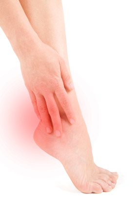
The patient is a 55-year-old male, who reports severe pain on the bottom of his left foot when he gets out of bed in the morning. He also notices pain under his left heel whenever he stands for thirty minutes or more. Over-the-counter pain medications provide some temporary relief, but his condition does not seem to be improving, even though he has been avoiding extensive walking and standing. He has been unable to work out at his local gym for the past sixty days, due to heel pain. There is no history of prior injury to his left foot or ankle.
Exam Findings
Vitals. This 5’10’’ financial analyst weighs 196 lbs, which means that he is overweight (BMI of 28). He demonstrates a thickened waist (44 in.), confirming that his excess weight is due to abdominal fat deposition. He has never smoked, and his blood pressure and pulse rate are within normal range, probably because of his regular involvement in physical exercise (gym activities).
Posture and gait. Standing postural evaluation finds generally good alignment, but a decreased lumbar lordosis. He has bilateral pes planus (flat foot), with no medial arches and bilateral calcaneal eversion. These findings are somewhat more pronounced on the left side. Both feet tend to toe out during walking.
Chiropractic evaluation. The patient’s lumbar spine is moderately tender throughout, and he demonstrates a generalized loss of vertebral mobility, with few specific fixations. Orthopedic and neurological provocative testing of the spine and pelvis is negative.
Primary complaint. Examination of the left foot reveals exquisite tenderness to palpation over the antero-medial aspect of the calcaneus. All ranges of motion for the left foot are full and pain-free, and manual muscle testing finds no evidence of weakness when compared to the right side.
Imaging
A lateral x-ray of the left foot demonstrates a small bony outgrowth from the anterior aspect of the calcaneus.
Clinical Impression
Chronic irritation at the insertion point of the plantar fascia into the calcaneus, with radiographic evidence of a heel spur. This irritation is apparently secondary to long-standing biomechanical stress associated with poor foot function, and excessive loading from strenuous exercise activity and too much body weight.
Treatment Plan
Adjustments. Mobilization and adjustments were provided to the lumbopelvic region. The left calcaneus was adjusted anteriorly and both navicular bones were adjusted superiorly.
:quoteright_open:SYMPTOMATIC HEEL SPURS ARE CHALLENGING CASE PRESENTATIONS, AND THEY REQUIRE APPROPRIATE PATIENT EDUCATION.:quoteright_close:
Stabilization. Individually designed stabilizing orthotics with shock-absorbing materials were provided to support his arches and to reduce calcaneal eversion. In addition, a calcaneal “divot” was ordered for the area under the left heel, in order to decrease the pressure on the bone spur.
Rehabilitation. A series of foot exercises was recommended to the patient, to improve the coordination and strength of his foot intrinsic muscles. After receiving his orthotics, he also performed standing Achilles tendon stretches, keeping his feet in forward alignment.
Response to Care
At first this patient’s heel pain was somewhat slow in responding. However, he was diligent with his exercises, and was eventually able to walk in the morning with no foot pain. At that point, he was advised to return to his regular exercise program at the gym, and he had no further heel pain. He was released to a self-directed home stretching program after a total of eight visits over six weeks.
Discussion
Radiographic evidence of a heel spur does not always correlate with heel pain. However, it frequently indicates chronic biomechanical stress to the insertion of the plantar fascia. Symptomatic heel spurs are challenging case presentations, and they require appropriate patient education.
If this overweight 55-year-old man had been less active, or if he had inherited feet with better arches, he would have been less susceptible to heel pain. However, if he is able to follow through on his decision to drop 22 – 25 pounds of abdominal adipose tissue (which is necessary for him to be considered in the normal weight range for his height), he will be less likely to suffer future recurrences.