
History and Presenting Symptoms
:dropcap_open:A:dropcap_close: 47-year-old female presents with recurrent, sharp pain in her low back. These episodes, which usually resolve within a few days, concern her because they are becoming more frequent. Using a Visual Analog Scale, she describes the usual pain level in her lower back as around 40mm. She doesn’t recall any injury to her back, and cannot identify any specific cause for her pain. She states that she just “tries to relax” for a few days, until the pain resolves.
 Exam Findings
 Vitals. This patient is 5’6’’ tall and she weighs 136 lbs, which is a BMI of 21.9; she is not overweight. Her blood pressure is 124/76 mmHg, with a pulse rate of 76 bpm. She reports that she has never used tobacco products, and averages 2-3 glasses of white wine per week.
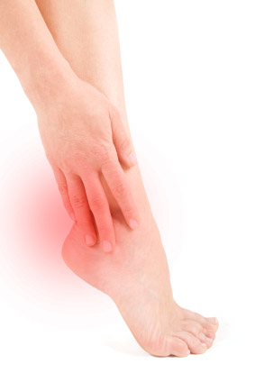
Postural examination. Standing postural evaluation finds basically good alignment throughout her pelvis and spine, except for an accentuated lumbar lordosis. She has a mild bilateral knee valgus and moderate calcaneal eversion, with hyperpronation bilaterally. During gait, both feet demonstrate a tendency to toe-out. An examination of her shoes reveals scuffing and wearing of the lateral aspect of both heels. She states that she usually wears shoes with higher heels for work, and that she has noticed that all her shoes wear out quickly.
Chiropractic evaluation. Kemp’s testing produces sharp pain localized to the lumbar spine when performed to both sides. Motion palpation identifies functional limitations in extension at the L3/L4 and L4/L5 levels, with moderate tenderness and loss of endrange mobility. Neurological tests are negative for nerve root impingement.
Imaging
A-P and lateral lumbopelvic x-rays in the upright position are taken during relaxed standing.The sacral base angle is 48° and the lumbar lordosis measures 62°, and the lumbar gravity line (from L3) falls anterior to the sacrum. There is evidence of chronic facet imbrication, with sclerosis seen at L3/L4 and L4/L5. There is no discrepancy in femur head or iliac crest heights, and no lateral listing or lateral curvature is noted.
Clinical Impression
Chronic facet syndrome with lumbar hyperlordosis and increased sacral base angle. This postural stress is being exacerbated by regularly wearing high heels, and by her tendency to overpronate during gait.
Treatment Plan
Adjustments. Flexion distraction and side posture adjustments for the lower lumbar region were provided as needed, with good response.
Stabilization. Individually designed stabilizing orthotics were supplied, and she was told to limit her heel height to 1” maximum. She was found to be wearing shoes that were too small for her feet, and was instructed to increase one full size for proper fit.
Rehabilitation. She was instructed in a daily core strengthening program, to be done at home using elastic exercise tubing.The focus was on activation of her transverse abdominis musculature, for improved spinal stability.
Response to Care
This patient responded rapidly to her spinal adjustments. She had very little difficulty in adapting to the flexible orthotics, and she reported that the slightly larger shoes with lower heels were much more comfortable. She was consistent with her home exercise program, as demonstrated by her exercise log. After six weeks of adjustments (eight visits) and daily home exercises, including wearing the orthotics in properly fitted shoes with lower heels, she was released to a self-directed home stretching program.
Discussion
This woman’s case reinforces the importance of investigating all sources of underlying biomechanical stress, especially when a spinal condition is chronic or recurrent. Shoe-related postural problems are not uncommon, especially in women. Many women don’t check their shoe size for many years, and they often wear shoes th at are too small for their feet. Heel height can complicate spinal facet syndromes, resulting in a poor response to chiropractic care.
Dr. John J. Danchik, the seventh inductee to the ACA Sports Hall of Fame, is a clinical professor at Tufts University Medical School and formerly chaired the U.S. Olympic Committee’s Chiropractic Selection Program. Dr. Danchik lectures on current trends in sports chiropractic and rehabilitation.

 :dropcap_open:V:dropcap_close:
 Vitamin D deficiency is a serious medical condition that has been associated with an increased risk of developing cardiovascular disease, type 2 diabetes, hypertension, various cancers and autoimmune diseases. Vitamin D insufficiency occurs at epidemic levels in many industrialized countries, where exposure to sunlight tends to be limited and diets tend not to include sufficient amounts of foods naturally rich in vitamin D. During 2009, Dr. Guillory tested more than 1,200 of his patients and found that roughly 90 percent had sub-optimal vitamin D levels, as determined by serum 25(OH) vitamin D levels below 32 ng/mL. Dr. Guillory achieved great success in treating this with Bio-D- Mulsion Forte®, a microemulsified preparation made by Biotics Research Corporation.
 20 full-time firefighters of the Aurora Fire Department were selected on a volunteer basis to participate in an eight-week study during the winter/spring months of 2009. Upon initiation of the study, the 20 subjects were advised to stop consuming multivitamins, cod liver oil and other supplements containing vitamin D. The subjects filled out a medical symptom questionnaire aimed to assess subjective indications of mood, energy level and digestive complaints. All subjects had blood drawn (at the Care Group, PC, office of Gerard Guillory MD in Aurora, Colorado ) and serum levels of 25- hydroxyvitamin D (25(OH)) vitamin D tested through Laboratory Corporation of America (Lab. Corp) via an assay developed by DiaSorin. The subjects were advised to take 4,000 IU/day (2 drops) daily of the liquid emulsified preparation produced by Biotics Research Corporation. After eight weeks of daily supplementation the study subjects 25(OH) vitamin D levels were retested by Laboratory Corporation of America. The subjects also filled out the same medical symptom questionnaire and the data was compiled.
 At the beginning of the study, the average baseline 25(OH) vitamin D blood level was 27.02 ng/mL. Current medical guidelines suggest that vitamin D insufficiency begins when blood levels are below 32 ng/mL and optimal disease prevention occurs when blood levels are above 60 ng/mL (REF). Only five study subjects had serum levels above the 32 ng/mL level and two subjects had blood levels less than 11 ng/mL. The majority of subjects had levels in the low to mid 20s (See Table 1). Prior to supplementation 75% of the subjects were deficient in vitamin D and 10% of the subjects were severely deficient (as defined by blood levels below 10 ng/mL).
 History and Presenting Symptoms
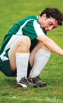
 History and Presenting Symptoms
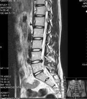
 Based on the examination it was apparent that patient DH had S1 inhibition, which was being expressed by a marked fibularis longus and brevis weakness. Patient DH was compensating for this by distributing more weight on the anterior talofibular ligament and calcaneofibular ligament to check ankle inversion. As such, there was no antagonist control of the stirrup effect created by the tibialis anterior and the fibularis longus. Without this being controlled the tibialis anterior, along with the extensor hallux longus, were overly dorsiflexing the first ray in relationship to the foot upon stance. The excessive lateral wear pattern on the patient’s shoe was associated with a lack of antagonist control of the deep posterior compartment maintaining the foot in an inverted position throughout the gait cycle and stance.
 :dropcap_open:I:dropcap_close:got a call from someone who sounded terrible.


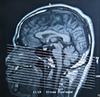
 awareness of the rate of traumatic brain injuries (TBI) or concussion in today’s youth. This information is mainly directed toward the adult population. Football has always been the sport of primary focus, but recent injuries to NHL star Sydney Crosby has made even more people sit up and take notice. For purposes of this article, our emphasis will be more on the adolescent.