 :dropcap_open:L:dropcap_close:ast month, I started a three-part series on the short leg syndrome and the absolute necessity of not only properly identifying it, but finding the exact cause, and tracing the effects up the leg into the pelvis and onto the spine.
I discussed the frequency of short leg syndrome in the population and its relevance to low back pain. We described the Allis Test for determining when the short leg was produced by structural misalignment above the knee, in the hip, and when the problem was below the knee, in the ankle. Either way, restricted joint range of motion was the result and, unfortunately, often goes undetected.
This month, we move down the leg to the feet and ankles. I’ll begin by describing what I believe is the most overlooked and underrated stress responsible for prolonging recurring and chronic structural problems.
Morton’s Syndrome—Long 2nd Metatarsal
Morton’s Toe is the presence of a second toe being longer than the first toe. This may occur in one or both feet and is not an unusual finding, since it is estimated that 40% of the population has a 2nd toe that is longer than the big toe.
This condition causes the weight bearing surface of the foot to shift laterally from the 1st metatarsal to the 2nd metatarsal. This creates a knife-edge rolling effect and can be seen as the lateral heel and medial sole wear out on shoes. Since the big toe is designed to bear weight when walking, there is a profound instability in weight bearing. The only possible answer is to support the 1st toe. The patient should be fitted with orthotics to correct this mechanical problem and failure to do so will result in a continued major mechanical stress for the patient and render your therapeutic efforts ineffective.
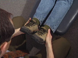
Ankle Fixations
Have the patient straighten their legs and examine for limited plantar flexion and dorsiflexion.
Ankle Dorsiflexion
Examiner stands at the patient’s feet: Passively dorsiflex and slightly evert the patient’s feet. Notice if there is restriction, especially on one side. When positive, search the Gastrocnemius muscle for evidence of muscle contraction.
Ankle Plantarflexion
Passively plantar flex and slightly invert the patient’s feet. Notice if there is restriction, especially on one side more than the other. When positive, palpate the reflex point for the anterior tibial muscle.
There are a number of adjusting techniques available for correction of these restrictions; however, because of the stress of weight-bearing, orthotics are often necessary.
The Prone Examination
With the patient in the prone position, we examine for restricted knee flexion, restricted dorsiflexion (heel tension), and restricted hip extension.
Prone Knee Flexion
This test is conducted by lifting the lower leg, and gently pushing the heels towards the pelvis. You are looking for a restriction in one or both legs. It is very important to place your hands closer to the knee than ankles, as you gently test for equal “springing” in the knees and do not force the legs toward the buttocks. In other words, we are checking unequal range of motion in the two legs and muscle contraction in the Quadriceps muscle.
- Restriction may also indicate cartilage degeneration damage in the knee joint.
- Very often when conducting the test, the patient complains of pain or soreness in the lumbo-sacral area. This is caused by the stretching of the ilo-lumbar ligament that runs from the Ilium to the 5th lumbar and the increase of the A-P lumbar curvature.
- When this occurs there will be muscle contraction and subluxation in the dorso-lumbar area, especially involving T11 to L1. Correction here will relieve the pain in the lumbo-sacral area much to the patient’s delight.
Ankle Dorsiflexion with Knee Flexion
Bend the lower legs off the table to 90o. Press downward on the balls of the feet pushing the toes toward the floor. The test is positive when there is unequal resistance. This indicates the presence of a stress point in the Soleus muscle. The Soleus and Gastrocnemius muscles are used to plantar flex the ankle joint. However, they can be examined separately because the Gastrocnemius is disabled when the knee is flexed to 90 degrees.
The Soleus is critical for running and jumping. When it is contracted, the patient often complains of painful heels and difficulty walking up and down stairs, as well as sacroiliac pain on the same side of the body. The Soleus is often referred to as the “2nd heart” because of its role in returning blood up the legs. Large venous sinuses are found in the calves covered by a tough fascia with large veins above that.
Next month, I conclude this series by describing restricted hip extension and its significance. Then, we’ll conclude by summarizing the flow of kinetic energy when walking, from the feet up the leg to the pelvis and lower back, and its importance to our profession.