Intertrochanteric Fracture Of The Proximal Femur
by Dr. Terry R. Yochum, D.C.; D.A.C.B.R.; Fellow, A.C.C.R. and Dr. Chad J. Maola, D.C.
CASE HISTORY
This elderly female patient slipped getting out of the bath tub. She heard a crack and felt immediate pain.
DIAGNOSIS
Fractures around the proximal femur are relatively uncommon in young to middle-aged patients with a sharp increase in the geriatric patient.1 Severe forces are necessary to fracture the proximal femur in the young and middle years, while only moderate to minimal trauma may induce a fracture in the osteoporotic bone of the elderly. Certain predisposing factors may allow fractures to occur, such as the presence of Paget’s disease, fibrous dysplasia, benign or malignant bone tumors, osteoporosis, osteomalacia and radiation-induced osteonecrosis.1
The overall incidence of all types of fractures of the proximal femur shows a two to one female-to-male ratio. A five to one female predominance exists with intracapsular fractures. The average age is approximately seventy years. It has been estimated that 10 percent of white females and 5 percent of white males will sustain fracture of the proximal femur by the age of eighty years. The incidence by the age of ninety years increases to 20 percent for women and 10 percent for men. Many elderly patients with fractures of the proximal femur die within six months of the original injury. This occurs secondary to pulmonary or cardiac complications. Therefore, fracture of the proximal femur and their attendant sinister1 complications are of such proportions that they represent a major health hazard to the elderly and constitute a significant public health issue because of their frequency, morbidity, and cost.

The standard radiographic examination of the hip joint includes an anteroposterior (AP) full pelvis, AP hip spot (involved side) and an oblique or frog-leg projection.1
Types of Hip Fractures
The types of hip fractures are divided into intracapsular and extracapsular, as determined by the relationship of the fracture line to the joint capsule. In general, intracapsular fractures have a high incidence of nonunion and avascular necrosis due to probable disruption of the tenuous blood supply. 1
Intracapsular Fracture. Any fracture involving the femoral head or neck proximal to the trochanters is classified as being intracapsular. These are then named according to the fracture location:
a) subcapital (involving the junction of the femoral head and neck;
b) midcervical (through the midportion of the fermoral neck);
c) basicervical (traversing ) the base of the femoral neck and its junction with the trochanters.
Most femoral neck fractures are subcapital; midcervical and basicervical fractures are uncommon.
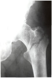
Extracapsular Fracture. This type of fracture occurs outside of the joint capsule and includes intertrochanteric, subtrochanteric and avulsion fractures of the greater or lesser trochanters. Avascular necrosis and nonunion are uncommon complications in extracapsular fractures.
The intertrochanteric fractures are usually comminuted, with the greater or lesser trochanter, or both, forming separate fragments. The oblique fracture line usually splits the trochanters, separating the femur into two components. The proximal component consists of the head and neck, and the distal component includes the shaft and the remainder of the trochanter.
The subtrochanteric fracture is found in the area two inches below the lesser trochanter. This is an uncommon type of fracture of the proximal femur. Middiaphyseal fractures follow severe trauma and are prone to malalignment unless treated appropriately. Pathologic fractures of the proximal femur often occur in the subtrochanteric region. Paget’s disease and metastatic lesions in the proximal femur may be predisposing factors to the development of a subrochanteric fracture; thus the presence of a subtrochanteric fracture should be a signal to the observer to look closely for roentgen signs of adjacent bone disease.
Dr. Terry R. Yochum is a second generation chiropractor and a Cum Laude Graduate of National College of Chiropractic, where he subsequently completed his radiology residency. He is currently Director of the Rocky Mountain Chiropractic Radiological Center in Denver, Colorado, and Adjunct Professor of Radiology at the Southern California University of Health Sciences, as well as an instructor of skeletal radiology at the University of Colorado School of Medicine, Denver, CO. Dr. Yochum can be reached at 1-303-940-9400 or by e-mail at [email protected].
Dr. Chad J. Maola is a 1990 Magna Cum Laude Graduate of National College of Chiropractic. Dr. Maola is a Chiropractic Orthopedist and is available for post-graduate seminars. He may be reached at 1-303-690-8503 or e-mail [email protected].
References
1. Yochum TR, Rowe,LJ: Essentials of Skeletal Radiology, 3rd ed., Williams & Wilkins, Baltimore, Maryland., 2005