I’ve spent a lot of time and ink over the past year, writing about the importance of, and how to rehabilitate, the normal cervical lordosis. This month, I want to spend a bit more time on the lumbar lordosis.
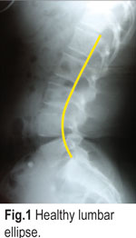
Recall that, in the cervical spine, the normal lordosis should be a simple arc of a circle. Unfortunately, for us, the healthy configuration of the lumbar spine is just a bit more complex. Ideally, the lumbar lordosis should be an ellipse with more curve in the lower segments than in the upper ones. ( Fig. 1. ) shows a healthy (not perfect) lumbar ellipse. I have traced George’s line in yellow to help visualize the accentuated curve in the lower segments.

The procedure for measuring the amount of lordosis on the lateral lumbar radiograph is similar to the cervical spine with just a few differences. We can obtain an accurate, quantitative value for the lumbar lordosis by using Jackson’s angle, just as we did for the cervical spine in an earlier article. We can construct Jackson’s angle using three simple steps.
Step One: Place dots on: a) the posterior-INFERIOR and b) posterior-SUPERIOR, corners of the vertebral bodies of L-1 and L-5. (See Fig. 2.) When complete, you should have drawn a total of four dots (shown here in red) on the film.

Step Two: Construct lines for both L-1 and L-5 by connecting the dots with a ruler. Be sure to Step Three – Using a protractor, measure Jackson’s angle to determine the amount of lordosis. Simply lay the base line of your protractor along the L-5 tangent line, center the protractor at the point where the lines intersect, and measure the acute (smaller) side of the angle formed between the L-1 and L-5 tangents. See Fig. 3. to see which side of the angle to measure.

What this tells us. So, what’s normal?
As measured here, Jackson’s angle gives us two very important pieces of information. First, it tells us the overall amount of lordosis which is present with a simple, accurate, and proven measurement. Second, it tells us where the apex of the lumbar lordosis is located. All of which sounds good, but we still need some kind of normal value to compare it to. After all, it does us little good to know how much lordosis is present if we don’t know what constitutes normal.
The best paper I’ve read to date was published in 1997 by Troyanovich, et al., in the Journal of Spinal Disorders. The paper, entitled Radiographich mensurations charactersistics of the sagittal lumbar spine from a normal population with a method to synthesize prior studies of lordosis, studied 552 asymptomatic subjects, in a wide range of age groups. The results lead the authors to conclude that there seems to be “an ideal sagittal lumbar curvature that may tend to protect holders of the geometric configuration against nociceptive tendencies. In other words, patients who fall within a certain range of lordotic values are less likely to experience back pain.
Here’s the short version. The normal value for Jackson’s angle as measured from L-1 to L-5 is from 35 to 47 degrees of lordosis. In pain free subjects, approximately 65% of the lumbar lordosis occurs between L4/5 and L5/S1 with the remainder (35%) of the lordosis occurring above L-4. Patients with acute low back pain, tended to be HYPERlordotic while patients with chronic LBP tended to be HYPOlordotic.
Now take a moment to refer back to Fig. 3. This patient presented with a Jackson’s angle of approximately 46 degrees (at the very upper range of “normal”) and complaining of mild lower back pain of short duration. This patient was treated successfully with a brief round of adjustments followed by a home treatment program consisting primarily of low back stretches and abdominal strengthening exercise to help reduce the lordosis back toward the middle range of normal.

Of course, every case isn’t so clean cut. There will always be some folks who fall within the “normal” values and yet are still symptomatic. Wouldn’t it be great, if everything in chiropractic was so picture perfect! Nevertheless, using simple radiographic analysis to accurately determine the postural status of your patient can be a great tool in determining how to best approach management of the case. Once you start analyzing your films in this manner, I think you will be surprised at the accuracy of Troyanovich’s conclusions. If you are interested in learning more about how to accurately analyze the lateral lumbar radiograph, I have prepared a FREE REPORT entitled, Measurement of the Lateral Radiographs, which explains the process in much more detail. Interested doctors may call our office for a free copy.
 Dr. Mark Payne is the president of Matlin Mfg., a manufacturer and distributor of postural rehab products since 1988. For a free, unabridged copy of Measurement of the Lateral Radiographs as well.