History
This young adult patient has had a motor vehicle accident with a “whiplash” type injury. Painful hyperextension of the cervical region is experienced by the patient.
 |
|
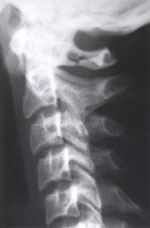
Figure 1. Note the unilateral fracture of the posterior arch of the atlas. |
Discussion
The radiolucent unilateral defect in the posterior arch of this patient’s atlas represents a complete acute vertical fracture.
Fractures of the posterior arch of the atlas are the most common of all C1 fractures.1,2,3 They account for at least 50% of all atlas fractures.1,2 The fracture is usually a bilateral vertical fracture through the neural arch, through or close to the junction of the arch to the posterior surface of the lateral masses. This fracture occurs as a result of the posterior arch of the atlas being compressed between the occiput and the large posterior arch of the axis during severe hyperextension. Almost 80% will have another cervical spine fracture. It is best seen on the lateral projection and can easily be overlooked.3 Serious complications are unusual, though associated cervical fractures may precipitate spinal cord injury. Close anatomic proximity of the vertebral artery to the fracture site may occasionally impact serious vascular injury.1,2
References
1. Yochum TR, Rowe LJ. Essentials of Skeletal Radiology, 3rd ed., Lippincott, Williams & Wilkins, Baltimore, Maryland, 2005.
2. Levine, AM, Edwards CC: Fractures of the Atlas: J Bone Joint Surg (Am) 73:680, 1991.
3. Landells CD, Van Peteghem PK: Fractures of the Atlas: Classification, Treatment and Morbidity. Spine 13:45, 1988.
Dr. Terry R. Yochum is a second generation Chiropractor and a Cum Laude Graduate of the National College of Chiropractic, where he subsequently completed his radiology residency. He is currently Director of the Rocky Mountain Chiropractic Radiological Center in Denver, Colorado and Adjunct Professor of Radiology at the Southern California University of Health Sciences, as well as an instructor of skeletal radiology at the University of Colorado School of Medicine, Denver, CO. Dr. Yochum’s 3rd edition textbook, Essentials of Skeletal Radiology, was released in the fall of 2004 and is now available for purchase. Dr. Yochum can be reached at 303-940-9400 or by e-mail at [email protected].
Dr. Chad J. Maola is a 1990 Magna Cum Laude Graduate of the National College of Chiropractic. Dr. Maola has co-authored five chapters in Dr. Yochum’s 3rd edition textbook and is rendering post-graduate lectures with Dr. Yochum and separately throughout the United States. Dr. Maola is a Chiropractic Orthopedist and is available for post-graduate seminars. Dr. Maola may be reached at 303-690-8503 or e-mail [email protected].