Spinal Cord Injury without Radiographic Abdnormalities SCIWORA
by William J. Owens, D.C., D.A.A.M.L.P. & Mark E. Studin, D.C., F.A.S.B.E.(C), D.A.A.P.M., D.A.A.M.L.P.
When evaluating the traumatically injured patient, correlation of all aspects of the diagnostic process is paramount to proper care. The diagnostic process starts with eliciting a complete and thorough history of the trauma and the resulting symptoms. “Eliciting” a history is different than “taking” a history. Eliciting a history involves the clinician asking pointed questions about each and every part of the patient’s body, to ensure subtle complaints are not overlooked. In many cases, these subjective complaints help to direct objective testing. Correlating the patient’s history with physical examination, lab results and imaging studies helps to complete the diagnostic picture and provide the doctor with an accurate causal relationship, prognosis and treatment plan.
What if the history, objective and radiological testing do not correlate? Did the patient have an accident? Go to the ER for evaluation only to be released with radiographs and CT being normal? Is it your policy to evaluate the patient or do you rely on the testing of others to diagnose your patients?

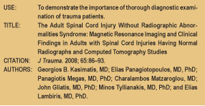
This very recent paper, published in The Journal of Trauma in 2008, by Kasimatis, et al., discusses in depth the presentation of patients with cervical spine injury that had a “radiologic-clinical mismatch.” The authors state, “This retrospective review provides information on adult patients with cervical spinal cord injuries whose radiographs and computed tomography studies were normal.” (Kasimatis, et al., 2008, p. 86) By definition, SCIWORA consisted of negative radiologic investigation with plain radiographs and CT scans, but not MRI. It is important to note that the authors reported, “No patient with an upper cervical spine injury did have any radiological and clinical mismatch.” (Kasimatis, et al., 2008, p. 90) It was shown that, in the cases presented in this paper, the highest level of involvement was the C4-5 level. In all patients reviewed that had SCIWORA, it was the non-boney components of the spinal cord, intervertebral disc or ligamentum flavum, that caused injury to the spinal cord, or it was an epidural hematoma. The epidural hematoma was particularly observed in the patients with a fused spinal column, such as ankylosing spondylitis or cervical spondylosis. The authors go on to say, “SCIWORA was first described by Pange, et al. (1982), and is a well known entity in pediatric populations, but its incidence in adults varies significantly among studies.” (Kasimatis, et al., 2008, p. 90). When imaging studies are reviewed, they are often read as having, or not having, radiological evidence of trauma. This is an important point of discussion since all structures in the spine must be evaluated. The physical examination plays a very important role, as does MRI. Since SCIWORA is an entity that evolved because of the lack of soft tissue imaging in the trauma patient, MRI becomes a very important diagnostic tool prior to the chiropractic adjustment.
The mechanism of injury in Spinal Cord Injury (SCI) is also important to understand, since two phases have been identified. In the first phase, it is the primary injury or mechanical insult that results in the damage to spinal cord tissue. This is particularly important in patients with bone spurs, as the cord can be stretched across these structures. Secondly, there is a physiological and histochemical response to the initial trauma that causes further damage to the spinal cord, “…among which free radical formation, vascular disturbances and apoptosis are the principal ones.” (Kasimatis, et al., 2008, p. 91).

This study reports some interesting final points:
1. “A rate of around 5% may be considered as representative of the true incidence of cervical SWIWORA.” (Kasimatis, et al., 2008, p. 90).
2. “Children below 8 years and the elderly above 60 are mostly affected, but the two populations have different characteristics.” (Kasimatis, et al., 2008, p. 90). In children, the disproportionate size of the head related to the neck causes excessive injury, while in the elderly it has more to do with pre-existing degenerative changes and loss of disc height. Since the pre-existing changes in the spine are the issue, consideration of this phenomenon in any patient that has pre-existing changes in the spine, regardless of age, is appropriate.
3. “….the physician dealing with a possible SCIWORA patient should be aware that the correlation of physical examination with the radiological studies is mandatory, and he should perform MRI in all cases.” (Kasimatis, et al., 2008, p. 92)
When evaluating patients that are traumatically injured, correlation of all clinical findings is mandatory, especially prior to chiropractic adjustment. In the case of spinal cord compromise, direct and speedy neurosurgical referral is critical.
Each issue, a clinical topic will be provided by Drs. Mark Studin & William J. Owens of the American Academy of Medical Legal Professionals (AAMLP), which is a national non-profit organization comprised of doctors and lawyers. The purpose of the organization is to provide its members with current research in trauma and spinal-related topics to keep the professional on the cutting edge of healthcare. Members may also sit for a Diplomate examination and be conferred a DAAMLP. The organization also offers support to the individual member’s practice. To learn more, go to www.aamlp.org or call 1-716-228-3847.
