History and Presenting Symptoms
A 56 year-old female describes a history of numerous episodes of lower back pain and disability. She reports that she has previously had physical therapy and chiropractic care, and has been evaluated by an orthopedic surgeon. None of the prior treatments has provided any long-term relief, since her low back pain returns, in spite of treatments and exercises. She says that she has evidence of spinal degeneration on her X-rays, but an MRI ordered by the orthopedic surgeon found no disc herniation or spinal stenosis. On a 100mm Visual Analog Scale, she rates her low back pain at about 45mm, with an occasional 80mm.
 Exam Findings
Vitals. The patient is a petite woman, who stands just over 5’ and weighs 122 lbs, resulting in a BMI of 23–she is not overweight. She has never smoked, and her blood pressure is 116/78 mmHg with a pulse rate of 68 bpm. She drinks wine and beer occasionally, and exercises regularly by walking briskly each morning with a neighborhood friend.
Posture and gait. Standing postural evaluation finds a lower iliac crest on the left, and a low left greater trochanter. The right shoulder is somewhat lower than the left, with no history of fracture or surgery. Her knees are well-aligned and there is no significant calcaneal eversion, foot flare, or low medial arch. Inspection of her shoes finds some scuffing and wearing at the lateral aspect of both heels.
Chiropractic evaluation. Motion palpation identifies several limitations in spinal motion at the right SI joint, the lumbosacral junction, L2/3, and at the cervicothoracic junction. Except for the right SI joint and the right piriformis and gluteus medius muscles, palpation elicits no significant tenderness, and all active spinal ranges of motion are full and pain-free. Her hip ranges of motion are also full and pain-free bilaterally.
Imaging
AP and lateral lumbopelvic X-rays in the upright, standing position are taken with the patient weight-bearing, heels aligned directly under the femur heads, and both knees extended. A substantial discrepancy in femur head heights is noted, with a measured difference of 8mm (left low femur head). A moderate left convex lumbar curvature (9°) is noted, and both the sacral base and the iliac crest are lower on the left side. There is noticeable loss of the L5/S1and L4/5 discs, with moderate osteophyte formation involving both motion segments. The sacral base angle and measured lumbar lordosis are within normal limits.
Clinical Impression
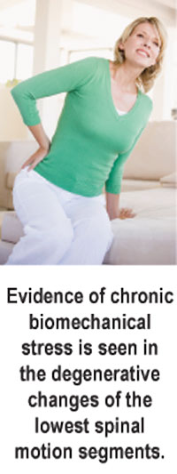
Multiple chronic lumbopelvic fixations, with an apparent anatomical leg length discrepancy (left short leg) and associated pelvic tilt and lumbar curvature. Evidence of chronic biomechanical stress is seen in the degenerative changes of the lowest spinal motion segments.
Treatment Plan
Adjustments. Specific chiropractic adjustments for the lumbosacral and sacroiliac joints were provided. Side-posture techniques were well tolerated in the lumbopelvic region, and resulted in very good articular releases.
Support. She was fitted with shock-absorbing, flexible custom-made orthotics based on imaging the foot in mid-stance (weightbearing). On a trial basis, she was temporarily fitted with a 4mm heel lift. A 6mm heel lift was permanently built onto the left orthotic. The stabilizing supports were introduced immediately, following the first week of regular adjustments. She had no difficulty in adapting to the heel lift or the orthotics.
Rehabilitation. She was instructed in a daily, at-home core strengthening program using elastic exercise tubing. The focus was on activation of her transverse abdominis and quadratus lumborum muscles, for improved spinal-pelvic stability.
Response to Care
This patient responded well to her spinal adjustments, and adapted well to her custom-made orthotics with the heel lift. After six weeks of adjustments (eight visits) and daily home exercises, she was released to a self-directed maintenance program.
Discussion
This patient had chronic lumbopelvic fixations, caused by her anatomical discrepancy in leg length, that had not been previously identified. In addition to spinal adjustments, her treatment included correction of her left short leg using a heel lift.
Dr. John J. Danchik, the seventh inductee to the ACA Sports Hall of Fame, is a clinical professor at Tufts University Medical School and formerly chaired the U.S. Olympic Committee’s Chiropractic Selection Program. Dr. Danchik lectures on current trends in sports chiropractic and rehabilitation.