History:
This patient is an adult male who injured his low back during a golf outing two months prior. His initial complaints included low back pain with radiation into his left lower extremity. At the time of presentation, he rated his pain as a ten on a visual analog pain scale. He had also already completed several epidural steroid injections without relief.
Clinical Examination: Height: 6’6″; weight: 260 lbs.
Neurological Examination:
Deep Tendon Reflexes: 2/2 and symmetrical. Dermatomal sensation was intact. Myotomal evaluation revealed 4+/5 weakness to the left anterior tibialis muscle. Long tract signs show no beats of ankle clonus. Babinski’s sign was flexor. Heel and toe walking revealed foot drop weakness/fatigue to the left anterior tibialis.
Orthopedic Examination:
Straight Leg Raising maneuver was 25 degrees left and 35 degrees right, with classic root tension signs on the left. The sciatic notch was exquisitely tender on the left side. Springing maneuvers showed pain over the spinous processes at L3 and L4. Sitting Bechterew’s performed with neck flexion and Valsalva’s maneuver revealed leg pain into the left foot and toes. Femoral nerve stretch test did not reveal L4 root tension signs.
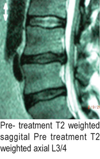
Radiology:
MRI evaluation of the lumbar spine was reviewed. This study consisted of both T1 and T2 weighted images in the saggital and transaxial planes. There was a moderate central disc herniation at L3/4. Mild eccentric stenosis of the left intervertebral foramina was also present (see pre treatment images).

Pre-treatment T2 weighted saggital; pre-treatment T2 weighted axial Pre treatment T2 weighted axial L3/4

Treatment plan:
The patient underwent flexion/extension provocation testing. This shows clinically which position removes the most pressure from the disc, thecal sac, and nerve root. Complete centralization/ removal of symptoms from his left lower extremity was achieved in neutral hyperextension of the lumbar spine. Clinically there was also a significant reduction in tenderness at the left sciatic notch. He was subsequently treated in this static position with High Power Laser Therapy (7.5 watts for 10 minutes) to each of the following: L3/4 disc space, left sciatic notch, and left posterior tibial/common peroneal nerve, total dosage was 13,500 Joules per session. This patient received a total of ten treatment sessions with the high power laser. At the end of the treatment plan, he was asymptomatic with normal neurological and orthopedic findings. His foot drop had also resolved completely.

Post treatment T2 weighted saggital post treatment T2 weighted axial L3/4.

Discussion:
High Power Laser Therapy has the ability to penetrate deeply and “bio stimulate” tissue to heal.
Laser photonic stimulation has been shown to increase blood flow and lymphatic drainage while, at the same time, stimulating endorphin and enkephalin release for pain management.8
Biostimulation translates into reduction of inflammation, and nerve regeneration.5,6 Regarding patients’ suffering from back and leg pain, the High Power Laser is thought to decrease inflammation of the disc and nerve root as well as aid in nerve regeneration.5 Healing of annular defects in the outer 1/3 of the disc (which is vascularized) have the ability to heal.9 Of interest in this particular case study is the fact that this patient has been followed for over 18 months. During that timeframe, he has not experienced any exacerbations of back or leg pain and has returned to all previous activities without pain!
Summary:
High Power Laser Therapy has the ability to reach deep within the body when compared to Low Level Laser Therapy.8 Results achieved seem to be long term in nature when compared to all other forms of treatment for disc pathologies. This may also be due to the fact that laser energy appears to also biostimulate collagen and fibroblast growth, which would enhance the tensile strength of the annular fibers of the degenerative disc patient.1,2,3,4 Clearly, further investigation regarding a large scale blind study is necessary to further correlate patient’s clinical findings with pre and post imaging studies.
 Dr. Costello is a board certified chiropractic orthopedisit. He is also board certified in quality assurance and utilization review with a sub-specialty certification in risk management. He currently serves as Chief of Clinical Services for Avicenna Laser Technology, Inc. For more information you can reach him via email at [email protected]