Description. When two adjacent vertebrae are osseously fused from birth, this joined unit is called a congenital block vertebra. Embryologically, this is the result of failure of the normal segmentation process of the somites during the period of differentiation at 3-to-8 fetal weeks.1 The block vertebra, by itself, is clinically insignificant. As there is no motion allowed at the fused level, there is no potential for degenerative disease of the disc or posterior apophyseal joints. The foramina at the blocked level may be smaller than normal, normally sized, or enlarged, but have not been shown to cause nerve compression. However, because of the lack of a motion segment, the free articulations above and below the block segment are stressed and usually result in premature degenerative discogenic spondylosis and arthrosis at the fully articulated levels, especially below the fusion site. Fusions are partial (i.e., do not completely involve the anterior and posterior spinal units) and may result in abnormal spinal curvature, usually scoliosis, because of a unilateral bar. Block vertebrae are most commonly found at C5-C6, C2-C3, T12-L1, and L4-L5, in decreasing order of incidence.1,2 
A recent report suggests that long-standing congenital or acquired fusion of upper cervical vertebrae may lead to stretching and laxity of the ligaments between the occiput and the atlas, resulting in excessive motion and brainstem or cord compression.2
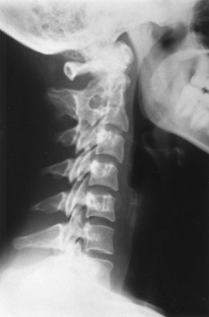
Radiologic Features. A typical congenital block vertebra will demonstrate the following roentgen signs: a diminished AP diameter of the vertebral body; a hypoplastic or rudimentary disc space that may show faint calcification; possible fusion of the apophyseal joints (50% of cases); and possible malformation or fusion of the spinous processes.
The anterior margins of the involved vertebrae form a concave surface, because of the decreased AP diameter at the fusion that is visible on plain film and MRI. This “wasp waist”3,5 or “C” shape can serve as a mnemonic device, to indicate that this fusion is “congenital.” Another helpful sign of this congenital anomaly is osseous fusion of the neural arches, that is almost never associated with infectious, traumatic processes or other causes of block vertebrae. 4,5 TAC
Dr. Terry R. Yochum is a second-generation chiropractor and a cum laude graduate of the National College of Chiropractic, where he subsequently completed his radiology specialty. He is currently Director of the Rocky Mountain Chiropractic Radiological Center, in Denver, CO, an Adjunct Professor of Radiology at the Los Angeles College of Chiropractic, as well as an instructor of Skeletal Radiology at the University of Colorado School of Medicine, Denver, CO. Dr. Yochum is, also, a consultant to Health Care Manufacturing Company that offers a Stored Energy system. For more information, Dr. Yochum can be reached at: 303-940-9400 or by e-mail at [email protected]. Magna Cum Laude graduate of National College of Chiropractic.
Dr. Chad Maola is a 1999
Magna Cum Laude graduate of National College of Chiropractic.