Evaluation and Management of Lumbar Discopathy with Lumbar Radiculopathy

Introduction

:dropcap_open:H:dropcap_close:ealth care costs have been rising for several years. Expenditures in the United States on health care surpassed $2.3 trillion in 2008, more than three times the $714 billion spent in 1990, and over eight times the $253 billion spent in 1980. Stemming this growth has become a major policy priority as the government, employers, and consumers increasingly struggle to keep up with health care costs.1   A study performed by a large managed care system demonstrated that systematic access to chiropractic care for neuromusculoskeletal conditions might be clinically beneficial and reduce the overall costs of medical care.2

lumbardiscopathyradiculopathyceaccreditedBack pain accounts for more than $100 billion in annual US health care costs and is the second leading cause of physician visits and hospitalizations. 3  Low back pain is a very common condition, accounting for 2.3% of physician visits in the United States.4   While lumbar disc prolapse, protrusion, or extrusion accounts for less than 5% of all low back problems, it is the most common cause of nerve root pain and surgical interventions.5

Chiropractors and their patients recognize the value of chiropractic interventions for the treatment of low back pain and now orthopedic surgeons are acknowledging the effectiveness of chiropractic care.  According to an orthopedic surgeon at the Texas Back Institute, chiropractic spinal manipulation is a safe and effective treatment for spinal pain.

It reduces pain, decreases medication needed, rapidly advances physical therapy, and requires very few passive forms of treatment, such as bed rest.  Jack Zigler, MD, orthopedic spine surgeon with the Texas Back Institute, states, “There are a lot of myths about chiropractic care. I decided to look into each of these myths, and what I found is that chiropractic education, side-by-side, is more similar to medical education than it is dissimilar.

Chiropractors work for us as screeners for surgical pathology. They can do the same work-up and send the patient who has already gone through his conservative treatment and had all his diagnostic work done to the surgeon.” 6

The National Prevention Strategy promulgates a vision of an interdisciplinary care system with chiropractors and medical doctors working together to improve the health and quality of life for individuals, families, and communities by moving the nation from a focus on sickness and disease to one on prevention and wellness.7

This putative case report provides the reader with a clinical learning opportunity that might improve treatment of low back pain.  The discussion addresses certain aspects of the evaluation and management process and proper documentation of the intervention.  I will demonstrate a reasonable approach to the examination and differential diagnosis of lumbar sprain/strain with resultant lumbar discopathy and lumbar radiculopathy and offer a prudent treatment plan.

Subjective:

Chief Concern:  “I have low back pain and numbness in the leg

Two weeks ago, I was lifting a freezer and hurt my back.”  The patient described the incident as follows: “I was in the back of a pickup truck and my friend was on the ground holding the other end of his new freezer.  I lost my balance while holding the freezer and felt a sharp pain in my back.  The freezer dropped and then I jumped off the back of the truck.  When my feet hit the ground, I almost passed out from the terrible pain.”  The patient’s wife attended and assisted with the history taking process.  He was a bit confused and agitated.

He complains of sharp, shooting pain in the lower back that shoots down the back of the right lower extremity to the calf with coughing, sneezing or having a bowel movement.  Prolonged sitting increases the pain in his back and causes numbness of the right calf and the dorsum of the foot. Taking NSAIDS, sitting in his reclining chair and alcoholic beverages reduce the pain.  For the past week, he has noticed that he stubs his toes of the right foot when walking for more than a block.  The worst pain experienced occurs with sitting and bending down to pick up his shoes.  He rated this pain at 9/10.  He has difficulty sleeping through the night.  Movement in bed increases back pain.  Upon waking, he experiences stiffness in the back.  He uses medications and ice on his back at night to sleep.  His pain reduced to 3-4/10 while resting and increased to 7-8/10 with cough or sneeze.

His primary care provider (PCP) told him that he “pulled” his back muscles.  The three view lumbar radiographic study (A-P, lateral and spot of L5) impression by the radiologist was “Moderate disc space narrowing at L5-S1 but negative for fracture or pathology.”  This study dated last week included the radiographs and the written report.  The PCP prescribed Ibuprofen and muscle relaxers.  He has never been to a chiropractor but his wife told him to make the appointment.  There is no history of back problems.  His review of systems did not reveal any history of serious illness or surgical interventions.  He has smoked one pack of cigarettes per day for the past 35 years and drinks 2-6 beers per night.

Objective:

General appearance:

This 35-year-old Caucasian male appeared to be in severe pain and was overweight and disheveled.  He was cooperative but seemed a bit confused and agitated during the history/interview process.  He was aggravated when he could not remember the date of injury when asked.  He blamed the confusion on the medications (muscle relaxers).

Posture:

Minor’s sign was present when the patient rose from the chair to stand.  The patient’s painful listing to the left revealed the antalgia sign.

Gait:

Painful behavior noted with limping of the right lower extremity (RLE).  Heel-walk revealed toe drop of right foot.  The toe walk did not demonstrate any weakness.

Palpation:

He demonstrated severe pain reaction with palpation of the right L 5-S1 region on the right and moderate pain reaction with palpation of the L5-S1 supraspinous ligament.  Myospasia of the lumbar paravertebral muscles was evident.  Pain was present with palpation at the right sciatic notch and at the area of the fibular nerve below the right knee.

Range of motion:

All active range of motion reduced due to the severe pain in the lumbar spine and myospasia of the paraspinal muscles.

Passive range of motion with flexion, extension, right rotation and right lateral flexion produced pain at L5-S1 on the right.

Resisted range of motion increased pain in the lumbar paravertebral muscles with flexion and extension.

Orthopedic tests:

O’Donoghue maneuvers revealed strain of lumbar paraspinal muscles and sprain of the L5-S1 joint/ligaments.

Kemp maneuver demonstrated reduced range of motion and radiating pain down the RLE to the lateral calf.

Valsalva maneuver produced pain at L5-S1 midline and on the right with radiation down RLE to posterior thigh.

Lindner sign present with radiating pain down RLE to the lateral calf.

Straight leg raise (SLR) tests:

Supine SLR RLE produced severe pain at L5-S1 on right to the lateral calf at 45 degrees.  Supine SLR LLE at 85 degrees reduced the pain in the RLE.  RLE SLR presented Lasegue, Braggard, and Sicard’s signs.  Turyn sign absent.

Neurological examination:

Sensory: Sharp, dull and light touch stimulation revealed the upper and lower extremity dermatomes to be intact except for hypesthesia of the right L5 dermatome.

Motor: All upper and lower extremity myotomes tested 5/5 bilaterally except 4/5 of right L5.

Deep tendon reflexes (DTR): 2+ bilaterally for the upper and lower extremities except 1+ with reinforcement of right hamstring reflex.

Babinski sign absent with stimulation of the plantar reflex (toes down going).8

Assessment:

Acute, moderate lumbar sprain/strain with resultant lumbar discopathy and lumbar radiculopathy

Plan:

  1. Spinal decompression therapy with distraction and manipulation in order to reduce pain and promote function (daily for five treatments).
  2. Cryotherapy (ice massage) to reduce pain and myospasia to be performed at home.
  3. Electrotherapy to reduce edema and pain.
  4. Patient was advised to avoid prolonged sitting and to ambulate frequently.
  5. He was advised to sleep on back or sides with pillow support of lower extremities.
  6. Spinal extension and stabilization exercises will be implemented in the subacute phase.
  7. A smoking cessation and weight loss program was suggested in order to improve overall health and possibly slow the disc degenerative process.
  8. A neurosurgical consultation is appropriate if he does demonstrate positive response to conservative care within 2 weeks.

Discussion:

An orthopedic test is most often a provocative maneuver with stretching, compressing, and contracting of tissues in order to duplicate the patient’s pain and identify the involved tissues.  Clinicians perform tests and observe for signs of dysfunction, pathology or disease.

You may reveal Dejerine signs during the history taking process by posing this simple question: “Do you have the pain with coughing, sneezing or bowel movement?”  You may perform a three-part test that requires the patient to sneeze, cough, and perform the Valsalva maneuver.  Normally, I do not perform the snuff test (pepper under the nose) to induce sneezing but I do ask the patient to cough. The presence of these three signs is termed a Dejerine Triad.  The testing for Dejerine signs determines the presence of nerve root compression, which can arise from osseous foraminal encroachment, disc protrusion (bulging), prolapse (herniation) or severe sprain/strain of the spine.

O’Donoghue maneuvers differentiate sprain from strain or reveal the presence or absence of such soft tissue injuries.  Muscle strain injuries will be painful with resisted motion.  Ligament sprain or joint injuries will be painful with passive motion.

Antalgia sign with anterior and lateral listing to the left with pain radiating down the RLE suggests the possibility of a posterior lateral disc herniation with a lesion located lateral to the nerve root.  If the posterior lateral disc herniation were located medial to the nerve root, the patient would list toward the side of leg pain.

The Kemp maneuver produced radiating pain down the RLE to the lateral calf with extension, rotation and lateral flexion to the right, which also suggests the possibility of disc lesion, which lies laterally to the nerve root.

Straight leg raise testing demonstrated three different nerve-root tension signs (Lasegue, Braggard, and Sicard) and the possibility of a disc lesion located lateral to the nerve root.

A three-part peripheral nervous system examination enables the clinician to determine the presence of upper motor neuron or lower motor neuron lesions.  It involves the testing of deep tendon reflexes, sensory function and motor function.  In this particular case, the patient presented with weakness of the dorsiflexor muscles of the right foot indicating motor deficit in the L5 myotome.  Sensory testing revealed hypesthesia or decreased sensory function in the L5 dermatome.

The 1+ DTR of the right medial hamstring tendon indicated a decrease in response to stroking of the tendon with a reflex instrument, which indicates dysfunction at the level of L5 nerve root.   These neurological findings and the absence of the Babinski sign indicate a lower motor neuron lesion involving the L5 nerve root rather than an upper motor neuron lesion, which would involve the spinal cord or brain.

Assessment:

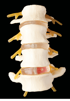
Diagnosis is the key to successful treatment.9   When treating a patient with discopathy and radiculopathy, the clinician must determine the stage of the healing process, the type of injury and tissue involved, and the site of lesion. This patient was determined to be in the acute phase due to the timing of the injury and the major complaint being pain.  The lower lumbar muscles and ligaments were overstretched and torn.  The L5-S1disc herniation is compressing the L5 nerve root.

Plan:

I prefer the use of spinal decompression equipment that permits manual control and manipulation when treating patients with discopathy and radiculopathy.  It is essential to determine if the posterior lateral disc herniation is located lateral or medial to the involved nerve root prior to administering manual methods and spinal decompression.  The spinal decompression intervention addressed the multi-directional disc dynamics in order to relieve compression of the spinal nerve roots.

Smoking and being overweight precipitate spinal disc disease.10  A causal relationship exists with nicotine and disc degeneration;11 and smoking with heavy use of alcohol are associated with suicidal ideation.12  I prescribed smoking cessation, weight loss, and alcohol cessation recommendations to improve the health and quality of life for this patient. A caring clinician will promote disease prevention and wellness.

Conclusion:

Chiropractic evaluation and management of lumbar discopathy with radiculopathy requires appropriate examination procedures in order to complete a differential diagnosis and develop a comprehensive treatment plan.  If you are interested in providing the most current evidence-based conservative spinal care, I strongly recommend the seventh edition of Low Back Pain: Mechanism, Diagnosis, and Treatment by James M. Cox.  I suggest any clinician interested in performing spinal decompression investigate the training offered by experts.13 14


This article may be used to earn CE Credits through the Postgraduate Department at the University of Bridgeport.  To learn if your state is eligible, and to register contact Anne Nilson at 203-576-4880 or register on the web at www.bridgeport.edu/tac


James J. Lehman, D.C., M.B.A., D.A.B.C.O. is an Associate Professor of Clinical Sciences at the University of Bridgeport College of Chiropractic.  Please remit any questions or comments to [email protected]


References:

  1. Centers for Medicare and Medicaid Services, Office of the Actuary, National Health Statistics Group, National Health Care Expenditures Data, January 2010.
  2. Antonio P. Legorreta, MD, MPH, R. Douglas Metz, DC, Craig F. Nelson, DC, MS, Saurabh Ray, PhD, Helen Oster Chernicoff, MD, MSHS, Nicholas A. DiNubile, MD. Comparative Analysis of Individuals With and Without Chiropractic Coverage Patient Characteristics, Utilization, and Costs. Arch Intern Med. 2004;164:1985-1992.
  3. Katz, J.N. Lumbar disc disorders and low-back pain: socioeconomic factors and consequences. J Bone Joint Surg Am. 2006;88: 21-24.
  4. Deyo, R.A., Mirza, S.K., Martin, B.I. Back pain prevalence and visit rates: estimates from U.S. national surveys, 2002. Spine. 2006;31:2724-2727.
  5. Manchikanti, L., Derby, R., Benyamin, R.M., Helm, S., and Hirsch, J.A.  A Systematic Review of Mechanical Lumbar Disc Decompression with Nucleoplasty.  Pain Physician 2009; 12:561-572 • ISSN 1533-3159.
  6. Zigler, J. Time to recognize value of chiropractic care? Science and patient satisfaction surveys cite usefulness of spinal manipulation. Orthopedics Today 2003 Feb; 23(2):14-15.
  7. National Prevention Council, National Prevention Strategy, Washington, DC: U.S. Department of Health and Human Services, Office of the Surgeon General, 2011. [Cited December 3, 2011]  Available from: http://www.healthcare.gov/prevention/nphpphc/strategy/report.pdf.
  8. Russell, S. & Triola, M. NYU School of Medicine. The Precise Neurological Exam. [Cited December 4, 2011] Available from: http://cloud.med.nyu.edu/modules/pub/neurosurgery/reflexes.html.
  9. Miller, K.J. Physical assessment of lower extremity radiculopathy and sciatica. J Chiropr Med. 2007 Spring; 6(2): 75–82.
  10. Kaila-Kangas, L., Leino-Arjas, P., Riihimäki, H., Luukkonen, R., Kirjonen, J. Spine (Phila Pa 1976). Smoking and overweight as predictors of hospitalization for back disorders. 2003 Aug 15; 28(16):1860-8.
  11. Kim, K.S., Yoon, S.T., Park, J.S., Li, J, Park, M.S., Hutton W.C. Inhibition of proteoglycan and type II collagen synthesis of disc nucleus cells by Nicotine. J Neurosurg. 2003 Oct; 99(3 Suppl):291-7.
  12. Fishbain, D.A., Lewis, J.E., Gao, J., Cole, B., Steele Rosomoff R. Are chronic low back pain patients who smoke at greater risk for suicide ideation? Pain Med. 2009 Mar;10(2):340-6. Epub 2009 Feb 25.
  13. Cox, J.M. Cox Technic (Flexion Distraction / Cox Technique / Cox Method). [cited December 4, 2011] Available from: http://www.youtube.com/watch?v=sqgNZj25bUk.
  14. Cuccia D. Spinal Decompression for Herniated Disc with ExtenTrac Technology.  [Cited December 4, 2011] Available from: http://video.google.com/videoplay?docid=-6907766059201864758#.

Headaches and Neck Pain

Introduction

:dropcap_open:T:dropcap_close:he World Health Organization (WHO) declared that headache disorders generate a substantial disability burden and suggested health classification of headaches amongst major public health disorders.  WHO also points out that there is a specific lack of public and professional awareness of the epidemiology of headache disorders and their impact on individual sufferers, their careers, family and colleagues, and on society.1

headachesandneckpainceaccreditedWhile health care professionals continue the controversial debate over the classification of cervicogenic headaches, a comprehensive multidisciplinary pain treatment program provides the greatest opportunity for overall clinical improvement.2  Recent evidence suggests that chiropractic care, including spinal manipulation, improves migraine and cervicogenic headaches.3 

The cervicogenic headache (CGH), first introduced by Sjaastad in 1983,4 had been discussed between 1860 and 1862 and described as a pain in the scalp mediated through the trigeminal nerve or the occipital nerves due to disease of the spine as documented in John Hilton’s classical text, Rest and Pain5.  A more recent review defines CGH and describes the nociceptive pathways: A chronic, hemicranial pain syndrome in which the sensation of pain originates in the cervical spine or soft tissues of the neck and is referred to the head.

The trigeminocervical nucleus is a region of the upper cervical spinal cord where sensory nerve fibers in the descending tract of the trigeminal nerve converge with sensory fibers from the upper cervical roots. This convergence of nociceptive pathways allows for the referral of pain signals from the neck to the trigeminal sensory receptive fields of the face and head.6

While science attempts to define CGH, the public suffers with headaches.  In fact, twenty-five percent of all headaches are actually “rebound” episodes triggered by the overuse of common pain medications, which include both prescription and over-the-counter drugs.7 This putative case report provides the reader with a clinical learning opportunity.  The discussion addresses certain aspects of the evaluation and management process and proper documentation of the intervention. I will demonstrate a reasonable approach to the examination and differential diagnosis of a patient with cervicogenic headaches, back and neck pain and offer a prudent treatment plan.

SUBJECTIVE

Chief Concern: I have headaches, back and neck pain

A woman claims that headaches have occurred 1-2 times per week for the past two months and that sitting at the computer for extended periods usually produces the headaches.  She confirms that she leans forward while typing on the laptop and experiences the headaches after 2-3 hours of work.  She demonstrated her slouching work posture while using the computer, which displayed a rounding of the shoulders and significant forward head posture. Then she pointed to the right occipital and temporal areas as the areas of her pain.  She denied any morning headaches, loss of vision, weakness or loss of consciousness with the headaches.

A dull ache in the neck has been bothering her for the past six months.  The daily neck ache occurs with prolonged work at the computer. The neck pain becomes a sharp, localized pain in the lower neck on the right side when she turns her head to the right. She also experiences stiffness in the neck upon waking in the morning.  A hot shower reduces the discomfort. Throbbing and aching in neck and shoulder blades occurs during the afternoons.

For the past three months, she has experienced lower back pain with prolonged sitting.  She described the low back pain as a burning pain across her lower back.  She experiences lower back pain and difficulty rising to stand due to stiffness in the back and hips after sitting for a prolonged time (3-5 hours).  Severe tightness in the back of her thighs and legs bother her while gardening. She experienced 50 percent relief of lower back pain after receiving one chiropractic adjustment last year.   She denied weakness or radiating pains in the extremities.  She rated her head pain on an 11-point numerical pain scale at between 3-6/10 and the neck and lower back pain rated at 0-4/10.

NSAIDS, relaxation, hot bath or shower and massage usually provide relief of her pains.   Her past medical history included numerous spinal strains and sprains as a young gymnast.  She consumes two sodas and eleven glasses of water per day.  She has gained 40 pounds over the past 4 months following treatment of an infection with steroids. She would like to have her neck and back adjusted to gain relief of the headaches and spinal pain.  This young woman insisted that adjustments “help a lot” and provide a “quick fix.” 

OBJECTIVE

This pleasant but distressed 35-year-old Hispanic female appears to be her stated age.  She is well-developed, obese, alert, cooperative and a good historian.

Vital signs: Height: 65” Weight: 183#, B/P: 169/123, Pulse: 78, Temperature: 97.35 degrees Fahrenheit aural. Allergies: milk, sulpha, codeine. Tobacco: She smokes three cigarettes during the day.  If she is “stressed out”, she smokes another three at night. Alcohol: denies. Recreational drugs: denies.

Posture: She exhibited a forward head posture and rounded shoulders while seated.  An inferior left iliac crest and right shoulder inferiority was observed while she was standing.

Leg length mensuration: She demonstrated a left functional leg length deficiency in the supine position and a right functional leg length deficiency while seated.  Anatomical measurement of the leg lengths demonstrated the lengths to be symmetrical at 33 inches.

Palpation: Pain produced at the occipital nerves on the right.  Pinching of the right upper trapezius muscles demonstrated a positive jump sign, taut bands, painful nodules and hypertonicity.  Myofascial trigger points revealed in the sternocleidomastoideus and posterior scalene muscles on the right.  Lumbar multifidi muscles were hypertonic with painful nodules at the levels of L4-5 bilaterally.  Palpation produced pain over the ligamentum nuchae at C2-3, C4-5–6 and the supraspinous ligaments of L5-S1.

Cervical range of motion: Active range of motion was full, symmetrical and without pain with flexion, extension, lateral flexion and rotation except for reduced right rotation and lateral flexion with pain at C2-3 and C5-6 on the right.  Passive range of motion produced pain at C2-3 and C5-6 right with right rotation and lateral flexion.

Dorso-lumbar range of motion: Active range of motion was symmetrical without pain with flexion and lateral flexion but hyperextension demonstrated hypermobility and pain at the lumbosacral joints bilaterally.

Posterior joint dysfunction revealed at L5-S1, C2-3 and C 5-6.  There was a reduced range of motion with right lateral flexion and rotation with pain on right at C2-3 and C5-6.  Palpation revealed cervical paravertebral hypertonicity on the right and pain at C2-3 and C5-6 over the ligamentum nuchae and the right posterior cervical muscles. Hypermobility and localized pain demonstrated at L5-S1 bilaterally with extension.

Cervical compression: Active and passive maximal foraminal compression produced pain at C 5-6 on right.

Cervical distraction: Positive with relief of pain in neck.

Kemp maneuver: Negative without sciatica but produced pain at L5-S1 bilaterally.

Modified Gillet: Demonstrated SIJ fixation left with a left thumb up going while the right thumb was down going.

One-leg hyperextension: Test produced pain at L5-S1 on right with flexion of left lower extremity.

Lumbar stabilization test: Negative and without pain upon extension of legs while prone.

Neurological examination: Deep tendon reflexes were 2+ bilaterally and brisk for the upper and lower extremities. Motor testing revealed the upper and lower extremities to be 5/5 without signs of atrophy. Sensory testing revealed the trigeminal nerve and extremity dermatomes to be intact for sharp and light touch. There were no signs of upper or lower motor neuron lesions. 

RADIOLOGY REPORT

Impressions:

  1. Decreased disc spacing at C5-6 and L5-S1
  2. Osteophytic formations at the anterior margins of the vertebral bodies at C5-6
  3. Imbrication of L5 and S1 zygapophyseal joints
  4. No signs of fracture, dislocation, or bone destruction 

ASSESSMENT

  1. Hypertension
  2. Obesity
  3. Cervicogenic headaches due to postural strain with resultant active myofascial trigger points and posterior joint dysfunction
  4. Lumbar facet syndrome
  5. Postural imbalance due to pelvic obliquity
  6. Suspected cervical and lumbar degenerative joint and disc disease 

PLAN

  1. Consult with the primary care provider prior to commencing conservative chiropractic management of her condition.  Suggest follow-up with her primary care physician due to the hypertension and sudden weight gain.  Discuss co-management of patient due to her hypertension, obesity and chronic musculoskeletal pain conditions.
  2. Upon approval by her primary care provider, I would like her to begin chiropractic care, a regular walking program and yoga in order to reduce weight and improve flexibility.
  3. Ordered radiographic study of the cervical and lumbar spine to determine status of discs and joints and determine indications of spinal manipulation.
  4. Will recommend chiropractic manipulation treatments and soft-tissue treatments three times per week for a period of two weeks to reduce pain and improve spinal function.  Follow-up exam after she receives the initial six treatments.  Advised 6-12 chiropractic treatments might be necessary to provide relief of her pain.
  5. Will recommend massage therapy once per week for 6 weeks to relieve stress and provide muscle relaxation.
  6. Recommended postural exercises to reduce forward head posture and rounded shoulders.
  7. We discussed the advantages and complications of chiropractic care.  She understands the diagnosis and treatment plan.  She consents to this treatment plan. She has been a patient in this office in the past and is very familiar with the recommended treatments.  She will follow-up with her primary care provider this week prior to commencing chiropractic care.

DISCUSSION

Hypertension

Her current reading of 169/123 classifies her condition as stage 2 hypertension.8  Hypertension,9 which causes arteriosclerosis10 and predisposes patients to heart disease,11 peripheral vascular disease, and cerebrovascular attacks,12 is the most common cause of death in the United States.13 As patient-centered clinicians, we must appreciate the need to evaluate this most serious condition and refer the patient to the primary care provider 

Cervicogenic headaches

Many family physicians consider headaches, which are common primary care conditions, to be enigmatic.14  Cervicogenic headaches arising from cervical vertebrae or myofascial tissues or other soft tissues of the neck and shoulders15 may be resistant to care if not properly diagnosed.16  Bogduk has described the anatomical basis for cervicogenic headaches, which might explain the rationale for chiropractic interventions with upper cervical spine manipulation: The trigeminocervical nucleus is a region of the upper cervical spinal cord where sensory nerve fibers in the descending tract of the trigeminal nerve (trigeminal nucleus caudalis) are believed to interact with sensory fibers from the upper cervical roots. This functional convergence of upper cervical and trigeminal sensory pathways allows the bidirectional referral of painful sensations between the neck and trigeminal sensory receptive fields of the face and head.17

Chiropractors who use spinal manipulation and prescribe cervical exercises for cervicogenic headaches are likely to render the most effective treatment.18  Yet, clinicians must perform a detailed neuromusculoskeletal evaluation in order to identify the involved tissues responsible for cervicogenic headaches.  Haldeman confirms my statement by saying that most pathologies affecting the cervical spine have been implicated in the genesis of cervicogenic headaches because of convergence of sensory input from the cervical structures within the spinal nucleus of the trigeminal nerve.19

Co-management

As a Board Certified Chiropractic Orthopedist, I am confident with my assessment of this patient for neuromusculoskeletal disease.  Many prescription and nonprescription drugs can cause or exacerbate hypertension, which includes corticosteroids and NSAIDS.20  Consequently, I am not comfortable evaluating and managing a patient presenting with hypertension and sudden weight gain following the use of corticosteroids.  I will consult with the primary care provider, express my concerns and discuss my neuromusculoskeletal evaluation and management.

The Affordable Care Act and the National Prevention Strategy expect health care providers to enhance coordination and integration of clinical, behavioral, and complementary health strategies.21 Since integrated health care describes a coordinated system in which health care professionals are educated about each other’s work and collaborate with one another and with their patients to achieve optimal patient well-being,22 co-managing this patient’s conditions will benefit the patient and educate the involved health care providers. 

Treatment Plan

Chiropractic physicians should develop treatment plans based upon patient needs, diagnoses, and response to care.  A study by Haas et al. suggests that multiple treatments are necessary to manage patients with cervicogenic headaches:

A large clinical trial on the relationship between pain relief and the number of chiropractic treatments is feasible. Findings give preliminary support for the benefit of larger doses, 9 to 12 treatments, of chiropractic care for the treatment of cervicogenic headache.23

CONCLUSION

Chiropractic physicians are responsible for evaluation and management of patients presenting with headaches.  The evaluation requires the use of orthopedic and neurological examination procedures that develop a differential diagnosis and a reasonable treatment plan.


James J. Lehman, D.C., M.B.A., D.A.B.C.O. is an Associate Professor of Clinical Sciences at the University of Bridgeport College of Chiropractic.  Please remit any questions or comments to [email protected]


Reference:

  1. Headache Disorders and Public Health: Education and Management Implications. This document results from the Meeting on Headache and Related Disorders, held at WHO headquarters, Geneva, 13-14 March 2000.  [Cited October 22, 2011]  Available from: http://www.migraines.org/new/pdfs/who.pdf
  2. Yadka, S., Gehret, J., Campbell, P., Mandell, S., & Ratliff, J.K. A Pain in the Neck: Review of Cervicogenic Headaches and Associated Disorders. JHN Journal.  [Cited October 22, 2011] Available from: http://jdc.jefferson.edu/cgi/viewcontent.cgi?article=1043&context=jhnj
  3. Bryans, R., Descarreaux, M., Duranleau, M., Marcoux, H., Potter, B., Ruegg, R., Shaw, L., Watkin, R., & White, E. Evidence-based guidelines for the chiropractic treatment of adults with headache. J Manipulative Physiol Ther. 2011 Jun;34(5):274-89. [Cited October 22, 2011] Available from: http://www.ncbi.nlm.nih.gov/pubmed/21640251
  4. Sjaastad, O., Saunte, C., Hovdohl, H., Gronbaek, E. ’Cervicogenic’ Headache, An Hypothesis. Cephalgia, 1983, 249-256.
  5. Hilton J. Rest and pain (1950:77). Walls, E.W. & Phillips E.E., eds. London: Bell.
  6. Biondi JM. Cervicogenic Headache. JAOA, September 2000; Vol 100, No. 9. Supplement to September 2000.  S7-14.  [Cited October 22, 2011] Available from: http://www.jaoa.org/content/100/9_suppl/7S.full.pdf
  7. Diener, H.C. & Limmroth, V. Medication-overuse headache: a world-wide problem. The Lancet Neurology, Volume 3, Issue 8, Pages 475 – 483, August 2004. [Cited October 22, 2011] Available from: http://www.thelancet.com/journals/laneur/article/PIIS1474-4422(04)00824-5/fulltext Chobanian, A.V., Bakris, G.L., Black, H.R. et al. (December 2003). Seventh report of the Joint National Committee on Prevention, Detection, Evaluation, and Treatment of High Blood Pressure. Hypertension, 42(6), 1206–52.
  8. Chobanian, A.V., Bakris, G.L., Black, H.R. et al. (December 2003). Seventh report of the Joint National Committee on Prevention, Detection, Evaluation, and Treatment of High Blood Pressure. Hypertension, 42(6), 1206–52.
  9. Novo, S., Lunetta, M., Evola, S., & Novo, G. (January 2009). Role of ARBs in the blood hypertension therapy and prevention of cardiovascular events. Current Drug Targets, 10(1), 20–5.
  10. Riccioni, G. (2009). The effect of antihypertensive drugs on carotid intima media thickness: an up-to-date review. Current Medicinal Chemistry, 16(8), 988–96.
  11. Agabiti-Rosei, E. (September 2008). From macro- to microcirculation: benefits in hypertension and diabetes. Journal of Hypertension, 26 Suppl 3, S15–21.
  12. Singer, D.R., Kite, A. (June 2008). Management of hypertension in peripheral arterial disease: does the choice of drugs matter?. European Journal of Vascular and Endovascular Surgery, 35(6), 701–8.
  13. Novo, S., Lunetta, M., Evola, S, & Novo, G. (January 2009). Role of ARBs in the blood hypertension therapy and prevention of cardiovascular events. Current Drug Targets, 10(1), 20–5.
  14. Sierpina, V., Astin, J., Giordano, J. Mind-body Therapies for Headache. Am Fam Physician. 2007 Nov 15; 76(10):1518-1522. [Cited October 23, 2011] Available from: http://www.aafp.org/afp/2007/1115/p1518.html
  15. Biondi, D.M. Cervicogenic Headache: A Review of Diagnostic and Treatment Strategies. J Am Osteopath Assoc April 1, 2005 vol. 105 no. 4 Suppl 16S-22S. [Cited October 23, 2011] Available from: http://www.jaoa.org/cgi/content/full/105/4_suppl/16S
  16. Edmeads, J. The cervical spine and headache. Neurology, 1988;38:1874-1878.
  17. Bogduk, N. The anatomical basis for cervicogenic headache. J Manipulative Physiol Ther. 1992;15:67-70.
  18. Jull, G., Trott, P., Potter, H., Zito, G., Niere, K., Shirley, D. et al. (2002). A randomized controlled trial of exercise and manipulative therapy for cervicogenic headache. Spine, 27(17), 1835-1843.
  19. Haldeman, S., Dagenais, S. Cervicogenic headaches: a critical review. Spine J. 2001 Jan-Feb;1(1), 31-46.
  20. Onusko E. Diagnosing Secondary Hypertension. Am Fam Physician. 2003 Jan 1;67(1):67-74.  [Cited October 22, 2011] Available from: http://www.aafp.org/afp/2003/0101/p67.html
  21. Onusko E. Diagnosing Secondary Hypertension. Am Fam Physician. 2003 Jan 1;67(1):67-74.  [Cited October 22, 2011] Available from: http://www.aafp.org/afp/2003/0101/p67.html
  22. U.S. Preventive Services Task Force. Integrating Evidence-Based Clinical and Community Strategies to Improve Health. Available at http://www.uspreventiveservicestaskforce.org/uspstf07/methods/tfmethods.htm. Accessed May 17, 2011.; Institute of Medicine. Crossing the Quality Chasm: A New Health System for the 21st Century. Washington, D.C.: National Academies Press; 2001. [Cited October 22, 2011] Available from:
  23. Haas, M., Groupp, E., Aickin, M., Fairweather, A., Ganger, B., Attwood, M., Cummins, C., & Baffes, L.J. Manipulative Physiol Ther. Dose response for chiropractic care of chronic cervicogenic headache and associated neck pain: a randomized pilot study.2004 Nov-Dec; 27(9):547-53. [Cited October 23, 2011] Available from: http://www.ncbi.nlm.nih.gov/pubmed/15614241

Low Back Pain

Introduction

lowbackpainceaccredited1
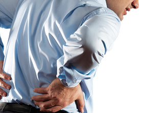
:dropcap_open:C:dropcap_close:hiropractors and primary care providers commonly see patients suffering with acute low back pain.  An episode of acute low back pain is the fifth most common reason for all physician visits.  This musculoskeletal malady affects 90% of the adult population in the United States at some time in life.1

The direct costs of low back pain total $20 billion per annum while indirect costs reach $50 billion each year in the United States.  Employers must account for this significant economic impact because one-half of the working population endures low back pain each year.  Fortunately, the vast majority (90%) of disabled workers return to work within 90 days.2

Although spinal manipulation provides relief of low back pain (LBP)3,  it has been my clinical experience that diagnosis is the key to successful treatment with chiropractic interventions. Murphy concurs with his published statement; There is great need for more accurate diagnosis in patients with spinal pain.4  Primary care researchers claim that LBP continues to be one of the most common and challenging problems in primary care, which requires a comprehensive understanding of the diagnosis and treatment of LBP.5

This putative case report provides the reader with a clinical learning opportunity.  The discussion addresses certain aspects of the evaluation and management process and proper documentation of the intervention. It is my intent to demonstrate a reasonable approach to the examination and differential diagnosis of a patient with low back pain and offer a prudent treatment plan.

Case report

Subjective:

A 28-year-old professional baseball player presented with a chief concern of “I have back pain and tightness when I run.”  The back pain began several hours after a motor vehicle accident that took place 6 months earlier.  He denied any history of low back pain prior to the motor vehicle accident.  The most recent episode of increased sharp low back pain began 5 days ago.  The patient denied any history of back pain prior to the motor vehicle accident and he believes that the car accident caused the back pain.  He mentioned that his father has seen a chiropractor for treatment of low back pain.

He was a front seat passenger, wearing a seat belt in a vehicle that rear-ended another vehicle, which was at a stop.  His vehicle was traveling about 35 miles per hour.  At the time of impact, he was conversing with the driver and turning to the left.  The patient denied loss of consciousness or head trauma. He declined any medical care at the scene of the accident.

He has experienced tightness and sharp back pain while running.  At my request, he pointed specifically to the right lumbosacral spine as the area of sharp pain with movement.  He has not experienced pain with batting or throwing.  The back has ached at night and frequently interfered with his sleep.  Normally, he has gained some relief with NSAIDS and massage.  To date, he has never received chiropractic care.

When asked to point to the area of the dull, aching pain he moved his finger up and down on the right side of the low back and was unable to identify a specific spinal level.   The patient denied any pain shooting down the legs but mentioned that sometimes he experienced discomfort in the right groin with prolonged sitting in the dugout.  The severity of the sharp pain at the lumbosacral spine with running was rated at 7 of 10 and the aching was rated at 3-5 of 10.

There was no history of other health problems.  He mentioned that his diet was good, with ample amounts of fruits and vegetables along with red and white meats.  When questioned about hydration, he said that he drinks about 20 ounces of water during a game, but very little water when not playing.  He preferred carbonated beverages and beer.  His total liquid intake has been 60 ounces per day.

He asked if he might anticipate complete relief of the low back pain with chiropractic care.  He has been worried that the pain may interfere with his athletic career.

Objective:

This young, healthy, Caucasian professional athlete appears his stated age.  He is an alert, cooperative and well-developed mesomorph.  He walks without a limp.

Vital indices: Height 74 inches, weight 205 pounds, blood pressure 110/64, pulse rate 66/minute and respirations of 12/minute

Postural evaluation demonstrated an inferior iliac crest on the right, an inferior right shoulder and a mild “S” type curvature.  His left occiput appeared inferior and posterior compared to the right occiput.    Anterior and posterior Adam’s positions presented a negative sign. I measured the leg lengths in supine and sitting positions. The mensuration demonstrated functional leg length inequalities with the right lower extremity appearing short while supine and longer with sitting.

Palpation of the area of the right lumbosacral joint was painful and graded as a grade 2 of 4 due to his wincing.  There was pain with palpation of the supraspinous ligaments at L5 and S1, and the area of the L5 facet joint on the right.  Palpation of the sacroiliac joints did not produce pain.   Hypertonicity of the multifidi muscles with taut bands and painful nodules bilaterally were present.  The patient demonstrated taut bands and painful nodules with a grade 3 of 4 pain reaction upon palpation of the right iliopsoas muscle.

Active range of motion of the thoraco-lumbar spine demonstrated full and pain-free motion with flexion and left lateral flexion but right lateral flexion and extension did produce pain at the lumbosacral spine on the right.  He mentioned that forward flexion at the waist felt good.  Passive range of motion of the thoraco-lumbar spine produced pain at the L5-S1 level on the right with right rotation and lateral flexion.  Resistive range of motion testing of the thoraco-lumbar spine produced pain in the right paravertebral muscles at the levels of L3-4-5-S1 upon right rotation and lateral flexion.

The Kemp test or maneuver was negative for radicular pain but did produce pain at the right L5 level of the lumbosacral spine with extension and rotation to the right.6   The straight leg test (SLR) produced pain at the right lumbosacral spine with extension of the right hip and lower extremity at 80 degrees.  There was no pain with the left SLR at 80 degrees.

Lumbar stability testing produced sharp pain at the lumbosacral spine on the right when the patient stood on one foot and extended his spine.  He also experienced pain with flexion of the legs while supine.  This pain reduced with manual compression over the lumbosacral spine while repeating the extension of the legs while supine.

The modified Gillet test performed while standing demonstrated hypomobility of the right sacroiliac joint.  The Gaenslen, distraction and sacroiliac thrust tests did not produce pain at the sacroiliac joints.  The Adams Supported Belt test and the drop test produced pain at L5 right.

Neurological evaluation

The sensory examination of the upper and lower extremities revealed an intact system for sharp and dull stimulations.  Motor testing of the upper and lower extremities were normal and graded at 5/5.  The deep tendon reflexes were within normal limits and graded at 2+ and brisk for the upper and lower extremities.  There were no signs of pathologic reflexes.

Assessment:

  1. Acute low back pain episode
  2. Post-traumatic lumbo-sacral sprain/ strain with resultant posterior joint dysfunction and myofascial pain
  3. Pelvic obliquity with resultant mild “S” type functional scoliosis
  4. Spondylolysis at L5 without spondylolisthesis
  5. Subclinical dehydration

Plan:

  1. I ordered a five-view lumbar spine radiographic examination to evaluate the lumbar spine for separation  of the pars interarticularis at the lower lumbar spine.
  2. Treatment is scheduled three visits per week for a period of four weeks in order to reduce pain and improve function.
  3. Treatment will consist of spinal manipulation, myofascial trigger-point release and massage therapy.
  4. I prescribed 140 ounces of fluid per day.7
  5. Re-evaluation will follow the four weeks of care
  6. A full explanation of the risks and benefits of treatment was provided to the patient.  He understood that he may elect to receive no care or seek another opinion with another provider.  The patient decided to have the radiographic examination and then return for the prescribed series of treatments.

Radiology report

Findings:

  1. Unilateral separation of the L5 pars interarticularis on the right
  2. No signs of anterolisthesis of lumbar vertebrae
  3. No signs of decreased disc spacing in lumbar spine
  4. No signs of degenerative joint changes

Impression:

1. Spondylolysis L5 right

Discussion

lowbackpainceaccredited2This putative case report demonstrates a focused history taking and physical examination process with a patient presenting post-traumatic low back pain.  I documented the patient encounter with SOAP notes (an acronym for subjective, objective, assessment, and plan).

A history of present illness (HPI) obtained during the interview with the patient investigated the onset, duration, and character of the present illness, as well as any acts or factors that aggravate or ameliorate the symptoms. The chief concern or presenting symptom prompted this detailed interview.8   The chief concern, “I have back pain and tightness when I run,” is the initial notation in the subjective section of the medical record.  It is significant that the chiropractic clinician identify the cause of the low back pain.  Since the patient experienced his first episode of low back pain on the day of the accident and he denied low back pain prior to the incident, it is my professional opinion that the motor vehicle accident caused his low back pain.

I prefer use of the mnemonic initialism, OPQRST, to facilitate the taking of the HPI.  This line of questioning includes discussion of the severity of the pain, timing and previous treatment for the present illness or injury.

The musculoskeletal examination focused on the spine with observation, inspection, palpation, range of motion testing, and orthopedic testing, followed by a three-part peripheral nervous system examination.

The negative Adam’s positions indicate a functional scoliosis rather than an anatomical or structural scoliosis.9   These positions involve observation of the patient’s flexed spine from both the anterior and posterior perspectives.  If a “C” or “S” curve is present, it will straighten with flexion of the spine.  Straightening of the spine in Adam’s positions indicates the presence of a negative sign, which indicates a functional curvature.  Chiropractors treat functional curvatures.10

It is common for a patient to present with pelvic tilting or pelvic obliquity due to muscle imbalances in the gluteal, quadratus lumborum, and/or the iliopsoas musculature,11  which may cause mild to moderate spinal scoliosis (“C” of “S”) due to lower limb length inequality.12 Static or flat palpation for pain is reliable  while movement restriction in the lumbar spine is not reliable.14   This patient presented pain reactions to palpation of specific lumbar joints/ligaments and myofascial trigger points,15  which enabled the identification of the painful tissues.

Chiropractic clinicians may identify and document the presence of a primary myofascial trigger point by locating a hyperirritable focus within a taut band of skeletal muscle.16   The resistive (isometric contraction) and passive ranges of motion findings indicate a sprain/strain injury of the lumbosacral spine (O’Donoghue maneuver).17 Kemp’s test assesses for intervertebral nerve root encroachment, muscular strain, ligamentous sprain, or pericapsular inflammation using a maneuver that involves flexion of the torso followed by extension and rotation of the lumbar spine while the examiner stabilizes the pelvis.18   You may perform this test in either the seated or the standing position, but I prefer the latter. This patient demonstrated pain with compression at the level of L5 on the right.

Assessment

The O’Donoghue maneuver assesses for muscular strain and ligamentous sprain.  The diagnosis of acute low back pain due to sprain of ligaments and strain of muscles was documented with the O’Donoghue maneuver for the lumbar spine.19  The differential diagnosis included pelvic obliquity due to the postural findings.20

Plan

In the absence of “red flag” findings or signs of cauda equina syndrome, four to six weeks of conservative care is appropriate for patients with acute low back pain.21  Chronic dehydration may promote low back pain.  Hence, it is reasonable to advise proper water intake to patients that experience low back pain when they do not consume adequate amounts of fluids.22

Note: Doctors interested in gaining continuing education units through the University of Bridgeport Health Sciences Postgraduate Education Department should complete the registration form and email it to Anne Nilson: [email protected].


James J. Lehman, D.C., M.B.A., D.A.B.C.O. is an Associate Professor of Clinical Sciences at the University of Bridgeport College of Chiropractic.  Please remit any questions or comments to [email protected]


Reference:

  1. Hart LG, Deyo RA, Cherkin DC. Physician office visits for low back pain. Frequency, clinical evaluation, and treatment patterns from a U.S. national survey. Spine 1995; 20:11-9.
  2. Deyo RA, Cherkin D, Conrad D, Volinn E. Cost, controversy, crisis: low back pain and the health of the public. Annu Rev Public Health 1991; 12:141-56.
  3. Bronfort G, Haas M, Evans RL, Bouter LM.  Efficacy of spinal manipulation and mobilization for low back pain and neck pain: a systematic review and best evidence synthesis.  Spine J. 2004 May-Jun; 4(3):335-56.
  4. Murphy DR, Hurwitz EL, and Nelson CF. A diagnosis-based clinical decision rule for spinal pain part 2: review of the literature. Chiropr Osteopat. 2008; 16: 7. (http://www.ncbi.nlm.nih.gov/pmc/articles/PMC2538525/)
  5. Borkan J, Van Tulder M, Reis S, Schoene ML, Croft P, Hermoni D. Advances in the field of low back pain in primary care: a report from the fourth international forum. Spine (Phila Pa 1976). 2002 Mar 1; 27(5):E128-32. (http://www.ncbi.nlm.nih.gov/pubmed/11880849)
  6. Evans RC. Illustrated orthopedic physical assessment. Mosby. Third edition.
  7. Hydration: Fluids for Life. ILSI North America, Monograph Series.  http://www.ilsi.org/northamerica/publications/hyd%20-%20hydration%20-%20fluids%20for%20life.pdf
  8. Mosby’s Medical Dictionary, 8th edition. © 2009, Elsevier.
  9. Evans RC. Illustrated orthopedic physical assessment. Mosby.Third edition.
  10. Cooperstein R. and Lew M. The relationship between pelvic torsion and anatomical leg length inequality: a review of the literature. J Chiropr Med. 2009 September; 8(3): 107–118.   (http://www.ncbi.nlm.nih.gov/pmc/articles/PMC2732247)
  11. Travell JG and Simons DG.  Myofascial Pain and Dysfunction: The Trigger Point Manual. Williams & Williams. Volume 2, 42 and 57.
  12. Knutson GA. Anatomic and functional leg-length inequality: A review and recommendation for clinical decision-making. Part I, anatomic leg-length inequality: prevalence, magnitude, effects and clinical significance. Chiropractic & Osteopathy 2005, 13:11.   (http://www.chiroandosteo.com/content/13/1/11)
  13. Haneline MT and Young M. A Review of Interexaminer and Interexaminer Reliability of Static Spinal Palpation: A Literature Synthesis. Journal of Manipulative and Physiological Therapeutics. June 2009. (http://w3.palmer.edu/young/Articles/JMPT_staticpalp.pdf)
  14. Keating JC Jr, Bergmann TF, Jacobs GE, Finer BA, Larson K. Interexaminer reliability of eight evaluative dimensions of lumbar segmental abnormality. J Manipulative Physiol Ther. 1990 Oct; 13(8):463-70.
  15. Travell JG and Simons DG.  Myofascial Pain and Dysfunction: The Trigger Point Manual. Williams & Williams Glossary, Volume 2, 1.
  16. Travell JG and Simons DG.  Myofascial Pain and Dysfunction: The Trigger Point Manual. Williams & Williams Glossary, Volume 2, 1.
  17. Evans RC. Illustrated orthopedic physical assessment. Mosby.   Third edition.
  18. Laslett M, McDonald B, Aprill CN, Tropp H, and Oberg B. Clinical predictors of screening lumbar zygapophyseal joint blocks: Development of clinical prediction rules.  The Spine Journal 6 (2006) 370–379. (http://fearonphysicaltherapy.com/_media/media/file/342138/Facet-clinical%20predictors-Laslett.pdf)
  19. Evans RC. Illustrated orthopedic physical assessment. Mosby. Third edition.
  20. Lehman JJ. Low Back Pain: Do you have a tilted pelvis? (http://backandneck.about.com/od/conditions/ss/tiltedpelvis.htm)
  21. Kinkade S. Evaluation and Management of Acute Low Back Pain. Am Fam Physician. 2007 Apr 15; 75(8):1181-1188.  (http://www.aafp.org/afp/2007/0415/p1181.html)
  22. Lehman JJ. Back pain and chronic dehydration.  (http://nutrition.about.com/od/hydrationwater/a/back_pain_water.htm)

The University of Bridgeport and THE AMERICAN CHIROPRACTOR: Bring Distance Learning to Your Doorstep

universityofbridgeport:dropcap_open:I:dropcap_close:n the hussle and bustle of running a practice, it can be hard to manage all of the continuing education credits that the states require.  With this in mind,  The American Chiropractor Magazine and the University of Bridgeport decided to work together, to help you get your CEU’s, without ever leaving home.

At least 6 continuing education credits are available over the next 3 months through articles that will be published in The American Chiropractor. Look for the “Distance Learning” tab at the top of the page to be certain that you are looking at an article that qualifies for CE credits. In the following interview with The American Chiropractor (TAC), Dr. James J. Lehman, Director of Health Sciences Postgraduate Education at the University of Bridgeport, explains the details.


TAC: What is the University of Bridgeport offering our readers?

DR. LEHMAN: The University of Bridgeport is offering The American Chiropractor readers an opportunity to learn and earn continuing education units by simply reading a clinical manuscript at their leisure within this issue, followed by completing and submitting a self-assessment.  This type of distance learning reduces costs of travel and tuition while enhancing convenience.


TAC: Please describe this innovative, distance-learning course.

DR. LEHMAN: This asynchronous learning mode of delivery permits all participants to access course materials on their own schedule and provides flexibility.

This distance education course can assist in meeting your demand for continuing education as a chiropractor, especially because it offers the possibility of a flexibility to accommodate your many time-constraints imposed by personal responsibilities and professional commitments. The course design reduces costs for participants while providing contemporary instruction to improve patient safety, promote patient-centered care, and meet the continuing education requirements of your state(s).  An asynchronous self-assessment process is available when required by certain states for CEU.


TAC: How much does this form of continuing education cost?

DR. LEHMAN: The six continuing education units (CEU) cost is $100 per participant.


TAC: How many states accept this form of distance learning?

DR. LEHMAN: Thirty-eight.


TAC: Which of the states do not accept this form of distance learning for continuing education credits?

DR. LEHMAN: Twelve states do not accept this form of distance learning.  They are New York, Texas, Florida, New Hampshire, Mississippi, Indiana, Kentucky, Louisiana, Maryland, Oklahoma, Wisconsin, and West Virginia.  Two states, Delaware and Utah require asynchronous self-assessment.  All other states have accepted this form of distance learning for CEU.


TAC: So all the rest accept it?

DR. LEHMAN:Yes


TAC: How does a chiropractor participate?

An interested chiropractor may contact the Health Sciences Postgraduate Department for advice.  The contact person is Anne Nilson.  She may be reached at 203-576-4880 or [email protected] You may pay the tuition with credit card, money order or check. The registration form may be located on the web at http://www.bridgeport.edu/tac.


TAC: Thank you, Dr. Lehman.

Turn the page to read the first CE accredited article.