Introduction
 :dropcap_open:C:dropcap_close:hiropractors and primary care providers commonly see patients suffering with acute low back pain. An episode of acute low back pain is the fifth most common reason for all physician visits. This musculoskeletal malady affects 90% of the adult population in the United States at some time in life.1
The direct costs of low back pain total $20 billion per annum while indirect costs reach $50 billion each year in the United States. Employers must account for this significant economic impact because one-half of the working population endures low back pain each year. Fortunately, the vast majority (90%) of disabled workers return to work within 90 days.2
Although spinal manipulation provides relief of low back pain (LBP)3, it has been my clinical experience that diagnosis is the key to successful treatment with chiropractic interventions. Murphy concurs with his published statement; There is great need for more accurate diagnosis in patients with spinal pain.4 Primary care researchers claim that LBP continues to be one of the most common and challenging problems in primary care, which requires a comprehensive understanding of the diagnosis and treatment of LBP.5
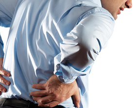
This putative case report provides the reader with a clinical learning opportunity. The discussion addresses certain aspects of the evaluation and management process and proper documentation of the intervention. It is my intent to demonstrate a reasonable approach to the examination and differential diagnosis of a patient with low back pain and offer a prudent treatment plan.
Case report
Subjective:
A 28-year-old professional baseball player presented with a chief concern of “I have back pain and tightness when I run.” The back pain began several hours after a motor vehicle accident that took place 6 months earlier. He denied any history of low back pain prior to the motor vehicle accident. The most recent episode of increased sharp low back pain began 5 days ago. The patient denied any history of back pain prior to the motor vehicle accident and he believes that the car accident caused the back pain. He mentioned that his father has seen a chiropractor for treatment of low back pain.
He was a front seat passenger, wearing a seat belt in a vehicle that rear-ended another vehicle, which was at a stop. His vehicle was traveling about 35 miles per hour. At the time of impact, he was conversing with the driver and turning to the left. The patient denied loss of consciousness or head trauma. He declined any medical care at the scene of the accident.
He has experienced tightness and sharp back pain while running. At my request, he pointed specifically to the right lumbosacral spine as the area of sharp pain with movement. He has not experienced pain with batting or throwing. The back has ached at night and frequently interfered with his sleep. Normally, he has gained some relief with NSAIDS and massage. To date, he has never received chiropractic care.
When asked to point to the area of the dull, aching pain he moved his finger up and down on the right side of the low back and was unable to identify a specific spinal level. The patient denied any pain shooting down the legs but mentioned that sometimes he experienced discomfort in the right groin with prolonged sitting in the dugout. The severity of the sharp pain at the lumbosacral spine with running was rated at 7 of 10 and the aching was rated at 3-5 of 10.
There was no history of other health problems. He mentioned that his diet was good, with ample amounts of fruits and vegetables along with red and white meats. When questioned about hydration, he said that he drinks about 20 ounces of water during a game, but very little water when not playing. He preferred carbonated beverages and beer. His total liquid intake has been 60 ounces per day.
He asked if he might anticipate complete relief of the low back pain with chiropractic care. He has been worried that the pain may interfere with his athletic career.
Objective:
This young, healthy, Caucasian professional athlete appears his stated age. He is an alert, cooperative and well-developed mesomorph. He walks without a limp.
Vital indices: Height 74 inches, weight 205 pounds, blood pressure 110/64, pulse rate 66/minute and respirations of 12/minute
Postural evaluation demonstrated an inferior iliac crest on the right, an inferior right shoulder and a mild “S” type curvature. His left occiput appeared inferior and posterior compared to the right occiput. Anterior and posterior Adam’s positions presented a negative sign. I measured the leg lengths in supine and sitting positions. The mensuration demonstrated functional leg length inequalities with the right lower extremity appearing short while supine and longer with sitting.
Palpation of the area of the right lumbosacral joint was painful and graded as a grade 2 of 4 due to his wincing. There was pain with palpation of the supraspinous ligaments at L5 and S1, and the area of the L5 facet joint on the right. Palpation of the sacroiliac joints did not produce pain. Hypertonicity of the multifidi muscles with taut bands and painful nodules bilaterally were present. The patient demonstrated taut bands and painful nodules with a grade 3 of 4 pain reaction upon palpation of the right iliopsoas muscle.
Active range of motion of the thoraco-lumbar spine demonstrated full and pain-free motion with flexion and left lateral flexion but right lateral flexion and extension did produce pain at the lumbosacral spine on the right. He mentioned that forward flexion at the waist felt good. Passive range of motion of the thoraco-lumbar spine produced pain at the L5-S1 level on the right with right rotation and lateral flexion. Resistive range of motion testing of the thoraco-lumbar spine produced pain in the right paravertebral muscles at the levels of L3-4-5-S1 upon right rotation and lateral flexion.
The Kemp test or maneuver was negative for radicular pain but did produce pain at the right L5 level of the lumbosacral spine with extension and rotation to the right.6 The straight leg test (SLR) produced pain at the right lumbosacral spine with extension of the right hip and lower extremity at 80 degrees. There was no pain with the left SLR at 80 degrees.
Lumbar stability testing produced sharp pain at the lumbosacral spine on the right when the patient stood on one foot and extended his spine. He also experienced pain with flexion of the legs while supine. This pain reduced with manual compression over the lumbosacral spine while repeating the extension of the legs while supine.
The modified Gillet test performed while standing demonstrated hypomobility of the right sacroiliac joint. The Gaenslen, distraction and sacroiliac thrust tests did not produce pain at the sacroiliac joints. The Adams Supported Belt test and the drop test produced pain at L5 right.
Neurological evaluation
The sensory examination of the upper and lower extremities revealed an intact system for sharp and dull stimulations. Motor testing of the upper and lower extremities were normal and graded at 5/5. The deep tendon reflexes were within normal limits and graded at 2+ and brisk for the upper and lower extremities. There were no signs of pathologic reflexes.
Assessment:
- Acute low back pain episode
- Post-traumatic lumbo-sacral sprain/ strain with resultant posterior joint dysfunction and myofascial pain
- Pelvic obliquity with resultant mild “S” type functional scoliosis
- Spondylolysis at L5 without spondylolisthesis
- Subclinical dehydration
Plan:
- I ordered a five-view lumbar spine radiographic examination to evaluate the lumbar spine for separation of the pars interarticularis at the lower lumbar spine.
- Treatment is scheduled three visits per week for a period of four weeks in order to reduce pain and improve function.
- Treatment will consist of spinal manipulation, myofascial trigger-point release and massage therapy.
- I prescribed 140 ounces of fluid per day.7
- Re-evaluation will follow the four weeks of care
- A full explanation of the risks and benefits of treatment was provided to the patient. He understood that he may elect to receive no care or seek another opinion with another provider. The patient decided to have the radiographic examination and then return for the prescribed series of treatments.
Radiology report
Findings:
- Unilateral separation of the L5 pars interarticularis on the right
- No signs of anterolisthesis of lumbar vertebrae
- No signs of decreased disc spacing in lumbar spine
- No signs of degenerative joint changes
Impression:
1. Spondylolysis L5 right
Discussion
 This putative case report demonstrates a focused history taking and physical examination process with a patient presenting post-traumatic low back pain. I documented the patient encounter with SOAP notes (an acronym for subjective, objective, assessment, and plan).
A history of present illness (HPI) obtained during the interview with the patient investigated the onset, duration, and character of the present illness, as well as any acts or factors that aggravate or ameliorate the symptoms. The chief concern or presenting symptom prompted this detailed interview.8 The chief concern, “I have back pain and tightness when I run,” is the initial notation in the subjective section of the medical record. It is significant that the chiropractic clinician identify the cause of the low back pain. Since the patient experienced his first episode of low back pain on the day of the accident and he denied low back pain prior to the incident, it is my professional opinion that the motor vehicle accident caused his low back pain.
I prefer use of the mnemonic initialism, OPQRST, to facilitate the taking of the HPI. This line of questioning includes discussion of the severity of the pain, timing and previous treatment for the present illness or injury.
The musculoskeletal examination focused on the spine with observation, inspection, palpation, range of motion testing, and orthopedic testing, followed by a three-part peripheral nervous system examination.
The negative Adam’s positions indicate a functional scoliosis rather than an anatomical or structural scoliosis.9 These positions involve observation of the patient’s flexed spine from both the anterior and posterior perspectives. If a “C” or “S” curve is present, it will straighten with flexion of the spine. Straightening of the spine in Adam’s positions indicates the presence of a negative sign, which indicates a functional curvature. Chiropractors treat functional curvatures.10
It is common for a patient to present with pelvic tilting or pelvic obliquity due to muscle imbalances in the gluteal, quadratus lumborum, and/or the iliopsoas musculature,11 which may cause mild to moderate spinal scoliosis (“C” of “S”) due to lower limb length inequality.12 Static or flat palpation for pain is reliable while movement restriction in the lumbar spine is not reliable.14 This patient presented pain reactions to palpation of specific lumbar joints/ligaments and myofascial trigger points,15 which enabled the identification of the painful tissues.
Chiropractic clinicians may identify and document the presence of a primary myofascial trigger point by locating a hyperirritable focus within a taut band of skeletal muscle.16 The resistive (isometric contraction) and passive ranges of motion findings indicate a sprain/strain injury of the lumbosacral spine (O’Donoghue maneuver).17 Kemp’s test assesses for intervertebral nerve root encroachment, muscular strain, ligamentous sprain, or pericapsular inflammation using a maneuver that involves flexion of the torso followed by extension and rotation of the lumbar spine while the examiner stabilizes the pelvis.18 You may perform this test in either the seated or the standing position, but I prefer the latter. This patient demonstrated pain with compression at the level of L5 on the right.
Assessment
The O’Donoghue maneuver assesses for muscular strain and ligamentous sprain. The diagnosis of acute low back pain due to sprain of ligaments and strain of muscles was documented with the O’Donoghue maneuver for the lumbar spine.19 The differential diagnosis included pelvic obliquity due to the postural findings.20
Plan
In the absence of “red flag” findings or signs of cauda equina syndrome, four to six weeks of conservative care is appropriate for patients with acute low back pain.21 Chronic dehydration may promote low back pain. Hence, it is reasonable to advise proper water intake to patients that experience low back pain when they do not consume adequate amounts of fluids.22
Note: Doctors interested in gaining continuing education units through the University of Bridgeport Health Sciences Postgraduate Education Department should complete the registration form and email it to Anne Nilson: [email protected].
James J. Lehman, D.C., M.B.A., D.A.B.C.O. is an Associate Professor of Clinical Sciences at the University of Bridgeport College of Chiropractic. Please remit any questions or comments to [email protected]
Reference:
- Hart LG, Deyo RA, Cherkin DC. Physician office visits for low back pain. Frequency, clinical evaluation, and treatment patterns from a U.S. national survey. Spine 1995; 20:11-9.
- Deyo RA, Cherkin D, Conrad D, Volinn E. Cost, controversy, crisis: low back pain and the health of the public. Annu Rev Public Health 1991; 12:141-56.
- Bronfort G, Haas M, Evans RL, Bouter LM. Efficacy of spinal manipulation and mobilization for low back pain and neck pain: a systematic review and best evidence synthesis. Spine J. 2004 May-Jun; 4(3):335-56.
- Murphy DR, Hurwitz EL, and Nelson CF. A diagnosis-based clinical decision rule for spinal pain part 2: review of the literature. Chiropr Osteopat. 2008; 16: 7. (http://www.ncbi.nlm.nih.gov/pmc/articles/PMC2538525/)
- Borkan J, Van Tulder M, Reis S, Schoene ML, Croft P, Hermoni D. Advances in the field of low back pain in primary care: a report from the fourth international forum. Spine (Phila Pa 1976). 2002 Mar 1; 27(5):E128-32. (http://www.ncbi.nlm.nih.gov/pubmed/11880849)
- Evans RC. Illustrated orthopedic physical assessment. Mosby. Third edition.
- Hydration: Fluids for Life. ILSI North America, Monograph Series. http://www.ilsi.org/northamerica/publications/hyd%20-%20hydration%20-%20fluids%20for%20life.pdf
- Mosby’s Medical Dictionary, 8th edition. © 2009, Elsevier.
- Evans RC. Illustrated orthopedic physical assessment. Mosby.Third edition.
- Cooperstein R. and Lew M. The relationship between pelvic torsion and anatomical leg length inequality: a review of the literature. J Chiropr Med. 2009 September; 8(3): 107–118. (http://www.ncbi.nlm.nih.gov/pmc/articles/PMC2732247)
- Travell JG and Simons DG. Myofascial Pain and Dysfunction: The Trigger Point Manual. Williams & Williams. Volume 2, 42 and 57.
- Knutson GA. Anatomic and functional leg-length inequality: A review and recommendation for clinical decision-making. Part I, anatomic leg-length inequality: prevalence, magnitude, effects and clinical significance. Chiropractic & Osteopathy 2005, 13:11. (http://www.chiroandosteo.com/content/13/1/11)
- Haneline MT and Young M. A Review of Interexaminer and Interexaminer Reliability of Static Spinal Palpation: A Literature Synthesis. Journal of Manipulative and Physiological Therapeutics. June 2009. (http://w3.palmer.edu/young/Articles/JMPT_staticpalp.pdf)
- Keating JC Jr, Bergmann TF, Jacobs GE, Finer BA, Larson K. Interexaminer reliability of eight evaluative dimensions of lumbar segmental abnormality. J Manipulative Physiol Ther. 1990 Oct; 13(8):463-70.
- Travell JG and Simons DG. Myofascial Pain and Dysfunction: The Trigger Point Manual. Williams & Williams Glossary, Volume 2, 1.
- Travell JG and Simons DG. Myofascial Pain and Dysfunction: The Trigger Point Manual. Williams & Williams Glossary, Volume 2, 1.
- Evans RC. Illustrated orthopedic physical assessment. Mosby. Third edition.
- Laslett M, McDonald B, Aprill CN, Tropp H, and Oberg B. Clinical predictors of screening lumbar zygapophyseal joint blocks: Development of clinical prediction rules. The Spine Journal 6 (2006) 370–379. (http://fearonphysicaltherapy.com/_media/media/file/342138/Facet-clinical%20predictors-Laslett.pdf)
- Evans RC. Illustrated orthopedic physical assessment. Mosby. Third edition.
- Lehman JJ. Low Back Pain: Do you have a tilted pelvis? (http://backandneck.about.com/od/conditions/ss/tiltedpelvis.htm)
- Kinkade S. Evaluation and Management of Acute Low Back Pain. Am Fam Physician. 2007 Apr 15; 75(8):1181-1188. (http://www.aafp.org/afp/2007/0415/p1181.html)
- Lehman JJ. Back pain and chronic dehydration. (http://nutrition.about.com/od/hydrationwater/a/back_pain_water.htm)