Pelvic and Hip Pain

History and Presenting Symptoms

A 27-year-old female describes a history of aching pain and tightness extending from her lower back into the pelvic region and both hips. She describes a recurrent pain that has bothered her since she was in a motor vehicle collision at the age of sixteen. As a passenger, she injured her right leg and hip during a frontal impact with a light post. No fractures were detected, but she had diffi culty walking for several months, and still gets very fatigued when walking or standing for more than 20 minutes. Her current pain is generalized to the posterior pelvis, but also involves her lower back and extends into both hips. On a 100mm Visual Analog Scale, she rates the pain in her lower back and pelvic region as varying from 30mm to 50mm.

Exam Findings

Vitals. This young woman weighs 152 lbs, which, at 5’9’’, results in a BMI of 22—she is not overweight. She reports that she attends yoga classes regularly and works out on a circuit training program at a local fi tness center. She has never smoked; she drinks beer occasionally; and her blood pressure is 118/76 mmHg with a pulse rate of 64 bpm.

Posture and gait. Standing postural evaluation fi nds a lower right iliac crest, and a low right greater trochanter. The left shoulder is somewhat lower than the right, but the spine demonstrates no signifi cant lateral curvature. Her knees are well aligned, but there is obvious medial bowing of the right Achilles tendon, with a lower medial arch on the right foot. During gait, the right foot toes-out and pronates excessively. Inspection of her shoes finds scuffing and wearing at the lateral aspect of the right heel.

Chiropractic evaluation. Motion palpation identifi es limitations in segmental motion at the right SI joint, with localized tenderness and loss of endrange mobility. Gaenslen’s and Yeoman’s tests for SI joint dysfunction both cause increased pain when the right side is stressed. Lumbar ranges of motion are within expected norms, and neurologic testing is negative for sensory, motor, and refl exive disorders.

Imaging

A lumbopelvic series (AP and lateral lumbopelvic views) is taken in the upright position during relaxed standing. An obvious discrepancy in femur head heights is noted, with the right side 6mm lower. A moderate lumbar curvature (5°) is convex to the right side, and the sacral base angle and measured lumbar lordosis are somewhat increased, but within normal limits. No loss of joint spacing or osteophyte formation is seen in the hip joints.

Clinical Impression

Chronic lumbopelvic misalignment, with mechanical dysfunction of the right sacroiliac joint complicated by leg length discrepancy. The difference in leg lengths is “functional,” since it is due to asymmetry of support in the lower extremities. The inequality results in a pelvic tilt and a slight lumbar curvature.

Treatment Plan

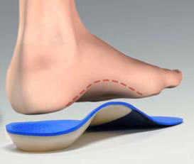
Adjustments. Specifi c chiropractic adjustments for the right sacroiliac joint and lumbar spine were provided as indicated. Manipulation of the right foot, including the navicular, cuboid, and calcaneal bones, was also performed. Support. Flexible, stabilizing orthotics were custom-made, and a pronation correction was added to the right side. The inserts provided support for her arches and included viscoelastic shock-absorbing material to decrease the biomechanical stress on her pelvis and sacroiliac joints.

Rehabilitation. She was shown a lumbopelvic muscle-training program to do in addition to her regular workouts. The “easy eight” exercises were performed daily at home using elastic exercise tubing.

Response to Care

The pelvis and foot adjustments were well tolerated, and the orthotics signifi cantly improved her postural alignment. After fi ve weeks of adjustments (eight visits) and daily home exercises, including wearing the orthotics, she was released to a self-directed maintenance program.

Discussion

This healthy and fi t young woman had a chronic pelvic misalignment caused by a prior injury. It was associated with pronation and biomechanical dysfunction in the right lower extremity. This asymmetry perpetuated her pelvic imbalance, in spite of a well-rounded fi tness program. She responded well to an appropriate combination of chiropractic adjustments, stabilization and support from custom-made orthotics, and specific exercises for the lumbopelvic support musculature.

Dr. John J. Danchik is the seventh inductee to the American Chiropractic Association Sports Hall of Fame. He is the current chairperson of the United States Olympic Committee’s Chiropractic Selection Program and lectures extensively in the United States and abroad on current trends in sports chiropractic and rehabilitation. Dr. Danchik is an associate editor of the Journal of the Neuromusculoskeletal System. He can be reached by e-mail at [email protected].

Asymmetrical Pronation Linked to Juvenile Injury

History and Presenting Symptoms

A 28-year-old male presents with recurring episodes of moderate low-back pain which always respond well to chiropractic adjustments, but eventually return. He recalls no specific back injuries, and cannot identify any triggering activities. On a Visual Analog Scale, he rates his low back pain currently at about 50mm. He has been able to avoid taking pain medication by getting regular chiropractic adjustments.


Exam Findings

Vitals. This active young man is 6 feet tall and weighs 176 lbs, resulting in a BMI of 24; his muscle definition indicates that he is not at risk of overweight. He doesn’t smoke, and his blood pressure is 116/78 mmHg with a pulse rate of 68 bpm. These findings are within the healthy range.

Posture and gait. Standing postural evaluation finds a lower right iliac crest and a low right greater trochanter. His knees are well aligned, but there is obvious medial bowing of the right Achilles tendon, and no medial arch on the right foot. When this is mentioned, he recalls a “bad sprain” injury to his right foot and ankle during high school football. He denies any persisting symptoms or current problems with the right foot or ankle.

Chiropractic evaluation. All active spinal ranges of motion are full and pain free, except that “aching stiffness” restricts left lateral flexion by 10°. Active palpation identifies a motion limitation in the right SI joint, which is also tender to direct pressure. Lumbosacral joint motion is restricted in left lateral flexion, with the feeling of generalized paraspinal muscle tightness. Provocative orthopedic and neurological tests for nerve root impingement and disc involvement are all negative.

Lower extremities. Closer examination reveals that he has no right medial arch when standing. His right calcaneus is noticeably everted when bearing weight. When seated and non-weightbearing, his right arch appears equal to the left, and manual testing finds no evidence of muscle weakness of the fibular or tibial muscles. The Navicular Drop test measures substantial asymmetry in excursion of the navicular bones when moving from sitting to standing (R = 3 mm of drop, L = 8 mm of drop from non-weight bearing to weight bearing). Palpation finds no significant tenderness in the right medial arch or plantar fascia.


Imaging


A lumbopelvic series (AP and lateral lumbopelvic views) reveals an obvious discrepancy in femur head heights, with a measured difference of 5 mm (right side lower). A moderate lumbar curvature (6°) is noted, convex to the right side, and both the sacral base and the iliac crest are lower on the right side. The sacral base angle and measured lumbar lordosis are within normal limits.


Clinical Impression


Asymmetrical pronation likely due to previous injury, with associated pelvic tilt and lumbar curvature resulting in chronic biomechanical stress and recurring subluxations in the lumbopelvic region.


Treatment Plan


Adjustments. Specific, corrective adjustments for the SI and lumbosacral joints were provided—with good response, as previously. Manipulation of the right foot, including the navicular, cuboid, and calcaneal bones, was also performed.

Support. Custom-made, flexible stabilizing orthotics were supplied, with a pronation correction added to the right side. He had no problems in wearing the orthotics, finding them “very comfortable.”

Rehabilitation. He was shown a series of lumbopelvic mobility exercises, using elastic exercise tubing at home. He was encouraged to continue his twice-weekly workouts at the local gym.


Response to Care


The lumbopelvic and foot adjustments were well tolerated, and the orthotics made a noticeable improvement in his postural alignment, both at the feet and the lumbopelvic region. After 6 weeks of adjustments (12 visits) and daily home exercises, including wearing the orthotics, he was released to a self-directed maintenance program.


Discussion

Excessive pronation and biomechanical asymmetries in the foot and ankle are often locally asymptomatic. In this active patient, the constant weightbearing stress to his SI and lumbosacral joints resulted in recurring spinal symptoms. Preventing chronicity is a vital aspect of chiropractic, and correction of this patient’s underlying pronation asymmetry was necessary. Having had no foot or ankle symptoms, he had not recognized that a previous lower-extremity sports injury could be a significant causative factor in his back problem. Fortunately, this was identified before any substantial degenerative changes developed.

Dr. John J. Danchik is the seventh inductee to the American Chiropractic Association Sports Hall of Fame. He is the current chairperson of the United States Olympic Committee’s Chiropractic Selection Program and lectures extensively in the United States and abroad on current trends in sports chiropractic and rehabilitation. Dr. Danchik is an associate editor of the Journal of the Neuromusculoskeletal System. He can be reached by e-mail at [email protected].


Q-Angle Problems – Increased Running Increases Pain

History and Presenting Symptoms

The patient is a 22-year-old recent college graduate, who reports a history of participation in many team sports in school, including field hockey, soccer, and softball. She admits that she has always had occasional knee problems, especially on the left side. She is now running regularly for fitness, and her left knee is noticeably sore and aching. More recently, she has developed stiffness and pain in her lower back, which is most noticeable after longer runs. She denies any specific injury to her knees or back, and reports that over-the-counter medications provide temporary relief.


Exam Findings

Vitals. This fit young woman weighs 126 lbs which, at 5’5’’, results in a BMI of 21—she is not overweight. She has never smoked tobacco, and her blood pressure and pulse rate are both at the lower end of normal range.

Posture and gait. Standing postural evaluation finds generally good alignment throughout her pelvis and spine. She has a mild bilateral knee valgus and moderate calcaneal eversion, with hyperpronation on the left side. Measurement finds a standing Q-angle of 27° on the left and 24° on the right (20° is normal for women). Evaluation of running gait identifies a tendency to hyperpronate, especially on the left.

Chiropractic evaluation. Motion palpation identifies a limitation in lumbosacral motion, with loss of endrange mobility to the left at L5/S1. Compensatory subluxations are noted at T10/T11 and T7/T8. Neurologic testing is negative.

Primary complaint. Clinical examination of the left knee finds no evidence of ligament instability, meniscal damage, or patellar tracking problems. All knee ranges of motion are full and pain-free, bilaterally. Manual muscle testing finds no evidence of specific muscle weakness or regional neurological dysfunction.


Imaging

No X-rays or other forms of musculoskeletal imaging were considered clinically necessary.


Clinical Impression

Excessive Q-angle on the left, associated with calcaneal eversion and hyperpronation. This is accompanied by lumbosacral and lower thoracic joint motion restrictions and compensatory subluxation.


Treatment Plan

Adjustments. Specific, corrective adjustments for the lumbosacral joint and the lower thoracic region were provided as needed. Manipulation of the left knee into external rotation was performed to decrease the internal rotation associated with her elevated Q-angle and hyperpronation. Both feet were adjusted as needed, especially the navicular bones.

Support. Custom-made, flexible stabilizing orthotics were provided to support all three arches and decrease the Q-angles. These included bilateral pronation corrections at the heel. As is necessary for most physically active patients, two pairs of stabilizing orthotics were ordered —one designed specifically for her running shoes and the other for her dress shoes at work.

Rehabilitation. Due to her active lifestyle, no specific rehab exercises were required. She was encouraged to perform a comprehensive stretching program after each run.


Response to Care

She reported a rapid response to the spinal and knee adjustments. After she began wearing her orthotics regularly during her runs, she reported a definite reduction in knee soreness, as well as a smoother stride and gait. Within three weeks of receiving her orthotics, she related that she had successfully increased her mileage in preparation for an upcoming 10K run, with no knee pain or back problems. She was released to a self-directed maintenance program after a total of ten treatment sessions over two months.


Discussion

The Q-angle is formed by the quadriceps muscle (primarily the rectus femoris) and the patellar tendon. This measurement quantifies the quadriceps muscle’s pull from the pelvis to the patella, and the patellar tendon’s pull from the tibia. Since large forces are transmitted through the patella during knee movement, any increase in the angle can result in a variety of symptoms, as well as problems in the pelvis or lumbar regions.

Because of their wider pelvic anatomy, women naturally have higher angles at their knees. The standing Q-angle is an objective method of measurement that includes the valgus stresses on the knee and internal rotation forces due to excessive foot pronation. Since we are most concerned with understanding how the knee functions during daily and sports activities, it makes more sense to obtain this important measurement while in a weight-bearing position.

Dr. John J. Danchik is the seventh inductee to the American Chiropractic Association Sports Hall of Fame. He is the current chairperson of the United States Olympic Committee’s Chiropractic Selection Program and lectures extensively in the United States and abroad on current trends in sports chiropractic and rehabilitation. Dr. Danchik is an associate editor of the Journal of the Neuromusculoskeletal System. He can be reached by e-mail at [email protected].


Increased Running Increases Pain

racewalkingHistory and Presenting Symptoms

A 33-year-old female presents with ongoing pain and discomfort in her left lower leg, which responds only temporarily to anti-inflammatory medications. She has been running regularly for the past six months, but develops left leg pain whenever she tries to increase her mileage. She ices her leg for fifteen minutes after each run, and performs stretches before and after she runs. She has tried wearing off-the-shelf shoe inserts to lessen the impact of heel-strike shock to her feet, but feels that they offered little protection. She describes her current level of leg pain as usually around 40mm on a Visual Analog Scale.


Exam Findings

Vitals. This physically active woman has taken up running in order to help control her weight. She has managed to lose about fifteen pounds over the past six months and now weighs 138 lbs, which, at 5’6’’, results in a BMI of 23; she is no longer overweight. She does not smoke and does not drink alcohol, and her blood pressure and pulse rate are both at the lower end of the normal range.

Posture and gait. Standing postural evaluation finds a lower iliac crest on the left, and a low left greater trochanter. The right shoulder is slightly lower than the left, with no history of fracture or surgery. The spinal curves are intact. She has substantial calcaneal eversion and hyperpronation on the left side, with a moderate outward flare of her left foot. The left medial arch is significantly lower than the right when standing, but it is not tender to direct pressure.  

Chiropractic evaluation. Palpation finds the left posterior tibialis muscle to be tight, tender and ropey, with the tenderness concentrated in the lower part of the muscle and extending into the tendon at the medial ankle. Pain is elicited when providing manual resistance to isometric contraction of the posterior tibialis muscle. There is also moderate tenderness in the right sacroiliac joint, and segmental dysfunction is found at the L4/L5 level on the left. Lumbar, knee, and ankle ranges of motion are full and pain-free, and circulatory and neurological tests are negative.


Imaging

Upright, weight-bearing X-rays of the lumbar spine and pelvis confirm a discrepancy in femur head heights, with the left side 7mm lower. A slight left convex lumbar curvature (5°) is noted, and both the sacral base and the iliac crest are lower on the left. The sacral base angle and measured lumbar lordosis are within normal limits.


Clinical Impression

Posteromedial shin splints caused by tibialis posterior tendinitis and excessive pronation. This is associated with calcaneal eversion and complete loss of the medial longitudinal arch on the left side. These lower extremity asymmetries result in a functional left short leg, which is being aggravated by her attempts to increase her running mileage.


Treatment Plan

Adjustments. Specific chiropractic adjustments for the lower extremities and the involved spinal regions were provided as needed. Transverse friction was applied to the tibialis posterior tendon to increase local circulation and stimulate collagen repair.

Support. Custom-made, flexible stabilizing orthotics were provided to support the left medial arch and decrease the asymmetrical stress on the tibialis posterior tendon and muscle. This included a varus wedge pronation correction for her left calcaneal eversion.

Rehabilitation. Dynamic resistance exercises for the left posterior tibialis muscle were performed daily, using exercise tubing. She continued her current running program, and was able to introduce a graduated increase in her training distance after four weeks of care.


Response to Care

She responded well to the spinal and extremity adjustments, and reported a rapid improvement with the friction massage and exercise. Within two weeks of receiving her orthotics, she returned to her progressive training program. She gradually increased her running mileage, with no return of her previous leg symptoms. She was released to a self-directed maintenance program after a total of ten treatment sessions over two months.


Discussion

Pain in the posteromedial aspect of the lower leg is a variant type of shin splints that is frequently associated with excessive pronation of the foot and ankle. Her previous use of generic shock-absorbing insoles had provided little benefit, since she required specific, customized support for her calcaneus and medial arch.

Dr. John J. Danchik is the seventh inductee to the American Chiropractic Association Sports Hall of Fame. He is the current chairperson of the United States Olympic Committee’s Chiropractic Selection Program and lectures extensively in the United States and abroad on current trends in sports chiropractic and rehabilitation. Dr. Danchik is an associate editor of the Journal of the Neuromusculoskeletal System. He can be reached by e-mail at [email protected].

Surgical Repair “Kneeds” Extra Care

History and Presenting Symptoms

The patient is a 49-year-old male who reports frequent aching and stiffness in his lower back. He denies any back injuries, but does relate an injury to his right knee that resulted in a full tear of his anterior cruciate ligament (ACL). Two years ago, he had a surgical reconstruction of the ACL using a patellar tendon graft. He says he still has occasional grinding and rare sharp pain in his right knee. Using a 100mm Visual Analog Scale, he rates the frequent pain in his low back at around 40mm, while the rare knee pain is a brief 65mm. He avoids the use of medications, but does get frequent massages, which provide some relief from his back pain.

Exam Findings

Vitals. This active male patient is 5’10″ tall and weighs 162 lbs. This results in a BMI of 23, which is within the normal range. He states that he works out twice a week at a local gym, and plays golf regularly. He is a non-smoker, and both his blood pressure and pulse rate are also within the normal ranges.

Posture and gait. Standing postural evaluation finds generally good alignment, with no lateral curvatures or listing of the spine. An increased lumbar lordosis is noted, and the pelvis tilts forward. His right knee demonstrates a mild valgus alignment and calcaneal eversion, and foot pronation is more prominent on the right side. Observation of gait identifies a tendency for the right foot to flare outward, and inspection of his shoes finds scuffing and wearing at the lateral aspect of his right heel.

Chiropractic evaluation. Motion palpation identifies a limitation at the lumbosacral joint, with moderate tenderness and loss of endrange mobility to the right at L5/S1. Additional subluxations are noted at T10/T11 and T5/T6. Lumbar ranges of motion are full and pain-free, except for extension, which is limited to 20° by pain localized to the right lumbosacral junction. This finding is confirmed by Kemp’s test, which is positive for localized pain when performed to the right side.

Lower extremities. Examination of the right knee finds no ligament instability, or evidence of patellar tracking problem. All knee ranges of motion are full and pain-free. Manual muscle testing finds no specific muscle weakness around the knees or ankles. Neurologic testing of the lower extremities is negative.

Imaging

No X-rays or other forms of musculoskeletal imaging were requested.

Clinical Impression


Mechanical dysfunction with postural imbalance of the lumbopelvic region. There is also evidence of poor biomechanical support from the previously injured right knee, which has undergone ACL reconstruction surgery.

Treatment Plan

Adjustments. Specific, corrective adjustments for the lumbosacral and thoracic spinal regions were provided as needed. No manipulation of the right knee or ankle was indicated. The cuboid bone of the right foot was adjusted several times.

Support. Custom-made, flexible stabilizing orthotics were provided to improve foot/ankle symmetry and to support the right knee.

Rehabilitation. This patient was shown a postural correction exercise using exercise tubing for his anteriorly rotated pelvis.

Response to Care

He responded well to his spinal adjustments, and was quite diligent in performing his corrective exercise. He also adapted well to his orthotics, and reported no more episodes of right knee pain. After four weeks of adjustments (eight visits) and daily home exercises, he was symptom-free and had regained full lumbar function. He returned to playing golf with no problems, and was then released to a self-directed maintenance program.

Discussion

In addition to a classic lumbopelvic postural problem, this patient demonstrated an asymmetry of lower extremity alignment associated with injury and surgical repair. Chiropractic adjustments to the lumbosacral and thoracic regions were combined with a specific postural correction exercise, and support for the foot and ankle. This patient had undergone successful post-surgical rehabilitation and the examination found no evidence of ligament instability or muscular weakness around the knee. However, close inspection and gait evaluation identified a lower extremity asymmetry that was contributing to his spinal problems. Custom-made stabilizing orthotics are frequently necessary after surgical repair procedures of the lower extremities, due to the persisting imbalances.

Dr. John J. Danchik is the seventh inductee to the American Chiropractic Association Sports Hall of Fame. He is the current chairperson of the United States Olympic Committee’s Chiropractic Selection Program and lectures extensively in the United States and abroad on current trends in sports chiropractic and rehabilitation. Dr. Danchik is an associate editor of the Journal of the Neuromusculoskeletal System. He can be reached by e-mail at [email protected].


Recurrent Ankle Sprains

History and Presenting Symptoms
The patient is a 38-year-old male, who plays soccer in an adult league on weekends.  He describes recurring episodes of pain and swelling along the outside of his right ankle for the past several years.  He presents for treatment of his lower extremity biomechanical faults, and wants to prevent future problems and improve his athletic performance with chiropractic care.

Exam Findings
Vitals.  This athletic male weighs 160 lbs., which, at 5’10’’, results in a BMI of 23; he is not overweight.  He is a non-smoker, and his blood pressure and pulse rate are at the lower end of the normal range.

Posture and gait.  Standing postural evaluation finds generally good alignment, with intact spinal curves, but a slightly lower iliac crest on the right, which is confirmed by a lower right greater trochanter.  He also demonstrates right calcaneal eversion and a low medial arch (hyperpronation).  A tendency to toe out (foot flare) on the right is noted during gait screening.

Chiropractic evaluation.  Motion palpation identifies a mild limitation in right sacroiliac motion, with moderate tenderness and loss of end range mobility.  Several compensatory subluxations are identified throughout the lumbar region.  Otherwise, all orthopedic and neurological testing is negative.

Lower extremities.  Examination of his right foot and ankle reveals slight general swelling of his ankle, which is moderately tender to palpation along the outer aspect.  All right ankle ranges of motion are full and pain-free, except inversion, which is limited by tightness and localized pain along the lateral foot and ankle.  Manual muscle testing finds mild weakness in the right peroneal muscle, when compared to the left side.

Imaging
No X-rays or other forms of musculoskeletal imaging were requested.

Clinical Impression

History of recurrent inversion ankle sprains associated with hyperpronation and foot instability.  This is accompanied by sacroiliac joint motion restriction and compensatory lumbar subluxations.

Treatment Plan

Adjustments.  Specific, corrective adjustments for the SI joints and lumbar region were provided as needed.  The right navicular bone was adjusted superiorly.

Stabilization.  Custom-made, flexible stabilizing orthotics were provided to support the arches and decrease calcaneal eversion, and to reduce the asymmetrical biomechanical forces being transmitted to the spine and pelvis.  Two pairs of stabilizing orthotics were ordered, one designed for his soccer shoes and the other for everyday shoe wear.

Rehabilitation.  He was initially instructed in daily self-mobilization and strengthening procedures, which included marble pick-up and towel-scrunching exercises.  After two weeks, daily strengthening of eversion and external rotation was introduced, using elastic exercise tubing.

Response to Care
The spinal and pelvic adjustments were well tolerated, and this active athlete required very few readjustments.  His compliance with the stabilization and exercise recommendations was very good, since he was quite motivated to improve his performance and to prevent future injuries to his ankle.  He adapted to and wore the orthotics without difficulty.  He faithfully filled out and brought in his exercise log at every visit, which provided an excellent opportunity to support his home-based efforts.

Within two weeks of receiving his orthotics, he completed several strenuous soccer practices without symptoms or swelling in the ankle.  He described a noticeable improvement overall in his athletic performance, saying that he felt “more stable.”  He was released from acute care to a self-directed maintenance program after a total of ten visits over two months.

Discussion
Interestingly, this athlete had been to several doctors before this encounter.  He was very frustrated by the lack of answers and recommendations.  His frequent and recurring inversion sprains occur in a foot and ankle that has poor medial support (a low medial longitudinal arch) and an everted calcaneus.  Biomechanical analysis found his right foot to be over flexible and unstable.

As is often found in these types of cases, the combination of specific adjustments, custom-made orthotic support, and strengthening of the lateral ankle support musculature brought about an excellent response.  This middle-aged athlete was very motivated to improve his sports performance, and he persisted with the recommended exercises.

Dr. John J. Danchik is the seventh inductee to the American Chiropractic Association Sports Hall of Fame.  He is the current chairperson of the United States Olympic Committee’s Chiropractic Selection Program and lectures extensively in the United States and abroad on current trends in sports chiropractic and rehabilitation.  Dr. Danchik is an associate editor of the Journal of the Neuromusculoskeletal System.  He can be reached by e-mail at [email protected] 

High Arches in a Yoga Instructor

History and Presenting Symptoms

A 44-year-old female patient is a yoga instructor, who reports the recent onset of pain under her left heel. The foot pain is most noticeable in the morning when she first starts to walk. When her foot bothers her, she notices that her entire back is stiff and less flexible during yoga poses. In addition to her frequent yoga classes and practice, she walks briskly for forty-five minutes every day and rides a bicycle about every other day. She rates her current level of left foot pain as getting up to about 75mm on a 100mm Visual Analog Scale, but subsiding as the day progresses.


Exam Findings

Vitals

This active and generally healthy female is 5’6’’ tall and weighs 142 lbs, which results in a BMI of 23; she is not overweight. Her blood pressure is 114/72 mmHg, with a pulse rate of 64 bpm. She reports that she has never used tobacco products and does not drink alcohol or soda pop. She has been a vegetarian for twelve years, and she watches her diet carefully for proper protein and nutrient intake.

Posture and gait.

Standing postural evaluation finds very good alignment throughout the pelvis and spine, with no lateral lists or spinal curvatures. No pronation or toe out is noted during screening evaluation of gait.

Chiropractic evaluation.

Motion palpation identifies a mild limitation in lumbosacral motion, with moderate tenderness and loss of endrange mobility to the left at L5/S1. A compensatory subluxation is noted at T10/T11. Neurologic testing is negative for nerve root impingement or peripheral nerve damage.

Lower extremities. 

Examination of the left foot and ankle finds no ligament instability, and all foot and ankle ranges of motion are full and pain-free. Palpation over the anteromedial portion of the plantar aspect of the left calcaneus elicits substantial “pinpoint” pain and discomfort. Manual testing finds no evidence of weakness in the ankle or foot muscles. Evaluation of foot alignment during mid-stance finds both medial arches to be quite high. Motion palpation identifies a mild limitation in lumbosacral motion, with moderate tenderness and loss of endrange mobility to the left at L5/S1. A compensatory subluxation is noted at T10/T11. Neurologic testing is negative for nerve root impingement or peripheral nerve damage. Standing postural evaluation finds very good alignment throughout the pelvis and spine, with no lateral lists or spinal curvatures. No pronation or toe out is noted during screening evaluation of gait.

Imaging

X-rays of the feet show a small bony spur on the left calcaneus, at the insertion of the plantar fascia. Otherwise, there is normal joint spacing and alignment, with no evidence of osteoarthrosis or other structural pathology.

Clinical Impression

High medial arches with resulting excessive supination (insufficient pronation). Increased biomechanical stress has been placed on the plantar fascia, resulting in the development of a chronic traction spur at the calcaneal insertion. There is moderate secondary spinal involvement, which has probably been minimized by her frequent yoga practice.


Treatment Plan

Adjustments.

Specific adjustments for the lumbosacral and thoracolumbar spinal regions were well-tolerated. Manipulation of the cuboid and calcaneus bones on both feet produced noticeable reduction of tension in the plantar fascia.

Support

Soft tissue mobilization of the plantar fascia was performed, along with cross-fiber friction to tolerance at the calcaneal insertion. Custom-made, flexible stabilizing orthotics were ordered to support the lateral and anterior arches. These orthotics included an additional layer of shock absorption materials to reduce impact stress throughout the entire gait cycle.

Rehabilitation.

She was encouraged to continue with her active lifestyle and yoga practice, including her frequent walking and cycling. Since she was in generally good condition, she did not require any specific corrective exercises.. Soft tissue mobilization of the plantar fascia was performed, along with cross-fiber friction to tolerance at the calcaneal insertion. Custom-made, flexible stabilizing orthotics were ordered to support the lateral and anterior arches. These orthotics included an additional layer of shock absorption materials to reduce impact stress throughout the entire gait cycle.


Response to Care

Once she began wearing her shock-absorbing orthotics, she noted a substantial decrease in tension and tenderness of the plantar fascia. Within two weeks of receiving the orthotics, she reported that she was able to perform all her exercise and personal activities with no foot or back pain or limitation. She was released to a self-directed maintenance program after a total of eight treatment sessions over two months.


Discussion

When the medial arches are high, the corresponding lateral and anterior arches are often low or collapsed, resulting in excessive strain and tension on the plantar fascia. Lack of pronation increases impact forces at heel strike; these forces are then transmitted up the kinetic chain to the spine. Excessive supination is much less common than excessive pronation, but is frequently an underlying factor in chronic stress on the plantar fascia at the calcaneal insertion. 

Dr. John J. Danchik is the seventh inductee to the American Chiropractic Association Sports Hall of Fame. He is the current chairperson of the United States Olympic Committee’s Chiropractic Selection Program and lectures extensively in the United States and abroad on current trends in sports chiropractic and rehabilitation. Dr. Danchik is an associate editor of the Journal of the Neuromusculoskeletal System. He can be reached by e-mail at [email protected]

The Aging Foot

History and Presenting Symptoms

A 64-year-old female presents with recurring, unresolving episodes of moderate pain in her lower back and left knee.  She recalls no specific back or knee injuries, and states that these problems have developed over the past couple of years, getting more noticeable in the past six months.  On a 100mm Visual Analog Scale, she rates her low back pain as usually 40mm, while her left knee varies from 20mm to 50mm.  The knee gets worse after walking, and she takes over-the-counter NSAID’s for relief.

Exam Findings

Vitals.  This 5’6’’ female weighs 164 lbs., which results in a BMI of 27.  She knows she is overweight and is following a sensible diet, but she is having difficulty doing the needed walking because of pain.  She quit smoking twelve years ago.  Her blood pressure is 124/84 mmHg, and her pulse rate is 80 bpm.  These findings are within the normal range.

Posture and gait.  Standing postural evaluation finds generally good alignment, with intact spinal curves, and no significant lateral listing of her pelvis or spine.  She demonstrates bilateral calcaneal eversion, worse on the left, with a lower left arch.  During gait, both feet pronate substantially, and both feet flare outwards (toe-out).

Chiropractic evaluation.  Motion palpation identifies numerous limitations in spinal motion: the left SI joint, the lumbosacral junction on the left, L2/L3 on the right, T11/12 generally, and at the cervicothoracic junction.  Palpation finds no significant local tenderness or muscle spasm in these regions, and all active thoracolumbar spinal ranges of motion are limited slightly by aging, but are pain-free.  Provocative orthopedic and neurological tests for nerve root impingement and/or disc involvement are negative.

Lower extremities.  Both knees demonstrate full and pain-free movements, and no provocative orthopedic tests are positive.  Closer examination finds a low medial arch on the right foot, and no arch remaining on the left when standing.  Her left calcaneus also demonstrates greater eversion when bearing weight.  Manual testing finds no significant muscle weakness in the fibular (peroneal) or anterior tibial muscles on either side.

Imaging

Lumbopelvic and knee X-rays in the upright, standing position are taken while weightbearing.  There is some loss of lumbar disc heights, most obvious at the lumbosacral joint, and decreasing cephalad.  A slight discrepancy in femur head heights is noted, with a measured difference of 4mm (left side lower).  A moderate lumbar curvature (4°) is also seen, convex to the left side, and both the sacral base and the iliac crest are slightly lower on the left.  The sacral base angle and measured lumbar lordosis are increased, but still within normal limits.  No significant loss of joint spacing or osteophyte formation is seen in the knee joints.

Clinical Impression

Moderate lumbopelvic imbalance and spinal dysfunction associated with generalized loss of arch height (worse on the left) and aging of the knee joints and feet.

Treatment Plan

Adjustments.  Specific, corrective adjustments for the SI joints and the lumbar and cervicothoracic regions were provided as needed, with good response.  Manipulation of the left foot and knee was also performed.

Support.  Custom-made, flexible stabilizing orthotics were supplied, which included bilateral pronation correction (varus wedges).  The patient described no problems in adapting to the orthotics, although close inspection found that the shoes she was wearing were one full size too small for her feet and she did need to purchase better-fitting shoes.

Rehabilitation.  She received instruction in a comprehensive spinal wellness exercise program using elastic resistance tubing.  She brought her exercise log to each visit so her adherence to the program could be encouraged.

Response to Care

The adjustments were well tolerated, and the orthotics made a noticeable improvement in her postural alignment, at the feet and the lumbopelvic region.  After six weeks of adjustments (twelve visits) and daily home exercises, including wearing the orthotics, she was released to a self-directed maintenance program.

Discussion

This patient’s history and physical examination are consistent with the commonly seen spinal effects of aging on the feet.  The combination of lower spinal symptoms with knee pain made worse by walking prompted an evaluation of the lower extremities during weight bearing.  The problem most in need of correction was the excessive pronation that was causing functional imbalance.

Dr. John J. Danchik is the seventh inductee to the American Chiropractic Association Sports Hall of Fame.  He is the current chairperson of the United States Olympic Committee’s Chiropractic Selection Program and lectures extensively in the United States and abroad on current trends in sports chiropractic and rehabilitation.  Dr. Danchik is an associate editor of  the Journal of the Neuromusculoskeletal System.  He can be reached by e-mail at [email protected].

Wellness Support for an Obese Patient

orthoticblueHistory and Presenting Symptoms

A 32-year-old male presents with a history of mild to moderate lower back pain, and aching tightness in his neck.  He states that his back pain comes and goes, with no specific triggering activities.  His neck tension is worse with long and stressful workdays.  He participates in no recreational sports or exercise activities, and recalls no back or neck injuries.  On a 100mm Visual Analog Scale, he rates the low back pain as usually 30-40mm, and his neck tightness as around 25mm.

Exam Findings

Vitals.  This man weighs 221 lbs, and is 5’10’’ tall.  This calculates to a BMI of 32—he is obese.  His waist measures 48 inches at the largest point above the ASIS, confirming that he has abdominal obesity, and is not just big and/or muscular.  His blood pressure is somewhat elevated at 142/90 mmHg, and his pulse rate is 80 bpm.

Posture and gait.  Standing postural evaluation identifies the effects of abdominal obesity, with a loss of the lumbar curve, and an accentuated thoracic kyphosis, with a forward head carriage.  There is no obvious lateral listing of the pelvis or lateral curvature of the spine.  He has mild bilateral knee valgus and bilateral calcaneal eversion, and very flat medial arches.  During gait evaluation, both feet flare outward and pronate substantially.

Chiropractic evaluation.  Motion palpation identifies several limitations in intersegmental spinal motion: the right SI joint, L4/L5 on the left, L2/L3 on the right, T11/12 generally, and several levels at the cervicothoracic junction.  There is no specific spinal tenderness or spasm of the paraspinal muscles, but adipose tissue is prevalent throughout.  All active thoracolumbar spinal ranges of motion are limited slightly by general stiffness.  Neurological tests are negative for nerve root impingement, but straight leg raise is limited bilaterally by hamstring shortening and tightness.

Imaging

Upright, weight-bearing X-rays of the lumbar spine demonstrate loss of intervertebral disc height at L4/L5 and L5/S1, with moderate osteophyte formation at those levels.  There is no discrepancy in femur head or iliac crest heights, and no lateral listing or lateral curvature.  The sacral base angle and lumbar lordosis are both decreased; this is consistent with the postural analysis.

Clinical Impression

Chronic spinal stress syndrome due to obesity.  This is accentuated by poor support from the lower extremities, with bilateral knee valgus, hyperpronation and calcaneal eversion.

Treatment Plan

Adjustments.  Specific, corrective adjustments for the identified subluxations were provided as needed, with good response.  Specific manipulation of both feet and knees was also performed.

Support.  Custom-made, flexible stabilizing orthotics were provided, to support the arches and decrease stress to the knees and back during walking.  A custom-made cervical traction pillow was ordered, based on four specific measurements.

Rehabilitation.  He was started on a localized spinal activation and strengthening program, using elastic resistance tubing for the multifidus muscles—first in extension, then into rotation and lateral flexion.  In addition, he was counseled on building up to sixty minutes a day of moderate physical activity for weight loss.  He chose to walk for thirty minutes during lunchtime and, after two weeks, added an additional thirty minutes before his evening meal.  He used an exercise log as part of his motivation for sustaining his healthy behaviors, which included returning to a previously successful healthy diet program. 

Response to Care

He tolerated his adjustments well, and adapted to the orthotics and cervical support pillow with no difficulty.  His low back symptoms resolved quickly and, shortly thereafter, his neck tightness disappeared.  His brisk walking program did not exacerbate his back pain, even when he increased to a total of sixty minutes daily.  After six weeks of adjustments (twelve visits) he was released to a maintenance program with instructions to continue his exercise and healthy diet program.

Discussion

Obesity is increasing in prevalence in all industrialized countries, since few of us must perform physical labor to obtain our food.  The accumulated weight places increased stress on the lower extremities and spine, resulting in chronic symptoms and accelerating degenerative changes.  Specific spinal adjustments and custom support for the spine and lower extremities need to be combined with individualized exercise instructions for best results.  An exercise/dietary log can improve patient adherence to recommended lifestyle changes.

Dr. John J. Danchik is the seventh inductee to the American Chiropractic Association Sports Hall of Fame.  He is the current chairperson of the United States Olympic Committee’s Chiropractic Selection Program and lectures extensively in the United States and abroad on current trends in sports chiropractic and rehabilitation.  Dr. Danchik is an associate editor of the Journal of the Neuromusculoskeletal System.  He can be reached by e-mail at [email protected].

Awkward Gait in a Young Runner

History and Presenting Symptoms

The patient is an eight-year-old boy being brought in by his parents, who are concerned about his awkward style of running. He has no symptoms or painful limitations, but his mother and father are both worried that his inefficient gait could lead to future problems. They have noticed that he is a less-effective participant on his soccer and baseball teams because of his running style. Upon further questioning, the father relates a personal history of difficulty running as a child, and an inability to excel in school and team sports. He wants his son to be spared these limitations, and be able to keep up with his friends and teammates.

Exam Findings

Vitals. This active young male weighs 66 lbs, which, at 4’4’’, results in a BMI of 17; this is considered a “healthy weight,” since he is within the 50-85th percentile for his age. His pulse is 92 bpm, and his respirations are 16/minute; all within normal ranges for his age.

Posture and gait. Standing postural evaluation finds generally symmetrical development, with intact spinal curves. There is no evidence of lateral curve or list, and his iliac crests are level. His knees are moderately valgus and both Achilles tendons demonstrate medial bowing. They insert into calcanei that are everted, and there is complete absence of both medial arches (pes planus). A distinct tendency to toe out is seen during gait screening, and he hyperpronates bilaterally. When performing a toe raise, however, the medial arches reappear, indicating that these are not rigid flat feet. Additional testing is performed on a treadmill at several speeds, while barefoot and wearing sports shoes. This confirms that, after heel strike, the feet roll medially and flare outwards, resulting in a very ineffective toe off.

Chiropractic evaluation. Motion palpation identifies a mild limitation in right sacroiliac motion, with moderate tenderness. A compensatory subluxation is identified at L4/5 on the left. No joint fixations are found in the feet and ankles. Manual testing finds moderate weakness of the posterior tibialis muscles bilaterally. Deep tendon reflexes in both lower extremities are somewhat sluggish, but all other neurological and orthopedic tests are negative.

Imaging

No imaging studies were performed.

Clinical Impression

Lack of development of the medial longitudinal arches with hyperpronation. This is associated with loss of medial support at the ankles and knees, and has resulted in sacroiliac joint motion restriction and a compensatory lumbar subluxation.

Treatment Plan

Adjustments. Specific, corrective adjustments for the SI joints and lumbar region were provided, as indicated.

Support. Custom-made, flexible, stabilizing orthotics were ordered to support the arches, decrease calcaneal eversion and knee valgus, and to reduce the asymmetrical biomechanical forces being transmitted to the spine and pelvis.

Rehabilitation. He was shown how to perform daily strengthening exercises for the posterior tibialis muscles, using elastic exercise tubing.

Response to Care

His father made sure that he did his exercises regularly, and he tolerated the spinal and pelvic adjustments with little difficulty. Once he began wearing the stabilizing orthotics, both he and his father noticed an immediate improvement in his running efficiency. He described it as feeling more “springy” when he ran; while his father noticed that he didn’t throw his arms out while running. He required only a few adjustments for his segmental dysfunctions, and was released from care after a total of eight visits over six weeks.

Discussion

This young man had apparently inherited his dad’s flat feet, and he was unable to run efficiently with his friends. While he didn’t have any symptoms, he was definitely at risk for eventual foot, ankle, knee, and/or spinal problems and injuries. The additional support from stabilizing orthotics helped realign his feet for more effective walking and running. Once the underlying biomechanical fault was minimized, he responded rapidly to the muscle strengthening exercises and chiropractic adjustments. Both he and his parents have been informed of the need to monitor his feet as he continues to grow, and to return for a new pair of flexible orthotics as soon as his feet have grown by one-and-a-half shoe sizes.

Dr. John J. Danchik is the seventh inductee to the American Chiropractic Association Sports Hall of Fame. He is the current chairperson of the United States Olympic Committee’s Chiropractic Selection Program and lectures extensively in the United States and abroad on current trends in sports chiropractic and rehabilitation. Dr. Danchik is an associate editor of the Journal of the Neuromusculoskeletal System. He can be reached by e-mail at [email protected].