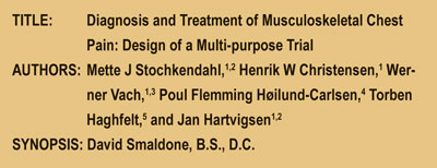
Diagnosis and Treatment of Musculoskeletal Chest Pain

From The Abstract:

Synopsis-DavdToday, hospitals are completely overwhelmed with patients who have acute chest pain, yet no cardiac abnormalities. Often patients find themselves with nowhere to go after cardiac pain has been ruled out as a diagnosis. This study was designed to possibly determine four things: If a multi-disciplinary approach may be used to treat these patients, “To investigate the diagnostic decision making process of a chiropractor…”, to compare manual adjustment procedure vs. home care, and if chiropractic care is cost effective for this population.

Key Points:

1) The study had a focus population of 300 people released from care after having no “…obvious cardiac or non-cardiac disease.” Out of the 300 possible people for the study, 120 people were randomized and were separated into two populations.

2) Discussion: “This study may potentially demonstrate that a chiropractor is able to identify a subset of patients suffering from chest pain predominantly of musculoskeletal origin among patients discharged from an acute chest pain clinic with no apparent cardiac condition.” 3) Baseline measurements included:

1. “semistructured interview,

2. A general health examination, and

3. A specific manual examination.”

4) MPS(Myocardial Perfusion Scintigraphy) was used for exclusionary/inclusionary measures: “In order to evaluate the population in terms of ischemic heart disease, all patients undergo MPS within two to four weeks following baseline evaluation. Using radionuclides, the myocardial perfusion is evaluated to determine the presence of regional areas with decreased blood flow because of coronary artery disease.”

5) For the comparison of chiropractic adjustments vs. self management the RCT (Randomized Clinical Trial) measure was used. “All CTA (Cervico Thoracic Angina) positive patients (estimated 120 out of the initial 300 patients) will be included in the RCT. The aim of this part of the study is to establish the effectiveness of chiropractic treatment, including spinal manipulation versus advice to promote self-management.”

6) The study was computer randomized and blinded to the patients to chiropractic vs. home management.

7) The chiropractic treatment included: The patient must have a high velocity low amplitude adjustment and could have other soft tissues techniques in conjunction. Also, “the protocol specifies up to ten treatment sessions of approximately 20 minutes, 1–3 times per week for four weeks, or until the patient is pain free, if this occurs within less than four weeks.” The other group received 15 minutes of advice and two or three exercises to do at home.

8) Outcome measures were: The primary outcome measure was an 11point measure (0-10) scale, 6 secondary outcome measures were utilized in this experiment as well.

9) “In summary, this article presents the rationale and design of a multi-purpose study consisting of a prospective diagnostic study, and an RCT, with a cost-effectiveness study alongside the central trial. It is anticipated to be completed in 2008, at which time the results will be made available. The first part of this study may potentially demonstrate that a chiropractor is able to identify a subset of patients suffering from chest pain predominantly of musculoskeletal origin among patients dismissed from an acute chest pain clinic with no apparent cardiac condition. The long term goal is to establish whether manual palpation may be used as part of the clinical examination to screen patients allowing for improvement in referral patterns. Furthermore, knowledge about the benefits of manual treatment in patients with musculoskeletal chest pain will inform clinical decision and policy development in relation to clinical practice.”

This is an Open Access article distributed under the terms of the Creative Commons Attribution License ( http://creativecommons.org/licenses/by/2.0 ), which permits unrestricted use, distribution, and reproduction in any medium, provided the original work is properly cited.


Dr.-David-Smaldone,-B.S.,-Dr. David Smaldone is a graduate of Palmer West Chiropractic College with an undergraduate in Athletic training. Dr. Smaldone has worked with thousands of Athletes in his career. He currently runs a practice in New Paltz, New York, and is an NYS licensed chiropractor. Dr Smaldone is also currently the Director of Chiropractic affairs for 123chiropractors.com.

Overtreating Back Pain

Key Points from Dr. Dan Murphy


1.     “Pain complaints are a leading reason for medical visits. The most common pain complaints are musculoskeletal, and back pain is the most common of these.”


2.    “The prevalence and impact of back pain have led to an expanding array of tests and treatments, including injections, surgical procedures, implantable devices, and medications. Each is valuable for some patients, but use may be expanding beyond scientifically validated indications, driven by professional concern, patient advocacy, marketing, and the media.”


3.     Although approximately 25% of US adults reported back pain during the past 3 months, this percentage has not changed for decades.


4.    Expanded testing and treatment for back pain have not improved outcomes, but have increased complications, including deaths.


5.       “Chronic back pain is among the most common patient complaints.”


6.    There are increases in Medicare expenditures for back pain diagnostics and treatments, as follows:

  1.   A 629% increase for epidural steroid injections
  2.   A 423% increase in expenditures for opioids for back pain
  3.   A 307% increase in the number of lumbar magnetic resonance images
  4.   A 231% increase in facet joint injections
  5.   A 220% increase in spinal fusion surgery rates


7.       These increases have not improved in patient outcomes or disability rates.


8.     Manufacturers aggressively promote new drugs and devices for the treatment of back pain, yet there is evidence of misleading advertising, kickbacks to physicians, and major investments by surgeons in the products they are promoting.


9.    The use of lumbar magnetic resonance imaging (MRI) has increased dramatically, and spinal surgery rates are highest where imaging rates are highest.


10.    Approximately 33–66% of spinal computed tomography (CT) imaging and MRI may be inappropriate.


11.   “Many factors probably underlie the growth of imaging, including patient demand, the compelling nature of visual evidence, fear of lawsuits, and financial incentives.”


12.   “One problem with inappropriate imaging is that it may result in findings that are irrelevant but alarming.”


13.    “Positive findings, such as herniated disks, are common in asymptomatic people.”


14.   Positive imaging findings result in more surgery and higher costs than those receiving plain X-rays, but the clinical outcomes are no better, including subsequent pain, function, quality of life, or overall improvement.


15.   Prescription opioid use is steadily increasing, especially for musculoskeletal conditions. Emergency department reports of opioid overdose parallel the numbers of prescriptions. Deaths related to prescription opioids are greater than the combined total involving cocaine and heroin.


16.   Cancer patients tend not to take opioids for long periods of time because they die. In contrast, patients taking opioids for back pain can do so for decades. More than half of the prescriptions for opioids are for back pain and, consequently, they constitute a major portion of those with opioid consumption complications.


17. The benefit of opioids drugs in clinical practice for the long-term management of chronic low back pain is questionable. 


18.    Ironically, “Opioid use may paradoxically increase sensitivity to pain.”


19.  Chronic use of opioid drugs may also cause hypogonadism, reduced testosterone levels, diminished libido, and erectile dysfunction.


20.  “Epidural corticosteroid injections may offer temporary relief of sciatica, but both European and American guidelines, based on systematic reviews conclude they do not reduce the rate of subsequent surgery.”


21.  “Facet joint injections with corticosteroids seem no more effective than saline injections.”


22. “For patients with axial back pain without sciatica, there is no evidence of benefit from spinal injections.”


23. Spine fusion surgery is limited when treating degenerative discs with back pain with no sciatica, yet they have increased 220% from 1990 to 2001 in the United States.


24.   “Higher spine surgery rates are sometimes associated with worse outcomes.”


25.   New and improved fusion techniques and devices, such as implants, increase the risk of nerve injury, blood loss, overall complications, operative time, and repeat surgery, but do not result in improved disability or reoperation rates.


26.  Increases in the rates of imaging, opioid prescriptions, injections, and fusion surgery might be justified if there were substantial improvements in patient outcomes; unfortunately, they are not. In fact, statistics indicate that disability from musculoskeletal disorders is rising, not falling.


27.    “Prescribing yet more imaging, opioids, injections, and operations is not likely to improve outcomes for patients with chronic back pain.”


28.    “There are no “magic bullets” for chronic back pain, and expecting a cure from a drug, injection, or operation is generally wishful thinking.”


29.    “Chronic back pain, like diabetes or asthma, is a condition we can treat but rarely cure,” and its management may “benefit from sustained commitment from health care providers; involvement of patients as partners in their care; education in self-care strategies; coordination of care; and involvement of community resources to promote exercise, provide social support, and facilitate a return to work.”


COMMENT FROM DAN MURPHY

In the 10 years that I have been doing my Article Reviews, we have seen a number of studies that show that spinal adjusting is highly effective, safe, cost effective, and results in long-termed stable outcomes in the treatment of chronic low back pain.


Daniel 20J.Murphy, D.C.Dr. Daniel J. Murphy D.C., D.A.B.C.O. graduated magna cum laude from Western States Chiropractic College in 1978. He received Diplomat status in Chiropractic Orthopedics in 1986. Since 1982, Dr. Murphy has served part-time as undergraduate faculty at Life Chiropractic College West, currently teaching classes to seniors in the management of spinal disorders. He has taught more than 2000 postgraduate continuing education seminars. Dr. Murphy is a contributing author to both editions of the book Motor Vehicle Collision Injuries and to the book Pediatric Chiropractic. Hundreds of detailed Article Reviews, pertinent to chiropractors and their patients, are available at Dr. Murphy’s web page, www.danmurphydc.com


The Whiplash Effect on Soft Tissues

TITLE: A Distinct Pattern of Myofascial Findings in Patients after Whiplash Injury

CITATION: Archives of Physical Medicine and Rehabilitation. Volume 89, Issue 7, July 2008, Pages 1290-1293. AUTHORS: Thierry Ettlin, MD; Corina Schuster, MPtS, PT; Robert Stoffel, PT; Andreas Brüderlin, PT; and Udo Kischka, MD


KEY POINTS FROM DR. DAN MURPHY

1. 85.1 percent of the patients with whiplash had positive trigger points in the semispinalis capitis muscle.

2. The patients with whiplash had a significantly higher prevalence of positive trigger points in the semispinalis capitis muscle than any of the control groups, which is a distinct pattern of trigger point distribution that differed significantly from other patient groups and healthy subjects.

3. In addition to neck pain and stiffness, whiplash patients often complain about “headache, brachialgia (pain radiating into one or both arms), vertigo or dizziness, chewing and swallowing problems, visuomotor disturbances such as blurred vision and reduced coordination, fatigue and reduced energy, neuropsychologic dysfunction, depression, irritability, and sleep disorders.”

4. Whiplash pain can be caused by injuries to the muscles, facet joints, ligaments, disks, and nerve roots.

5. Reduced cervical range of motion is a prominent finding in whiplash-injured patients.

6. Myofascial tension of the scalene muscles causes a functio-nal thoracic outlet syndrome that may explain brachialgia.

7. “The only consistent finding [to explain whiplash pain] reported in the literature is a painful facet joint dysfunction C1-2,” verified by anesthetic injection.

8. These authors main hypothesis was that “patients with whiplash disorder would display more trigger points in the semispinalis capitis muscle, which is localized in the upper neck,” consistent with a C1-C2 facet injury.

9. The semispinalis capitis is an easily locatable marker for the upper cervical spine. “The semispinalis capitis was included because its referred pain zone is parieto-occipital and periorbital, which is the most frequent pattern of referred pain to the head in patients with whiplash.”

10. The criteria for myofascial pain syndrome are:

  • Palpable hardening (trigger point and/or taut band) in the muscle belly
  • Pressure pain in the trigger point or taut band
  • Referred pain while manipulating the trigger point in the taut band
  • Recognition of the elicited pain as the patient’s known and familiar pain

11. “The prevalence of trigger points in the semispinalis capitis muscle was significantly higher in the whiplash injury group than in each of the other groups. Patients with whiplash syndrome, therefore, showed a distinct pattern of trigger point distribution that differed significantly from other patient groups and a healthy control group.”

12. “Our findings support the hypothesis that the most severe musculoskeletal pathology after whiplash is found in the upper part of the cervical spine.” They are consistent with the biomechanics of the injury and of a “painful C1-2 facet joint dysfunction. In contrast, trigger points in the other patient groups and in healthy people were predominantly found in the lower cervical spine and the shoulder girdle.”

13. Trigger points are a neuromuscular dysfunction at the motor endplate of a skeletal muscle fiber.

14. A mechanical trauma stimulates the release of excessive amounts of acetylcholine at the neuromuscular junction and increased intracellular calcium activates local muscle contraction. This causes increased metabolism [use of oxygen to produce ATP] and relative local ischemia; this leads to failure of the calcium pump which is required for the muscle to relax. “Consequently, the calcium continues to stimulate contraction, and a vicious circle develops.”

15. “The semispinalis capitis muscle was more frequently affected by trigger points in patients with whiplash, whereas other neck and shoulder muscles and the masseter muscle did not differentiate between patients with whiplash and patients with nontraumatic chronic cervical syndrome or fibromyalgia.”

Dr. Dan Murphy graduated magna cum laude from Western States Chiropractic College in 1978. He received Diplomat status in Chiropractic Orthopedics in 1986. Since 1982, Dr. Murphy has served part-time as undergraduate faculty at Life Chiropractic College West, currently teaching classes to seniors in the management of spinal disorders. He has taught more than 2000 postgraduate continuing education seminars. Dr. Murphy is a contributing author to both editions of the book Motor Vehicle Collision Injuries and to the book Pediatric Chiropractic. Hundreds of detailed Article Reviews, pertinent to chiropractors and their patients, are available at Dr. Murphy’s web page, www.danmurphydc.com 


Schmorl’s Nodes

TITLE: The Pathogenesis of Schmorl’s Nodes in Relation to Acute Trauma

CITATION: Spine, Volume 23(21), 1 November 1998, pp2272-2275. AUTHORS: Fahey, V. MBBS; Opeskin, K., FRCPA, MBBS; Silberstein, M. MD, FRACR; Anderson, R., FRCPA, MRCPath; Briggs, C., PhD, BSc.

Advanced imaging, such as CT and MRI, is often indicated in the traumatically injured patient. The question of whether findings pre-dated the injury is a common question in the medical-legal arena. One finding that can give the clinician difficulty is Schmorl’s nodes. This paper was published in Spine. Recognized internationally as the leading journal in its field, Spine is an international, peer-reviewed, bi-weekly periodical that considers for publication original articles in the field of spine care. It is the leading subspecialty journal for the treatment of spinal disorders. Only original papers are considered for publication, with the understanding that they are contributed solely to Spine. The journal does not publish articles reporting material that has been reported at length elsewhere.

This paper points out that, historically, most spine physicians accept that Schmorl’s nodes occur as a result of trauma. No studies have shown a direct causal relation between a traumatic episode and the formation of an acute Schmorl’s node. From a medical-legal perspective, particularly in the clinical setting, it is important that the association between trauma and acute Schmorl’s nodes be determined.

This study utilized 70 thoracolumbar spines that were obtained from individuals killed in motor vehicle accidents. The idea was to determine whether Schmorl’s nodes occur acutely as a result of trauma.

The manuscript points to the fact that Schmorl’s nodes can be caused by a single traumatic event (Fahey, & Silberstein, 1998, p. 2227). The diagnosis of an acute Schmorl’s node requires the clinician to dig deep into their knowledge of imaging techniques. It was determined that acute Schmorl’s nodes cannot be seen as such on plain radiographs, and must be visualized utilizing MRI. This is due to the lack of bony callus formation in the acute phase, as Schmorl’s nodes are vertical disc herniations that break through the endplates of the vertebral bodies. This is often the case when a patient receives plain film radiographs during an emergency evaluation post accident, which are reported negative for pathology. They then are referred for MRI of the spine, to determine the integrity of the intervertebral discs and surrounding soft tissue, which is then positive for Schmorl’s node formation. Chronic Schmorl’s node formation will show a callus formation on both plain film radiographs and the MRI.

The most common cause of acute Schmorl’s nodes is axial loading of the spine as a result of a traumatic event, especially in younger patients whose annulus fibrosis is intact. It has been shown that axial loading may be produced from a fall off a ladder or jumping from a height, but loading may also occur when an occupant hits his head on the roof of the vehicle after a rear impact, or hyper-flexes the thoracolumbar spine as a result of significant whiplash forces (Fahey, & Silberstein, 1998, p. 2224). The acute Schmorl’s nodes in this paper were detected in individuals 11-30 years of age, with a male to female ratio of 9:1, and were localized to the region of T8-L1. It was determined that the anatomical orientation of the facet joints in the thoracolumbar spine made it particularly susceptible to injury due to compressive forces.

When evaluating the traumatically injured patient, considering the difference in imaging techniques and how to use them can have a profound impact on a doctor’s ability to properly diagnosis a traumatic lesion. Obtaining all imaging studies and reviewing them is a critical step in the proper care of trauma victims, especially in the case of acute Schmorl’s nodes, since they are not present on plain film radiographs post accident.


Each issue, a clinical topic will be provided byDrs. Mark Studin & William J. Owens of the American Academy of Medical Legal Professionals (AAMLP), which is a national non-profit organization comprised of doctors and lawyers. The purpose of the organization is to provide its members with current research in trauma and spinal-related topics to keep the professional on the cutting edge of healthcare. Members may also sit for a Diplomate examination and be conferred a DAAMLP. The organization also offers support to the individual member’s practice. To learn more, go to www.aamlp.org or call 1-716-228-3847.


A Closer Look

KEY POINTS FROM DAN MURPHY

1. Because statin drug therapy is likely to continue for many years, or for a lifetime, the official written position of the National Cholesterol Education Program of the National Institutes of Health state, “the decision to add drug therapy to the regimen should be made only after vigorous efforts at dietary treatment have not proven sufficient.” “Vigorous” dietary efforts are defined as a minimum of six months of intensive dietary counseling before starting statin drug therapy.

2. Statin drug trials are not preceded by vigorous dietary efforts because to do so would help people and render statin drug therapy less effective in reducing deaths from coronary heart disease (CHD) and other causes of mortality.

3. In performing a clinical trial of a drug, “all-cause mortality” is the only endpoint measure not prone to diagnostic variance and is, therefore, not popular with the drug company studies. Most statin drug trials do not even look at all-cause mortality because of the probability that taking the drugs does not alter all-cause mortality.

4. Drug company study designers search for endpoints that are most apt to yield a positive result. “This would not be the scientific approach but would make sense if the aim was to make the study appear highly successful.”

5. “If a drug or other intervention neither extends life nor improves its overall quality, then it is of no value.”

6. “There is no rigorous reporting of all-cause morbidity, nor of measurement of changes in overall quality of life, in any of the [statin drug] studies.”

7. Statin drug trials show absolute differences of less than 1 percent to a maximum of 3.3 percent in all-cause mortality between the control and treatment groups. “These are not impressive results.”

8. However, drug companies make statin drug results look impressive “by expressing the results as relative difference rather than as absolute difference.” In a statin drug trial of patients with existing CHD, the difference in deaths between the statin group and the placebo group was 3.1 percent (14.1 percent of the placebo group died and 11 percent of the statin group). The benefit of such results can appear to be magnified by expressing them as relative differences, which would be 11/14.1 = 22 percent: “The statin drug lowered the risk of death by 22 percent (11 is 22 percent lower than 14.1).”

9. The small differences favoring statin drugs in published studies “have been magnified by the manner of presentation of results, most notably by the use of relative differences between statins and placebo groups rather than absolute differences.”

10. Another serious problem is that the study does not state the number needed to treat (NNT) for one patient to benefit, which is over 100 in primary prevention trials. This means that more than 100 patients would have to take the drugs for one patient to actually receive any benefit.

11. In a study where 100 patients take statins drugs, two will have a fatal heart attack. In 100 patients taking a placebo, three will have a fatal heart attack. The absolute risk reduction of a fatal heart attach is 1 percent. Yet the drug company spins the pathetic results by dividing 2/3 and publish the relative risk, which is a 33 percent reduction of a fatal heart attack. This is dishonest. These authors claim an honest disclosure would be to state “if you take statins then, in seven years’ time, there is a one chance in about 120 that your death will have been prevented.”

12. Using current available number needed to treat (NNT) data and assuming the cost of a year of statin drugs is $500, the cost of postponing one death by using statin drugs is $85,500 for patients with the highest risk, to more than $300,000 for those with the lowest risk.

13. “It is arguable that statins are cost-effective for the small minority of people at especially high risk of CHD.”

14. “Lowering the threshold to make much larger numbers of people eligible for drug therapy has the effect of making statins an extremely expensive means of preventing heart disease. The case for statin drugs, especially for primary prevention, has not been made.”


Dr. Dan Murphy graduated magna cum laude from Western States Chiropractic College in 1978. He received Diplomat status in Chiropractic Orthopedics in 1986. Since 1982, Dr. Murphy has served part-time as undergraduate faculty at Life Chiropractic College West, currently teaching classes to seniors in the management of spinal disorders. He has taught more than 2000 postgraduate continuing education seminars. Dr. Murphy is a contributing author to both editions of the book Motor Vehicle Collision Injuries and to the book Pediatric Chiropractic. Hundreds of detailed Article Reviews, pertinent to chiropractors and their patients, are available at Dr. Murphy’s web page, www.danmurphydc.com


Adverse Drug Rates Shocking!

Development, Testing, and Findings of a Pediatric-Focused Trigger Tool to Identify Medication-Related Harm in US Children’s Hospitals Pediatrics

April 2008; Volume 121, Number 4; pp. e927-e935

Glenn S. Takata, MD; Wilbert Mason, MD, MPH; Carol Taketomo, PharmDe; Tina Logsdon, MSf; Paul J. Sharek, MD, MPHg.


KEY POINTS FROM DAN MURPHY

1. Older data, using outdated methods of investigation indicate: “Data using the established Harvard Medical Practice Study methodology revealed a 2.3 percent rate of adverse drug events (ADE’s) in the pediatric inpatient population.”

2. This study, using the “trigger method” of investigation, reveals: “Use of this more sophisticated detection tool identified an 11.1 percent rate of ADE’s in pediatric inpatients.”

3.This study used the “trigger method” to identify adverse events to children in hospitals, noting that the “trigger method” identifies adverse event rates as “much as fifty times higher than hospital-based occurrence reporting strategies and identifying adverse rates in high risk populations as high as 112 per 100 patients, with adverse drug event rates of 20 per 100 patients.” [WOW!]

4.In this study: “Twenty-two percent of all adverse drug events were deemed preventable, 17.8 percent could have been identified earlier, and 16.8 percent could have been mitigated more effectively.”

5. Only 3.7 percent of adverse drug events were identified in existing hospital-based occurrence reports, which constitute traditional voluntary reporting methods. [Very Important: This indicates that traditional methods of reporting hospital adverse drug events only report the “tip of the iceberg.”]

6. “Adverse drug event rates in hospitalized children are substantially higher than previously described.”

7. “The Institute of Medicine concluded that between 44,000 and 98,000 lives are lost per year in US hospitals as a result of error.”

8. “Recent data from the Harvard group using more sophisticated detection methods revealed a 6.5 percent rate of adverse drug events (ADE’s) alone in the adult inpatient setting, with 33 percent of these events described as preventable.”

9. “The medication class that most frequently was associated with an adverse drug event was analgesics/opioids (51 percent).”

10. 2.8 percent of adverse drug events cause severe harm, from one of the following categories: [Important]

    • Contributed to or resulted in temporary harm to the patient and required initial or prolonged hospitalization

    • Contributed to or resulted in permanent patient harm

    • Required intervention to sustain life

    • Contributed to or resulted in the patient’s death


Dr. Dan Murphy graduated magna cum laude from Western States Chiropractic College in 1978. He received Diplomat status in Chiropractic Orthopedics in 1986. Since 1982, Dr. Murphy has served part-time as undergraduate faculty at Life Chiropractic College West, currently teaching classes to seniors in the management of spinal disorders. He has taught more than 2000 postgraduate continuing education seminars. Dr. Murphy is a contributing author to both editions of the book Motor Vehicle Collision Injuries and to the book Pediatric Chiropractic. Hundreds of detailed Article Reviews, pertinent to chiropractors and their patients, are available at Dr. Murphy’s web page, www.danmurphydc.com


Physical Exercise Is Key to Recovery

The effect of pre-injury physical fitness on the initial severity and recovery from whiplash injury, at six-month follow-up

Clinical Rehabilitation April 2008; 22: pp. 364–376Mark Geldman, Ann Moore and Liz Cheek

In this study:

• 58% of whiplash-injured patients had not reached functional recovery at 3 months.

• 30% of whiplash-injured had not reached functional recovery at 6 months.

These authors reference the following studies and results:

1. Gargan M, Bannister G, Main C, Hollis S. The behavioural response to whiplash injury. J Bone Joint Surg Br 1997; 79: 523–26. This study found that 71% of whiplash-injured patients had not recovered at 3 months.

2. Radanov BP, Sturzenegger M, Stefano GD. Long-term outcome after whiplash injury: a 2-year follow-up considering features of injury mechanism and somatic, radiologic, and psychosocial findings. Medicine 1995; 74: 281–97. This study found that 44% of whiplash-injured patients had not recovered at 3 months, and that 31% had not recovered at 6 months.

3. Sterling M, Jull G, Vicenzino B, Kenardy J, Darnell R. Development of motor system dysfunction following whiplash injury. Pain 2003; 103: 65–73. This study found that 62% of whiplash-injured patients had not recovered at 3 months.

4. Sterling M, Kenardy J, Jull G, Vicenzino B. The development of psychological changes following whiplash injury. Pain 2003; 106: 481–89. This study found that 62% of whiplash-injured patients had not recovered at 6 months.


CLINICAL MESSAGES FROM AUTHORS:

1. “Moderate to high levels of pre-injury physical fitness enhance the chances of early recovery following whiplash injury.”

2. “Aerobic-based fitness should be encouraged in frequent road users.”

3. “Low pre-injury physical fitness is highly predictive of poor recovery following whiplash injury.”

KEY POINTS FROM DR. DAN MURPHY

1. In the studies referenced, between 44% to 71% of whiplash-injured patients have not recovered by 3 months after being injured.

2. In the studies referenced, between 30% to 62% of whiplash-injured patients have not recovered by 6 months after being injured.

3. In this study:

• A) 58% of the patients had not reached functional recovery at 3 months.

• B) 30% had not reached functional recovery at 6 months.

4. 50% of whiplash-injured patients “require more than one month off work.”

5. “There is a great deal of evidence linking low levels of aerobic fitness to increased incidence of various injuries and diseases.”

6. “Individuals with medium to high levels of pre-injury physical fitness were much more likely to recover from whiplash injury than those with low levels of pre-injury physical fitness.”

7. “Pre-injury physical fitness did have a markedly significant effect on recovery at both three months post injury and six months post injury, with recovery being significantly better for the medium and high fitness groups.”

8. “These findings strongly suggest that individuals with low levels of recreational physical activity are at markedly greater risk of poor recovery from whiplash injury.”

9. “At three months, no (0 of 16) individuals with low fitness had functionally recovered compared with 51% (39 of 77) of individuals with greater levels of recreational physical activity.”

10. “At six months, 35% (6 of 17) of individuals with low fitness had functional recovery compared with 80% (53 of 67) of those with higher physical fitness.”

11. “Individuals who perform no regular recreational exercise recover less well than individuals who perform 10–60 minutes of moderate activity per week; thus, even moderate levels of activity confer some recovery benefits.”

12. Individuals who did more than 3 hours of heavy physical activity per week (e.g., running) recovered slightly less well than individuals with moderate levels of physical activity. “A possible explanation why individuals with very high levels of activity recover less well compared with those with moderate levels of activity may be due to the effect of over training, which can inhibit the immune system.”

13. “Individuals with medium/high pre-injury fitness were almost twice as likely to return to their usual work within the first three months as individuals with low fitness.”

14. “Low pre-injury physical fitness is a highly specific predictor of failure to recover from whiplash injury.”

15. “Moderate to high levels of pre-injury physical fitness enhance the chances of early recovery following whiplash injury.”

COMMENTS FROM DR. DAN MURPHY

This study makes it quite clear that many whiplash-injured patients have not recovered from their injuries at 3 and 6 months post trauma. This study also makes it clear that we should ask our whiplash-injured patients about how often they engage in physical exercise, as low levels of fitness are associated with a poor prognosis for recovery.

Dr. Dan Murphy graduated magna cum laude from Western States Chiropractic College in 1978. He received Diplomat status in Chiropractic Orthopedics in 1986. Since 1982, Dr. Murphy has served part-time as undergraduate faculty at Life Chiropractic College West, currently teaching classes to seniors in the management of spinal disorders. He has taught more than 2000 postgraduate continuing education seminars. Dr. Murphy is a contributing author to both editions of the book Motor Vehicle Collision Injuries and to the book Pediatric Chiropractic.


Plasticizers and Your Baby

Baby Care Products: Possible Sources of Infant Phthalate Exposure

Pediatrics

Official Journal of the American Academy of Pediatrics Vol. 121, No. 2, February 2008, pp. e260-e268

by Sheela Sathyanarayana, MD, MPH; Catherine J. Karr, MD, PhD;

Paula Lozano, MD, MPH; Elizabeth Brown, PhD; Antonia M. Calafat, PhD;

Fan Liu, MS; and Shanna H. Swan, PhD


KEY POINTS FROM DR. DAN MURPHY

1. “Phthalates are man-made chemicals found in personal care and other products.”

2. Phthalates can alter human male reproductive development.

3. Phthalate exposure is widespread in infants.

4. Infant exposure to lotions, powders, and shampoos is significantly associated with increased urinary concentrations of phthalates, and associations increased with the number of products used.

5. Young infants are more vulnerable to developmental and reproductive toxicity of phthalates because of their immature metabolic system capability and because of increased exposure dosage per unit body surface area.

6. Phthalates are synthetic, man-made chemicals that have toxic effects to the developing endocrine and reproductive systems.

7. Phthalates are used in the manufacturing of a wide variety of industrial and common household products.

8. Phthalate chemicals are found in plastic products such as children’s toys, lubricants, infant care products, chemical stabilizers in cosmetics, personal care products, and polyvinyl chloride tubing.

9. “Phthalates are not chemically bound to these products and are, therefore, continuously released into the air or, through leaching, into liquids, leading to exposure through ingestion, dermal transfer, and inhalation.”

10. “Children are uniquely vulnerable to phthalate exposures given their hand-to-mouth behaviors, floor play, and developing nervous and reproductive systems.”

11. Phthalates are associated with sperm DNA damage in male adults and have widespread effects on endocrine and reproductive systems.

12. Phthalate exposure through breast milk is associated with abnormal reproductive hormone levels in three-month-old infants, “suggesting that early human exposures may have an adverse impact on endocrine homeostasis.”

13. “Phthalates have also been found in food products and are thought to be contaminants that enter the food supply during processing and packaging.”

14. Mothers’ use of baby lotion was associated with an 80 percent increase in phthalate concentrations.

15. Infant powder use was associated with a 60 percent increase in infant urine phthalate concentration.

16. Infant shampoo use was associated with a 40 percent increase in infant urine phthalate concentration.

17. Mothers’ use of infant lotion, infant powder, and shampoo was significantly associated with higher phthalate metabolite urinary concentrations.

18. This study shows that dermal exposure is an important route of exposure for some phthalates, particularly for young infants.

19. Phthalate exposures come from multiple sources, including plastics, personal care products, and household products, and multiple exposure routes may be involved.

20. Oral ingestion of phthalates occurs through food, medicines, and indirect dust ingestion.

21. Infants are exposed to phthalates through oral ingestion of breast milk/formula, and dermal exposure to specific infant care products.

22. “In the United States, there is no requirement that products be labeled as to their phthalate content. Parents may not be able to make informed choices until manufacturers are required to list phthalate contents of products.”

23. These authors “recommend limiting amount of infant care products used and not applying lotions or powders unless indicated for a medical reason.”

24. “Phthalate toxicity is of increasing importance in the scientific and public community.”

Dr. Dan Murphy graduated magna cum laude from Western States Chiropractic College in 1978. He received Diplomat status in Chiropractic Orthopedics in 1986. Since 1982, Dr. Murphy has served part-time as undergraduate faculty at Life Chiropractic College West, currently teaching classes to seniors in the management of spinal disorders. He has taught more than 2000 postgraduate continuing education seminars. Dr. Murphy is a contributing author to both editions of the book Motor Vehicle Collision Injuries and to the book Pediatric Chiropractic.

Long-Term Prognosis of Soft-Tissue Injuries of the Neck

Historic Whiplash Article Review

FROM ABSTRACT   

We reviewed 43 patients who had sustained soft-tissue injuries of the neck after a mean 10.8 years. Of these, only 12% had recovered completely. Residual symptoms were intrusive in 28% and severe in 12%. Pain in the neck and lower back was the most common complaint and older patients had a worse prognosis. After two years, symptoms did not alter with further passage of time.

KEY POINTS FROM DR. DAN MURPHY

1. In patients who had sustained soft-tissue injuries of the neck after a mean 10.8 years:

A. Only 12% had recovered completely.

B. Residual symptoms were intrusive in 28%.

C. Residual symptoms were severe in 12%.

2. Neck and low back pain were the most common complaints 10.8 years after whiplash injury.

3. Older patients have a worse prognosis.

4. After two years, symptoms did not alter with further passage of time.

5. Residual symptoms at 10.8 years following whiplash injury include neck pain (74%), paraesthesia (45%), lower back pain (42%), headache (33%), dizziness (19%), auditory symptoms (14%), dysphagia (2%), visual symptoms (2%).

6. “Auditory symptoms comprised tinnitus and deafness.”

7. In this study, 40% of the whiplash injured patients suffered from significant disabling symptoms that required continued treatment 10.8 years after being injured.

8. Thirty-five percent of those injured in motor vehicle collisions will experience delayed symptoms. [Important]

9. “Seat-belts alone afford no protection [against hyperextension whiplash injuries].”

10. In the review of a number of whiplash studies, “it would seem that most patients have reached their final state within two years of injury.”

11. “Patients with objective neurological signs and restriction of neck movement were more likely to experience continuing symptoms.”

12. The fact that symptoms do not resolve even after a mean ten years supports the conclusion that litigation does not prolong symptoms.


Dr. Dan Murphy graduated magna cum laude from Western States Chiropractic College in 1978. He received Diplomat status in Chiropractic Orthopedics in 1986. Since 1982, Dr. Murphy has served part-time as undergraduate faculty at Life Chiropractic College West, currently teaching classes to seniors in the management of spinal disorders. He has taught more than 2000 postgraduate continuing education seminars. Dr. Murphy is a contributing author to both editions of the book Motor Vehicle Collision Injuries and to the book Pediatric Chiropractic.

Reference

Long-Term Prognosis of Soft-Tissue Injuries of the Neck

Journal of Bone and Joint Surgery (British)

Vol. 72-B, No. 5, September 1990, pp. 901-3 by M. F. Gargan and G. C. Bannister, University of Bristol, UK


Treating Fibromyalgia Nutritionally

 DISCUSSION FROM DR. DAN MURPHY

Oxygen is required to make adenosine 5’-triphosphate (ATP). Chronic hypoxia results in reduction of ATP. The body will respond by increasing anaerobic glycolysis, resulting in increased pyruvate production and increased lactic acid, both of which increase pain perception. The pain of fibromyalgia is caused, in part, by hypoxia (which is, in itself, painful) and the increase of lactic acid from increased anaerobic glycolysis.

Glucose is also required to make ATP. Therefore, the body will also respond by attempting to increase the genesis of glucose. The body can make glucose for the purpose of producing ATP by assembling it from smaller molecules, especially from amino acids. The process of assembling glucose from smaller molecules is termed gluconeogenesis. To make glucose from amino acids, the body has to break down proteins, a process termed proteolysis, and the best source of protein for this purpose is from the muscles. The pain of fibromyalgia is also caused by the proteolysis of muscle tissue that occurs in order for the required gluconeogenesis to increase ATP synthesis in these patients.


KEY POINTS FROM DR. DAN MURPHY:

1. “Fibromyalgia (FM) is a common clinical syndrome of generalized musculoskeletal pain, stiffness and chronic aching, characterized by reproducible tenderness on palpation of specific anatomical sites, called tender points.”

2. Fibromyalgia is primary when not associated with systemic causes, trauma, cancer, thyroid diseases and pathologies of rheumatic or connective tissues.

3. “FM is nine times more common in middle-aged women (between the ages of 30 and 50 years) than in men.”

4. The best proposed etiology for fibromyalgia is chronic hypoxia.

5. These authors propose that fibromyalgia symptoms are predominantly caused by enhanced gluconeogenesis with breakdown of muscle proteins, resulting from a deficiency of oxygen and other substances needed for ATP synthesis.

6. Magnesium and malate have a critical role in ATP production and, therefore, fibromyalgia symptoms may be caused by magnesium and malate deficiency.

7. Fibromyalgia is associated with irritable bowel syndrome, tension headache, primary dysmenorrhea, mitral valve prolapse and chronic fatigue syndrome.

8. Various treatment modalities have been tested in FM patients with poor results, including tryptophan, ibuprofen, and tricyclic drugs.

9. “Elevated catecholamines are observed in urine of FM patients.” [Important: elevate catecholamines (norepinephrine and epinephrine) are the result of increased sustained sympathetic tone. Increased sustained sympathetic tone can be the consequence of reduced mechanical integrity, such as a vertebral subluxation.]

10. Reduced oxygen reduces ATP synthesis. Oxygen is reduced by hypoxia, magnesium deficiency, malate deficiency.

11. Magnesium is reduced by excess aluminum and/or excess calcium.

12. ATP production is controlled by the vitamins thiamine (B1), riboflavin (B2), and pyridoxine (B6) because they are essential for the electron transport system, and all three vitamins require magnesium to become biologically active.

13. During anaerobic glycolysis, from glucose to acetyl-Co A, there are eleven distinct steps; nine of the eleven [82 percent] steps require magnesium.

14. The Krebs Cycle (Citric Acid Cycle) has nine steps, and three of them require magnesium.

15. An “adequate oxygen supply enhances ATP yield by 18-19 fold.”

16. Fibromyalgia symptoms improve following aerobic conditioning.

17. Aluminum has a high affinity for phosphate and blocks the absorption and utilization of phosphate for ATP synthesis, causing decreased mitochondrial ATP levels. Adequate levels of magnesium prevent this toxic effect of aluminum.

18. Malic acid is one of the most potent chelators of aluminum and is most effective in decreasing brain aluminum levels.

19. “The most common symptoms associated with FM—myalgia, chronic fatigue syndrome, irritable bowel syndrome, mitral valve prolapse, tension headache and dysmenorrheal—have been reported in patients with magnesium deficiency, and magnesium supplementation improves these symptoms.”

20. Malate deficiency is the cause of the ATP deficiency seen with exhaustive physical activity.

21. “In humans as well as in other animals tested, when there is increased demand for ATP, there is also an increased demand and utilization of malate.”

22. Malate demands are greater in hypothyroid fibromyalgia patients.

23. Fibromyalgia patients supplemented with a daily dose of 300-600 mg magnesium plus 1200-2400 mg of malic acid “all patients reported significant subjective improvement of pain within 48 h of starting.” Additionally, the fibromyalgia tender point scores were reduced by about 60 percent at four weeks and 66 percent at eight weeks.


COMMENTS FROM DR. DAN MURPHY


Each capsule of the malic acid plus magnesium supplement I use in the management of fibromyalgia patients contains 294 mg of malic acid and 59 mg of magnesium. Each capsule also contains a balance of compounds that have also been shown to benefit patients with fibromyalgia: grapeseed extract, betatine HCL, silymarin extract, bromelain, papain, trypsin, lipase, amylase, pancreatin, lecithin (phosphytidal choline), l-leucine, l-valine, glucosamine sulfate, N-acetyl glucosamine, and ornithine alpha keto-glutarate.

Another article [Article 17-07: Treatment of Fibromyalgia Syndrome with Malic Acid: Journal of Rheumatology, May 1995;22(5):953-8] suggests that the minimum dosage should be six tablets per day (two with each meal), which would supply 1,764 mg of malic acid and 354 mg of magnesium. For some patients, the optimal dosage should be nine tablets per day (three with each meal), supplying 2,646 mg of malic acid and 531 mg of magnesium.

Reference

“Management of Fibromyalgia: Rationale for the Use of Magnesium and Malic Acid” by Guy E. Abraham, M.D., and Jorge D. Flechas, M.D., M.P.H. Journal of Nutritional Medicine No. 3, 1992, pp. 49-59. 


Dr. Dan Murphy graduated magna cum laude from Western States Chiropractic College in 1978. He received Diplomat status in Chiropractic Orthopedics in 1986. Since 1982, Dr. Murphy has served part-time as undergraduate faculty at Life Chiropractic College West, currently teaching classes to seniors in the management of spinal disorders. He has taught more than 2000 postgraduate continuing education seminars. Dr. Murphy is a contributing author to both editions of the book Motor Vehicle Collision Injuries and to the book Pediatric Chiropractic.