Trigenics® Myoneural Medicine: A Neurological Treatment System

Defined, Trigenics® Myoneural Medicine is an energetic sensorimotor restoration system. It incorporates a neurologically-based, multimodal methodology for local or full-body assessment and treatment.  Simplistically, it restores optimum brain and body communication and is directed at treating the causative, aberrant neurology of neuromusculoskeletal dysfunction. Aberrant histology (adhesions) and arthrokinetic osteology (subluxations) are then far more effectively treated using soft tissue myofascial techniques and joint manipulation procedures.  Trigenics® can be applied as Western manual medicine to correct aberrant sensorimotor function or as Eastern meridian medicine to balance neurosomatic energetics.

Treatment procedures and outcome are based upon functional neurology. Trigenics® is not a soft tissue treatment technique, although the result of treatment using this technique is often the correction of many soft tissue conditions. From a manual medicine perspective, soft tissue myofascial techniques and osseous manipulation are still often needed to treat aberrant histology and arthrokinetic osteology. In some cases, Trigenics® works as a stand-alone system of care, although it is often used as an integral and critical component in a multidisciplinary approach to condition correction.
 
How Trigenics® Works

The myoneural procedures used involve the synergistic, simultaneous application of three main treatment techniques/modalities for a summative neurological effect: Reflex Neurology, Mechanoreceptor Manipulation and Cerebropulmonary Biofeedback. This mode of action works on the basis of integrating neurological convergence projection from both segmental (PNS) and suprasegmental (CNS) pathways.  After reviewing the methodologies used in Trigenics, Dr. Ted Carrick, DC, PhD, DACAN, DABCN, DACNB, DAAPM, FACCN, and founder of the ACA neurology diplomate program, stated that “the multimodal stimulation approach utilized in Trigenics® is consistent with the principles of neuroplasticity and enhanced corticoneural reorganization of the somatosensory and sensorimotor systems.”

Trigenics® is used on a very wide variety of patients, including infants and geriatrics, and can be used to treat many conditions. In addition to being used to accelerate rehabilitation and for structural correction, it can be used as an alternative no-force manipulation. Specific Trigenics® Sports Power Augmentation treatments clinically increase athletic power and performance to greater than normal levels. Other neurological applications can be used for restrictive capsular and bursal conditions, such as “frozen shoulder”.  Myoneural Exercises have also been developed to enhance patient recovery with neurologically enhanced exercise.

Trigenics® is often referred to as the “missing element” in neuromusculoskeletal care. In strictly using osseous manipulation to treat the vertebral subluxation complex, not correcting aberrant neurologic input (dysafferentation) to the muscular holding elements will lead to the frustrating outcome of chronic, recurring intervertebral dyskinesia. In treating musculoskeletal conditions, non-treatment of aberrant neural innervation and compensatory tone imbalances (short/weak muscles) will lead to the frustrating outcome of incomplete strength rehabilitation and functional restoration. Trigenics® provides the solution by firstly correcting aberrant proprioceptive neurology to provide for functional reafferentation and reefferentation. Treatment of aberrant histology (adhesions) and arthrokinetics (subluxation/dyskinesia) is subsequently addressed, using soft tissue myofascial techniques and chiropractic procedures.
 
Dystonia: A Case Study Using Trigenics® As the Primary Treatment

A 60-year-old male presented to the clinic on May 7, 2003, with complaints of bilateral neck discomfort over the two weeks prior.  The neck pain was primarily acute right-sided upper trapezius, without radiation into his arm.  The patient was also noticing increasing cervicogenic-type headache symptomatology into his right occipital area, with a tight hat-band feel at times. No recent trauma or accidents.  In 1981, the patient was diagnosed, specifically, with adult-onset cervical dystonia myoclonic variant symptomatology. He also stated that it took the doctors and neurology specialists 15 years to reach that diagnosis.  The patient feels better when he maintains an erect posture, and the symptoms of dystonia worsen when he turns his head to the right.  The patient is currently medicated with clonazepam for the dystonia symptoms.

Dystonia Brief

Dystonia is a neurological movement disorder affecting more than 300,000 people in North America. It is characterized by involuntary muscle contractions, which force certain parts of the body into abnormal, sometimes painful, movements or postures.1  Its pathophysiology is complex and is not fully understood.2 Dystonia can affect any part of the body including the arms and legs, trunk, neck, eyelids, face, or vocal cords.  It is not usually fatal, nor does it affect intellect.  If dystonia causes any type of impairment, it is because muscle contractions interfere with normal function.  Features such as cognition, strength, and the senses, including vision and hearing, are normal. Dystonia has numerous underlying etiologies,3 and is classified 3 ways: Age of onset, body distribution of symptoms or etiology. The classification of dystonia by etiology uses broad categories: Primary and secondary dystonia. Primary dystonia is defined by the existence of dystonia alone, without any underlying disorder.  Secondary forms of dystonia arise from and can be attributed to numerous causes, such as birth injury, trauma, toxins, or stroke.

Physical Examination

Orthopedic testing was negative for any structural pathologies.  Rhomberg’s, Houle’s, and Adson’s were also negative.  Vibration sense, light touch and temperature were unremarkable. Cervical range of motion was digitally measured using a dual function hand-held inclinometer/dynanometer.  Cervical range of motion findings are listed, comparing pre- and post-treatment measures. Manual muscle testing of the cervical and upper thoracic muscles was also digitally measured, using the  MicroFET 3.  The initial muscle strength test revealed equally bilateral weakness in nearly all the muscles tested.  On the last re-evaluation, the testing was done prior to treatment.

Treatment

The patient was seen two-to-three times per week for 12 weeks.  During each visit, the patient received 20 minutes of interferential nerve/muscle stimulation (IFC) on his cervical musculature, to relax muscles prior to the Trigenics® protocols.  The patient met with the kinesiologist on a monthly basis for exercise therapy, which was primarily designed for daily home utilization.  The patient was very diligent about keeping up with the at-home exercise regimen.  Three months after his last treatment, the patient reported that he was continuing to do well with the following findings: Cervical flexion had more than doubled, going from 20 to 45 degrees; cervical extension had more than tripled, going from 10 to 35 degrees, left head rotation increased from 60 to 75 degrees; right head rotation almost doubled, going from 40 to 70 degrees; left lateral head flexion almost doubled, going from 20 to 35 degrees; and right lateral head flexion more than tripled, going from 10 to 35 degrees.   

Digital muscle strength testing on the day of initial presentation of the cervico-thoracic musculature (including levator scapulae, upper trapezius, cervical extensor group, anterior scalenes, middle scalenes, sternocleidomastoid, suboccipitals, middle trapezius, rhomboids, pectoralis major middle head, pectoralis major lower head, pectoralis major upper head and pectoralis minor) revealed a summative strength measurement of 124.6 lbs of force on the right side of his body and 110.8 on the left side of his body. After 12 weeks of Trigenics® treatment, he had a summative measurement of 265.9 lbs on the right side and 220.0 on the left. This represents a 113.4% increase in his strength on the right side and 100% on the left.
 
Summary

Medical treatment of dystonia primarily relies on therapeutic agents, including anticholinergics, benzodiazepines, and botulinum toxin3 and neurofunctional surgery including deep brain stimulation.4,5

Trigenics® was recently redefined as a neuromanual sensorimotor restoration system.6 Its treatment protocols utilize a multimodal environmental stimulation, based upon a motoric window of change.  It works on the basis of neurological convergence projection from both segmental and suprasegmental pathways.  The myoneural procedures evoke a multimodal sensory motor activation at a rate specific to the patient, encouraging proper movement/muscle firing pattern, and beneficially altering the neurological input into the central nervous system.  Home exercises are important to reinforce the treatments.

The aforementioned case study demonstrates dramatic clinical results using Trigenics® for increasing range of motion and functional strength in focal dystonia in the cervical region.  In this case, significantly restricted ranges of motion were doubled or tripled. Also dramatic is the significant increase in strength, wherein the patient at least doubled his overall strength.

The brain can be guided to change its signal patterns through appropriate stimulation of the sensorimotor system. We look forward to further advancements and testing with other patients using the above and additional parameters. As chiropractic doctors, we have the skills and attainable knowledge to produce results such as these, without drugs or invasive measures.

References

1.  www.dystonia-foundation.org
2.  Trost M. Dystonia update.  Curr Opin Neurol. Aug. 2003; 16(4):495-500.
3.  Langlois M, Richer F, Chouinard S.  New perspectives on dystonia.  Can J  Neurol Sci. Mar. 2003; 30 suppl 1:S34-44.
4.  Kupsh A, Kuehn A, Klaffke S,  Meissner W, Harnack D, Winter C, Haelbig TD, Kivi A, Arnold G, Einhaupl KM, Schneider GH, Trottenberg T.  Deep brain stimulation in dystonia.  J Neurol Feb. 2003; 250 Suppl 1:I47-52.
5.  Lozano AM, Abosch  A.  Pallidal stimulation for dystonia.  Adv Neurol  2004; 94:301-8.
6.  Austin A.  Lecture at the annual American Board of Chiropractic Sport Physician Symposium Mar. 2004, Las Vegas, Nevada.

Dr. James Fung is the Clinical Director of the Absolute Health Clinic in Toronto, Ontario, Canada. He received his undergraduate degree in Medical Genetics and Molecular Biology from University of Toronto and following that attended the Canadian Memorial Chiropractic College. For more information, visit www.absolutehealthclinic.com

A registered Trigenics Physician and Trigenics Instructor, Dr. Philip McAllister is the Clinical Director of the Back To Basics Chiropractic & Rehabilitation Centre in Guelph, Ontario, Canada, a multi-disciplinary center with the inclusion of Trigenics Myoneural Medicine.  For more information, visit www.backtobasicscentre.com

Canadian-Estonian chiropractor, Dr. Allan Gary Oolo Austin, is the originator of Trigenics.  Over 400 doctors and therapists throughout North America, Australia and Europe have now taken the Trigenics RTP program.  For more information, call 1-888-514-9355 or  visit www.trigenicsinstitute.com.

The Webster Technique in Pregnancy for Safer, Easier Births

pregnantbellyChiropractic care is essential for the pregnant mother. Her systems and organs are now providing for two, and their optimal function is critical for the baby’s healthy development. The mother’s spine and pelvis undergo many changes and adaptations to compensate for the growing baby, and the risk of interference to her nerve system is substantially increased. Specific chiropractic care throughout pregnancy works to improve nerve system function, providing greater health potential for both the mother and baby.

Another important reason for care throughout pregnancy is to help establish balance in the mother’s pelvis. Because of a lifetime of stress and trauma to her spine and pelvis, her pelvic opening may be compromised, resulting in a less than optimum passage for the baby. Williams Obstetrics text tells us that, “Any contraction of the pelvic diameters that diminishes the capacity of the pelvis can create dystocia (difficulty) during labor.” They further state that the diameter of the woman’s pelvis is decreased when the sacrum is displaced. Dr. Abraham Towbin, medical researcher on birth, tells us that the bony pelvis may become “deformed” this way, by trauma.

Additionally, these compensations to her spine and pelvis during pregnancy are likely to cause an imbalance to her pelvic muscles and ligaments. The woman’s pelvis supports her growing uterus with specific ligaments. When the pelvic bones are balanced, the uterus is able to enlarge symmetrically with the growing fetus. If the bones of the pelvis are subluxated, this will directly affect the way the uterus will be supported. Unequal ligament support of the uterus will cause torsion to the uterus, reducing the maximum amount of room for the developing fetus. Reduced space for the fetus is called intrauterine constraint.

In some cases, this constraint restricts the fetus’ positions during pregnancy, adversely affecting his/her developing spine and cranium. Additionally, these limitations on the fetus’ movement during pregnancy may prevent him/her from getting into the best possible position for birth. Any birth position other than the ideal vertex, occipital anterior position of the baby may indicate the inhibiting effects of constraint. Such mal-positions lead to longer more painful labors with increased medical interventions in birth. Often c-sections result and both the mother and baby miss the many benefits of a natural vaginal birth.

The Webster Technique, developed by Dr. Larry Webster, founder of the International Chiropractic Pediatric Association (ICPA), is a specific chiropractic analysis and adjustment for pregnant mothers. Working to correct sacral subluxations, this technique balances pelvic muscles and ligaments in the woman’s pelvis, reduces intrauterine constraint and allows the baby to get into the best possible position for birth. For many years, the ICPA has instructed numerous doctors in this technique, and their clinical results show a high success rate in allowing babies in the breech position to go into the normal head down or vertex position.

Dystocia is the obstetric term defined as difficult or prolonged labor. Based on the four physiological causes of dystocia as defined by Williams Obstetrics text, specific chiropractic adjustments address each of these reasons, potentially eliminating the physiological causes of dystocia. This means that the doctor of chiropractic can play a vital role in the prevention of dystocia when the patient is seen throughout pregnancy. Reducing the potential for dystocia lowers the incidence of medical interventions associated with difficult labor and delivery.

The Webster Technique offers the doctor of chiropractic a means of analysis and correction to be used throughout pregnancy to facilitate easier, safer deliveries.  As in all techniques during pregnancy, the analysis must be specific and the thrust gentle. The sacral adjustment may be done side posture when the practitioner avoids any torsion to the pelvic and respiratory diaphragms. The adjustment may be done prone with the use of pregnancy pillows for comfort and safety. In either mode, correcting the sacral misalignment is paramount.

Specific sacral analysis is performed on each visit throughout pregnancy to determine the correction needed. Care continues throughout pregnancy to restore and maintain balance in the pelvis. There are no known contraindications to this adjustment throughout pregnancy.

The ICPA offers doctors of chiropractic the opportunity to learn this technique as originally instructed by Dr. Webster and with variations pertinent to the many specific cases that come into your office. Specifically, the ICPA Perinatal Class offers the attendee an understanding of the physiological causes of dystocia and the specific adjustments relevant to its prevention.

It is important that doctors of chiropractic actively embrace the pregnant population into their practices.  The potential elimination of dystocia, because of the positive effects the chiropractic adjustment has on birth outcome, is an important service chiropractic offers.

References

1. Cunningham FG, et al, Dystocia Due to Pelvic Contraction, Williams Obstetrics, Nineteenth Ed. 1989.
2. Towbin A, “Dystocia”, Brain Damage in the Newborn and its Neurologic Sequelle, 1998.
3. Netter F. Pelvic Viscera and Perineum.  Atlas of Human Anatomy, 1994.
4. Hellstrom B. Sallmander U.  Prevention of Spinal Cord Injury in Hyperextension of the Fetal Head. JAMA 1968, 204(12): 1041-4.
5. Anriig C, Plaugher G.  Chiropractic Management of In-Utero Constraint. Pediatric Chiropractic, 1998; Chapter 5, page 102.

Jeanne Ohm, DC, has  practiced Family Chiropractic since 1981. She is currently Executive Director of the International Chiropractic Pediatric Association and is the instructor on Perinatal Care in their 360-Hour Diplomate program. She can be contacted via their site at: www.icpa4kids.com.

Reduce Spinal Scoliosis Without Braces or Surgery

cobbangle
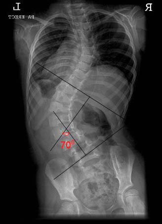
Dr. Hugh B. Logan, who founded Logan College of Chiropractic, first developed the basis for this procedure for the correction of scoliosis.  Over the last fifty years, I have modified, improved and updated the procedure to allow the chiropractor to produce consistent results. I established the International Scoliosis Research Center to provide research, consultation and seminars to the profession on this subject.  The doctor of chiropractic is the only health care professional capable of reducing a scoliosis without braces or surgery.

Theory

It has been confirmed clinically that subluxation of the sacrum, both anterior and inferior, predisposes to the development of a scoliosis. The body of the lowest freely movable vertebrae will always rotate to the low side of its foundation. Utilization of the Mawhiney Procedure, utilizing Logan Basic Technique, heel lift application, restrictions and exercise, will reduce the scoliosis in a prescribed period of time.

Treatment procedure

The treatment is set on a minimum three-month evaluation period, incorporating Logan Basic Technique, bilateral weight scale evaluation, heel lift application, exercises and restrictions, and specific vertebral adjusting. Vertebral adjusting is very specific, because each section of the scoliosis develops in a progressive manor, and the vertebra to be adjusted must be adjusted in proper sequence.

Patient is seen three times per week for the first three weeks and then reduced to twice weekly for the next nine weeks.  In cases of Grade II scoliosis and above, a hanging X-ray is taken at the initial exam, to determine the extent of scoliosis reduction that can be anticipated in ninety days.  The hanging X-ray is achieved by having the patient hang, by their hands, for a two-minute period, and the exposure is made while the patient is suspended.  It was proved, in clinical trials, that this procedure will show the actual amount of reduction that may be obtained, which vertebral segments will rotate, and how much reduction will be made in the Cobb’s angle.

Heel lift application is determined by the sacral inferiority, the lowest freely movable vertebrae and the bilateral weight scales.  The heel lift application has nothing to do with a measurable leg length, but only with the sacral inferiority.

Ninety-day evaluation should demonstrate a 15% reduction in the scoliosis and the next ninety-day period would have the patient seen twice weekly. In advanced cases, a traction/distraction table would increase the scoliosis reduction expected in ninety days.

Each visit would first have the patient checked on the bilateral weight scales, to determine if the lift placement is correct in thickness to support the sacrum. The patient will respond to scoliosis reduction best with the use of an adjustable table such as the Zenith Hy-LO. A flat table does not allow for AP changes in the prone posture necessary to encourage results. The patient will then receive an application of Logan Basic Technique, for approximately five minutes. The contact is always on the low side of the sacrum and is best applied after the patient is manually positioned anatomically in the prone position, to encourage curvature reduction and muscle relaxation.

The patient is restricted from all contact sports and any activity that may traumatize the pelvis or sacrum. The only exercise utilized the first thirty days is the hanging exercise, done twice daily. This exercise consists of having the patient hang, by their hands, twice daily for an accumulated time of two minutes. Other exercises, such as free weights, may be added later.

Once the heel lift is placed and monitored, the scoliosis will reduce, allowing the Logan Basic Contact, specific vertebral adjusting, restrictions and exercise to control the speed of the reduction.

Conclusion

Proper chiropractic care has demonstrated, over the last seventy years, to be the only profession to reduce spinal scoliosis without the use of braces or surgery. It is a specialty subject and any chiropractor not properly trained in a college sponsored post-graduate course risks a malpractice problem if the scoliosis increases while the patient is under their care.

R. B. Mawhiney, D.C, D.I.S.R.C., graduated from Logan College in September 1953 and had a family practice, specializing in scoliosis, for forty-eight years before retiring.  He has been on the post-graduate faculty of ten chiropractic colleges, written thirteen books, and has published numerous clinical and scientific articles.  He presently teaches scoliosis reduction seminars and serves as a consultant to the profession by providing treatment procedures to doctors.

Dr. Mawhiney may be reached at 25540 Belle Helene, Leesburg, FL  34748; or by email at [email protected].

*The scoliosis manual, Chiropractic Procedures in Spinal Distortion Cases, is available through the author.

Manipulation Under Anesthesia or Joint Anesthesia

Manipulation under Anesthesia (MUA) has been shown to be a viable approach in the treatment of chronic recalcitrant spinal pain.  The results have been remarkable in many patients who have exhausted efforts using conventional models of conservative management.

“Because of the advancement of new medications and the use of conscious sedation, the anesthesia element of the MUA makes the procedure one of the most easily adaptable neuromusculo skeletal treatment modalities that manual practitioners have at their disposal for chronic and certain acute neuromuscular skeletal problems.”1

This procedure has been widely used for centuries and recently has had major improvements in its efficiency by combining it with another mode of pain management, fluoroscopically guided intra- articular injection known as MUJA.

Since the combination of these two procedures, our team of physicians, which include anesthesiologists, osteopathic physicians, and chiropractors, has successfully performed over five thousand manipulations under joint anesthesia.

Discussion

Manipulation under joint anesthesia (MUJA) was presented in 1999, at the Conference of the World Federation of Chiropractic, as an alternative to office manipulation.  There is literature that dates back to 1938, in which manipulation was performed following an anesthesia injection of the sacroiliac joints.2

In 1997, Nelson, Aspegren, and Bova studied the benefits of the use of epidural steroid injection and manipulation on patients with chronic low back pain.3

As described by the CPT 2003 Code 22505, spinal manipulation under anesthesia is defined as an outpatient manipulation of the posterior motor units of the spine, requiring anesthesia and designed to reduce fibroblastic proliferation and restore range of motion and visco-elasticity to the per capsular connective tissue and para vertebral musculature in areas of the spinal segmental dysfunction not amenable in office manipulation.4

Protocol

Upon careful selection of the patient by the persons performing the injection and delivering the manipulation, the patient is then assessed as to what type of injection will be performed at precisely what level.

The procedure is then executed in a properly equipped surgical suite to allow for optimal setting of both injections and the manipulation being performed.  Upon completion of the injection, whether it is an epidural, facet block, trans-foraminal, or sacroiliac, the manipulation under joint anesthesia is then performed.  A series of facial lengthening, tendon stretching and ligamentous mobilization along with the realignment of the joint are carried through while the patient continues under conscious sedation.  These results are attained by using passive stretches, myofascial release, and specific articular and adjustment procedures.

“The basic concepts behind the mobilization, manipulation, and adjusting procedures, while the patient is under a sedative/hypnotic, is to increase ligamentous, tendinous, and muscular flexibility that has not been achieved in the office therapeutic routine.  Standard manipulative techniques are used, but the physiologic state of the patient is changed and the procedure is done in a controlled environment.  When used on properly selected patients, it is more cost effective and more productive to the patient’s return to normal lifestyle than prolonged conservative care or possible surgical intervention.”5

The rational for using sedation is to allow those patients who cannot tolerate any use of manual techniques due to muscle guarding, spasm, severe pain, and muscle contractors to regain their activities of daily living and begin a structured regimen of home exercise.

The medication of choice used for conscious sedation is Propofol.  “This medication allows the patient to not respond to the initial pain stimuli with an immediate muscle contraction.  The maneuvers could then be performed without losing their end range. The natural protective mechanism are present, but slowed down temporarily, and pain is perceived at a lowered threshold and not remembered.”6

Conclusion

As discussed throughout this paper, MUJA is an excellent choice for patients who continue to suffer from recalcitrant pain.  There is an over abundance of chronic cases which have shown no improvement with conventional approaches to neuromusculoskeletal injuries along with spinal axial pain.  For this patient population, a course of manipulation under anesthesia should be considered as the next phase in their treatment prior to surgical intervention.  Standardization of this protocol would allow more potential patients, who continue to suffer, a chance to get their lives back and return to the activities they enjoyed prior to their injury.

References

1. Gordon R. Commentary:  Manipulation Under Anesthesia, Journal of Manipulative and Physiological Therapeutics. 2001 Vol. 24 Number 9.
2. Haldeman KO, Soto Hall R., The diagnosis and treatment of sacroiliac conditions by injection of procaine (Novocain). J Bone Joint Surgery 1938; 20-A :675-86.
3. Nelson L. Aspegren D, Bova C.  The use of epidural injection and manipulation on patients with chronic low back pain.  J. Manipulative Physiology Ther. 1197; 20: 263-6.
4. CPT 2003 American Medical Association 47-C Procedure Musculoskeletal/Surgery
5. Gordon R. Conservative.  Chiropractic adjustive therapy versus MUA adjustive therapy.  Florida Chiropractic J 1993;1:22-3.
6. Gordon R. Commentary:  Manipulation Under Anesthesia. Journal of Manipulative and Physiological Therapeutics 2001 Vol. 24 Number 9. 

Dr. Peter M. Ferraro is a 1996 graduate of New York Chiropractic College. He began early in his career specializing in the treatment of herniated disc injuries by becoming certified in the Cox Distraction techniques at National Chiropractic College. In the past four years, Dr. Ferraro has become certified in Manipulation Under Anesthesia and has performed over 5,000 MUA/MUJA treatments.

For more information, Dr. Ferraro can be reached at 973-478-2212 or at [email protected].

The Evolution of Bloodless Surgery and Chiropractic

Bloodless Surgery or Chiropractic Manipulative Reflex Technique (CMRT) encompasses the  relationship between somatovisceral and viscerosomatic reflexes and, therefore, between the somatic and autonomic nervous systems. R. J. Last in his book, Anatomy: Regional and Applied, points out:

There is only one nervous system.  It supplies the body wall and limbs (somatic) and viscera (autonomic).  Its plan is simple.  It consists of afferent (sensory) and efferent (motor) pathways, with association and commissural pathways to connect and coordinate the two.  There is no more than this, in spite of the many pages devoted to its study.1

Bloodless Surgery has historically been used in chiropractic as a term describing soft tissue treatment affecting an organ and its related vertebral relationship or viscerosomatic and somatovisceral reflexes.2,3  Bloodless surgery has also been used to describe methods of manipulating joints and soft tissue without being related to the viscera.4

James F. McGinnis was a chiropractor that relocated to California in the early 1920’s, where he earned a naturopathic doctorate.  In the 1930’s, he became one of the best known of several chiropractic bloodless surgeons and traveled around the nation to teach his methods.2 Around this time, Major Bertrand DeJarnette, developer of Sacro Occipital Technique, was also practicing and teaching extensive methods of bloodless surgery. DeJarnette published a comprehensive book on the topic, entitled Technic and Practice of Bloodless Surgery, in 1939, which remains the most complete discussion on the topic to date.3

During this time, DeJarnette used chromotherapy, which was purported to affect the physiology of the patient.  The process involved the “filtering of white light through special screens or filters”5 through a mechanism called the chromoclast.  He would use this device to help with his bloodless surgery procedures and found that it appeared to have, among other therapeutic characteristics, anesthetic properties.  During the 1940’s, DeJarnette stopped teaching and selling the chromoclast, as he reported that those using the device were not using it properly and might cause the patient harm, for which he did not want to be held responsible.

He continued to teach and practice bloodless surgery through the 1940’s, and began its modification to use more reflex applications and referred pain indicators, as a method of affecting organ symtomatology. In the 1950’s, he furthered his investigations into reflexes and their effect on the viscera and related vertebra.  By the early 1960’s, DeJarnette modified the nature of Sacro Occipital Technique’s method of bloodless surgery from its 1939 procedures, which might take two-to-four hours of preparation and treatment, to procedures that could be practiced in a span of 15 minutes.6,7  For multiple reasons, he decided to change the name of his method of affecting referred pain pathways, viscerosomatic/somatovisceral reflexes, and direct organ manipulation to Chiropractic Manipulative Reflex Technique (CMRT).

CMRT is used as a method of treating the spine or vertebra visceral syndromes associated with viscerosomatic or somatovisceral reflexes,8-10 dysafferentation at the spinal joint complex,11 and visceral mimicry type somatic relationships.12  Treatment involves location and analysis of an affected vertebra in a reflex arc, by way of occipital fiber muscular palpation, similar to trigger point analysis or Dvorak and Dvorak’s spondylogenic reflex syndromes.13  Once specific vertebra reflex arcs are located, corroborated with referred pain pathways and clinical symtomatology, then the specific vertebra to be treated is isolated by pain provocation, muscle tension and vasomotor symtomatology.  Often times, if a vertebral dysfunction is chronic or unresponsive to chiropractic spinal manipulation, then a viscerosomatic or somatovisceral component is evaluated.14  Treatment of the viscerosomatic or somatovisceral component is performed using soft tissue manipulation, myofascial release techniques and reflex balancing methods.7

Bloodless Surgery, has been used and taught by Sacro Occipital Technique (SOT) chiropractors since 1939 and was practiced much more extensively in the 1930’s and 40’s.  Since 1960, it has been called CMRT, and focuses predominately on the vertebra and viscerosomatic/somatovisceral relationships. CMRT is listed as a chiropractic technique throughout the chiropractic literature.15-19 SOT clinicians using these methods of CMRT and bloodless surgery for years are beginning to publish their methods in the literature which is helping to further establish this successful method of care, used for decades by chiropractors.20-24

Presently, those interested in learning about SOT and CMRT, as taught by Major Bertrand DeJarnette, can attend seminars by Sacro Occipital Technique Organization–USA (SOTO-USA) and can visit the website for seminar information and research updates at www.soto-usa.org, or call (781) 237-6673.  Currently, SOTO-USA is the only organization that is teaching CMRT, specifically, as developed by DeJarnette.

Dr. Charles L. Blum is the President of Sacro Occipital Technique Organization–USA (SOTO-USA), PO Box 24936, Winston-Salem, NC 27114-4936.  For more information call 336-760-1618, or email [email protected].

For DeJarnette SOT, dentocranial co-treatment, and up to date integrated SOT treatments, SOTO-USA will be having its yearly clinical symposium in St. Louis, MO October 6-9th, 2005.

References

1. Last RJ, Anatomy: Regional and Applied, Sixth Edition, Churchill Livingstone: New York, 1978:20.
2. Keating JC James F. McGinnis, D.C., N.D., C.P. (1873-1947): Spinographer, Educator, Marketer and Bloodless Surgeon Chiropractic History, 1998; 18(2): 63-79.
3. DeJarnette MB, Technique and practice of bloodless surgery, Privately Published, Nebraska City, NB, 1939.
4. Taylor H, Sir Herbert Barker: Bone-Setter and Early Advocate of “Bloodless Surgery”   Journal of the American Chiropractic Association 1995  Jul; 32(7): 27-32.
5. DeJarnette MB, Chromotherapy, Privately Published, Nebraska City, NB, 1941.
6. DeJarnette MB, Chiropractic Manipulative Reflex Technique, Privately Published, Nebraska City, NB, 1964.
7. Blum CL, Monk R, Chiropractic Manipulative Reflex Technique, Sacro Occipital Technique Organization–USA, Winston-Salem, NC, 2004.
8. Budgell BS, Reflex effects of subluxation: the autonomic nervous system.  J Manipulative Physiol Ther 2000 Feb;23(2):104-6
9. Budgell BS, Spinal Manipulative Therapy and Visceral Disorder.  Chiropractic Journal of Australia  1999  Dec; 29(4): 123-8
10. Sato A  The reflex effects of spinal somatic nerve stimulation on visceral function. J Manipulative Physiol Ther.  1992 Jan;15(1):57-61.
11. Seaman DR, Winterstein JF, Dysafferentation: A Novel Term to Describe the Neuropathophysiological Effects of Joint Complex Dysfunction. A Look at Likely Mechanisms of Symptom Generation.  Journal of Manipulative and Physiological Therapeutics. 1998 May; 21(4):  267-80
12. Szlazak M, Seaman DR, Nansel D,  Somatic Dysfunction and the Phenomenon of Visceral Disease Simulation: A Probable Explanation for the Apparent Effectiveness of Somatic Therapy in Patients Presumed to be Suffering from True Visceral Disease, J Manip Physiol Therp. 1997 Mar; 20(3) :  218-24
13. Dvorak J, Dvorak V, Manual Medicine: Diagnostics, 3rd Edition, (Translated from German) George Theime Verlag, Stuttgart, Germany, 1988: 326-33
14. Heese N, Viscerosomatic Pre- and Post- Ganglionic Technique, Am Chiro, 1988 Mar:16-22.
15. Peterson DH, Bergman TF, Chiropractic Technique: Principles and Procedures (Second Edition) Mosby: St. Louis, MO, 2002: 493, 497
16. Gleberzon BJ, Chiropractic “name techniques”: a review of the literature. European Journal of Chiropractic 2002; 49: 242-3.
17. Gleberzon BJ, Chiropractic “Name Techniques”: A Review of the Literature. J Can Chiropr Assoc 2000;45(2): 86-99.
18. Bergmann TF, Various Forms of Chiropractic Technique, Chiropractic Technique May 1993; 5(2):53-5.
19. Cooperstein R, Gleberzon BJ, Technique Systems in Chiropractic:  Churchill Livingstone: New York, NY April 2004: 209, 211, 214, 217.
20. Courtis G, Young M, Chiropractic management of idiopathic secondary amenorrhœa: a review of two cases: British Journal of Chiropractic Apr 1998; 2(1):12-4.
21. Cook K, Rasmussen S, Visceral Manipulation and the Treatment of Uterine Fibroids: A Case Report, ACA Journal of Chiropractic Dec 1992; 29(12): 39-41.
22. Blum, CL, Role of Chiropractic and Sacro Occipital Technique in Asthma, Chiropractic Technique, Nov 1999; 10(4): 174-180.
23. Blum CL, Chiropractic care of diabetes mellitus? A Case History, Journal of Vertebral Subluxation Research, Accepted for publication April 2003.  
24. Blum CL, Resolution of gallbladder visceral or mimicry pain, subsequent to surgical intervention, International Research and Philosophy Symposium Sherman College of Chiropractic, Spartanburg, SC, Oct 9-19, 2004: 10-11.

Snoring and Sleep Apnea – Structural Implications

The uvula and soft pallet may come into partial or total contact on the back wall of the upper airway.  When the contact is partial or intermittent, snoring (a loud vibration of these soft tissues) may result.  The tongue may also drop posteriorly onto the back wall of the upper airway, coming into contact with the uvula and soft pallet, thus forming a tight blockage, preventing any air from entering the lungs.  Respiratory effort on the part of the diaphragm and chest may cause the blockage to seal tighter.  In order to breathe, the person must arouse or awaken, causing tension in the tongue and, thereby, opening the airway, allowing air to pass into the lungs.  Apnea, sleep apnea or obstructive sleep apnea is defined as the cessation of breathing for 10 or more seconds while asleep.1  Traditional methods of treatment usually involve Continuous Positive Airway Pressure (CPAP) devices.  Matrix Repatterning techniques were used in ten cases of long-standing, moderate to severe cases of upper airway obstruction.
 
Matrix Repatterning

Matrix Repatterning uses a manual scanning procedure to determine the location of primary structural restrictions, followed by mechanical testing to determine specific vectors of fascial tension.  Treatment is generally applied manually, with light force directed into the resistance barriers.  Matrix Repatterning is based on a revolutionary, new model of the underlying structure of organic tissue—the Tensegrity Matrix—which explains the complex interrelationship of all the structural components of the body.  It extends the basic concept of the tissue response to injury, beyond the level of joint, muscle and ligament, to include all structures of the body as potential sources of dysfunction.2

The tensegrity matrix model of the body, as elaborated by Stephen Levin, M.D.3 and Donald Ingber, M.D., Ph.D.,4 holds that the body tissues are composed of interconnected tension icosahedra (complex triangular trusses), which inherently provide a balance between stability and mobility.  This structural model explains many of the observed phenomena related to body support, movement, response to stress and trauma, as well as the effects of various therapeutic interventions.  This theory has been verified by several studies in recent years.  According to Ingber, a key investigator who has proven the existence of this structural model at the cellular level, “The principles of tensegrity apply at essentially every detectable size scale in the human body.  At the macroscopic level, the 206 bones that constitute our skeleton are pulled up against the force of gravity and stabilized in a vertical form by the pull of tensile muscles, tendons and ligaments.  In other words, in the complex tensegrity structure inside every one of us, bones are the compression struts, and muscles, tendons and ligaments are the tension-bearing members.”4
 
Mechanism of Upper Airway Obstruction

The upper airway is constructed of the hard and soft palate above, the posterior pharynx, the tongue, and the epiglottis at the level of the tracheo-esophageal junction below.  There are several mechanisms of partial or complete obstruction.

When the ability of the tissues to adapt or compensate becomes overwhelmed by mechanical or physiologic stress, the fascial system responds by altering the patterns of tension and elasticity.  The tensegrity matrix explains the physiologic changes, which manifest in injured or strained tissue.  The apparent fibrosis of muscle and fascia can be seen as an altered electro-mechanical relationship at the molecular level.  The matrix is, thus, converted from a neutral, flexible form to a strained, high-energy, linearly-stiffened mode as shown in Figure 1.

Obstruction may occur by approximation of several structures and tissues.  This may include the soft palate retracting toward the posterior pharyngeal wall, the soft palate descending to approximate the posterior aspect of the tongue, the tongue retracting toward the posterior pharynx, or descending to approximate the epiglottis.  These tissues may deviate from their functional positions within the upper airway due to a number of structural dysfunctions, including cranial vertex or occipital trauma, leading to descent and/or protraction of the cranial base, along with the maxillary portion of the roof of the upper airway.  Vertex compression, in our studies, has also shown a tendency to lead to radial expansion of the upper cervical vertebrae (an intraosseous deformation), leading to loss of anterior/posterior dimension of the upper airway.  Facial trauma may cause deviation of the maxilla or mandible.  Hyperflexion injury of the cervico-thoracic spine may also induce an approximation of the posterior tissues toward the tongue and epiglottis.  This is common in motor vehicle collisions (rear end or front end) and in falls onto the back of the head or upper back.  Several other mechanisms of structural dysfunction are also currently under investigation.

Assessment

In cases of upper airway obstruction, a specific airway obstruction test (AOT), developed by the author, was also used to verify partial or complete obstruction.  This involves placing the patient in an accentuated position of upper cervical hyperflexion or moderate extension, along with varying degrees of rotation or lateral flexion.  The ease or difficulty of breathing, along with the amount of airflow turbulence noise, was recorded for each position.  The dysfunctional structural patterns associated with snoring and sleep apnea (see above) were then evaluated using the standard Matrix Repatterning assessment.
 
Method of Treatment

A maximum of four treatments to resolve these patterns were administered over a maximum period of two months for ten patients with moderate to severe upper airway obstruction.  Two of these cases were previously diagnosed with significant sleep apnea, as verified by sleep studies. 

Results

AOT was improved significantly in 80 percent of the cases.  Patients (and spouses, or sleeping partners) reported a cessation or a significant improvement in snoring in 70 percent of the cases.  The two individuals, diagnosed with sleep apnea, reported they were able to sleep through the night without the assistance of a CPAP machine, on which they were previously dependant.  These findings suggest that structural dysfunction may play a role in the development of upper airway obstruction and that Matrix Repatterning procedures may be beneficial in the management of these conditions.  The findings suggest that a randomized controlled trial within a broader population base might be indicated.

Key Terms:
Matrix Repatterning,
Upper Airway Obstruction (UAO),
Airway Obstruction Test (AOT),
Continuous Positive Airway Pressure (CPAP).

References

1. Exar EN, Collop NA: The upper airway resistance syndrome. Chest 1999 Apr; 115(4): 1127-39
2. The Matrix Repatterning Program for Pain Relief, GB Roth, New Harbinger Publications, Oakland CA, 2005.
3. The Importance of Soft Tissues for Structural Support of the Body, SM Levin.  Positional Release Therapy: Assessment & Treatment of Musculoskeletal Dysfunction, K D’Ambrogio & GB Roth, Mosby-Elsevier, St. Louis, 1997.
4. The Architecture of Life, DE Ingber, Scientific American, Vol. 1, 1998

George B. Roth, BSc, DC, ND, is President of The Roth Institute, and on the Post-Graduate Faculty of Logan College of Chiropractic. He can be reached by phone at 416-977-6841, email at [email protected], or visit www.rothinsitute.com.

Techniques: Diversity of Art and Science

Chiropractic is an incredibly diverse profession, boasting more than 100 different techniques (one list puts the number closer to 120 and one online source even claims there are 200).

Some are extremely well known, such as Diversified or Atlas Orthogonal Technique. Others possess a smaller (but often extremely loyal) following, such as the McTimody Technique or the Columbia Technique. Most doctors incorporate several techniques into their practices and many attend frequent seminars to learn new techniques in the hope of improving the quality of care they provide patients.  While this array of techniques contributes to the rich texture of our profession, developing a technique requires more than merely modifying an adjusting table and giving seminars.

Craig Nelson, DC, in an article titled “The Nelson Method—Five Steps to Your Own Technique” (Journal of Manipulative and Physiological Therapeutics, vol. 16, No. 2 Feb. 93, pp.115-117), gave facetious advice on how to start a technique. After suggestions about coming up with a name and sounding scientific (“complicated mathematical equations work really well”), he advised, “Don’t attempt to test the reliability or effectiveness of your technique. You do not want to ask questions you do not know the answer to.” Luckily for chiropractic, and chiropractic patients, most developers ignored that last piece of advice and conducted at least some—often extensive—research on their techniques before introducing them to the profession.

But the lack of solid research into chiropractic, in general, and techniques in particular, is distressing and has not gone unnoticed by our critics. An online information page put out by AETNA insurance notes that, “There are more than 100 chiropractic and spinal manipulative adjusting techniques, and practitioners may vary in their approaches.” The page goes on to make such statements as, “Overall, the quality of studies has been poor. Better-quality research is necessary to make a clear conclusion,” (referring to neck pain). “However, because of weaknesses in this research, no clear conclusions can be drawn,” (referring to asthma). “Because there are a limited number of studies in humans and weaknesses in existing research, it is unclear if chiropractic techniques are beneficial….” (Referring to just about everything else).

The lack of high-quality research is a problem we must address, if chiropractic is to be accepted by the public; and technique developers are in an ideal position to help compile that research. According to the Council on Chiropractic Practice (CCP) Guidelines, “Techniques and methods for correcting subluxation must be judged on their intended outcome and most, if not all, chiropractic techniques have some physiological and/or structural outcome that measures their results.”

In fact, outcomes research is the key to establishing or choosing any technique. Our profession cannot afford to use or promote a technique out of blind loyalty or habit. As with every other aspect of chiropractic, we have to look at the literature and make sure the technique is the result of scientific research. Preferably, that research will have been published in a peer-reviewed journal, either by the developer or by doctors who use the technique in their practices.

Erin Elster, DC, whose research has been published in the Journal of Vertebral Subluxation Research, JMPT, and Today’s Chiropractic, has stated, “I encourage all doctors of chiropractic to publish their research using their chiropractic techniques and technologies, in order to expand the chiropractic knowledge base and to benefit the profession.”

The quality and amount of research on techniques is also critical in the development of chiropractic practice guidelines. Since chiropractors often define themselves and their services by the techniques they use, professional guidelines must address those techniques.

“The involvement of technique experts is crucial to the development of any chiropractic guideline,” states Matthew McCoy, DC, Vice President of the Council on Chiropractic Practice. “Unfortunately these dedicated and unsung heroes have effectively been shut out of other groups’ guideline development efforts and the research community. Instead, they need to be deeply involved in this process, since the art of chiropractic is the application of the philosophy and science.”

Involving technique experts in both research programs and guideline development has far-reaching benefits for the profession and for patients. Not only will it ensure the continued safety of chiropractic adjustments, but it will enable doctors to provide the evidenced-based care demanded by the public today.

Dr. Jackson is Chief Executive Officer, Research and Clinical Science (RCS), a private sector research program exploring issues of subluxation correction and chiropractic care as they relate to health and wellness. Previously, he served as president of Chiropractic Leadership Alliance and Creating Wellness Alliance. He was owner/operator of several private practice offices in California and Idaho that specialized in high volume, family wellness based care. Dr. Jackson may be reached at 800-909-1354; or, for more information, contact Barbara Bigham at 503-362-2145.

Where, When and When Not to Adjust

Today, with more and more studies concluding that the standard methods of chiropractic analysis are unreliable and not reproducible, many doctors of chiropractic are looking for a way to analyze the spine and extremities that can give them assurance.  Activator Methods Chiropractic Technique (AMCT) utilizes a protocol incorporating leg length analysis, pre- and post-adjustment, to determine exactly where, when and when NOT to adjust.  Therefore, this protocol not only helps to assure the doctor has found the affected spinal level but also enhances their confidence that the adjustment was successful.

Research has been conducted to evaluate interexaminer reliability using experienced chiropractors to measure reproducibility of prone leg length assessment and concludes that reliability of prone leg checks can be consistent.1 In comparison, static and motion palpation fall below what is clinically acceptable in terms of reliability.  AMCT utilizes prone extended leg length assessment (Position 1, Figure 1) to determine the functional short leg of the patient or pelvic deficient (PD) side, either the AS or PI ilium. Once the pelvic deficiency is corrected using precise contacts and lines of drive via the Activator adjusting instrument, the leg lengths become equal. (Figure 2).

ISOLATION TESTS: With the pelvis now balanced, screening for areas to adjust involves the use of active muscle tests called isolation tests. (Figure 3) Each isolation test corresponds to a specific vertebral segment.  The patient performs the requested motion and the doctor observes for any changes in leg length on the side that was originally the PD side.  If a change occurs, the doctor flexes the knees to 90 degrees (Position 2) and observes any change in the PD leg to determine which side of the spine to adjust.  If the PD leg lengthens in Position 2, the doctor adjusts the PD side of the corresponding segment on a specific contact (example, the mammillary or transverse process) with a precise line of drive utilizing the Activator adjusting instrument.  Correction will be observed in a post-adjustment leg check as the legs balance again.  If the patient’s leg lengths remain equal after the isolation test, no adjustment is required and the doctor moves on to test the next level of the spine.

AMCT conceives that the leg reactivity observed after an isolation test is due to facilitation as a result of nerve interference at the affected spinal level.  According to Malik Slosberg, DC, “normal muscles respond to normal, innocuous movements by appropriately contracting briefly to perform the requested movement and then relaxing.”

Therefore, in unaffected areas, these movements do not appear to alter relative leg lengths.  He goes on to say, “When a muscle group is facilitated, its response to stretch or contraction may be both excessive and prolonged.  Such alterations of muscle response apparently affect the functional leg length and result in alteration of relative leg lengths.”2

If facilitation is present, an exaggerated contraction of paraspinal muscles occurs and leg reactivity is observed.  Decades of documented clinical observation from doctors of chiropractic internationally have culminated into this protocol for analysis.   In one reliability study, 72 subjects were examined by two DC’s, for upper cervical subluxation, using a chin tuck isolation test.  Good reliability between examiners was found.3

Drs. Warren Lee and Arlan Fuhr founded instrument adjusting over 35 years ago.  Not only does an instrument allow the doctor to passively adjust the patient, but it also provides a specific contact, force, speed and line of drive so that you can be assured to affect the joint as efficiently as possible. The latest Activator adjusting instrument, Activator IV (Figure 4), has a reproducible preload and four precision force settings that have been studied in independent labora-tories.The minimal force setting starts at approximately 19 lbs and the instrument ranges up to a maximal force setting of approximately 55 lbs. Today, the Activator is still the fastest adjusting instrument. It is over 300 times faster than a manual thrust.4

At this speed, the doctor is affecting the involved joint before the muscle spindle reflex is initiated.  To compare the effects of manual adjusting to instrument adjusting, we can look at a bone movement study where a manual lumbar side-posture adjustment using 540N of force moved L4 1.1 mm.5  In a similar study, the Activator adjusting instrument moved L4 1.6 mm with a force of 140N.6

STUDY IN CLINICAL SETTING:  The following is a case series that will help illustrate the effectiveness of the AMCT protocol and adjustments in a clinical setting.  The purpose of this paper was to determine if there was a basis for the treatment of Temporomandibular disease (TMD) using the chiropractic protocol developed by Activator Methods, Inc.  The study was set in a private, solo practice of an Activator advanced proficiency rated chiropractor with 15 years experience.  Nine adult volunteers with articular TMD were recruited from the practice of the treating clinician.  Change was measured from baseline to follow-up using a Visual Analog Scale (VAS) for temporomandibular joint (TMJ) pain and maximum active mouth opening without pain. AMCT protocol for full spine and TMJ analysis and adjusting (Figure 5) was followed. Participants were typically seen three times per week for two weeks and, according to individual progress, thereafter for six more weeks. The results showed a median VAS decrease of 45 mm (range 21-71) and all experienced improvement. The median increase of mouth opening was 9 mm (range 1-15) with improvement in all.  The results of this prospective case series indicate that the TMD symptoms of these participants improved following a course of treatment using the AMCT protocol.7

AMCT is now taught in almost every chiropractic college in the United States.  With over 140 peer-reviewed articles and conference presentations published, doctors of chiropractic can be assured that, by choosing AMCT as their primary method of analysis and adjustment, they are offering their patients an effective and safe treatment and will have fewer doubts about where, when and when NOT to adjust!

For more information, contact Activator Methods, International, 2950 N. Seventh St. Suite 200, Phoenix, AZ 85014; or call (602) 224-0220.  www.activator.com

References

1. Nguyen, et al., JMPT 1999; 22(9):565-9.
2. Malik Slosberg DC, MS, in Today’s Chiropractic 17:17,1998. 
3. Youngquist, et al., JMPT 1989; 12:93-97.
4. Keller, et al., JMPT 1999; 22(2):75-86.
5. Maigne, Guillon, JMPT 2000; 23:531-536.
6. Nathan, et al., JMPT 1994; 17(7):431-44.
7. DeVocht, et al., JMPT 2003; 26(7): 421-425.

Case Study: Rotator Cuff Impingement Syndrome

History and Subjective Complaints:

A 24-year-old professional/international circuit tennis player presents with a right shoulder injury, 6 months post U.S. Open.

Objective Findings:

Decreased right shoulder flexion, abduction and external rotation.

X-Ray:

Absent 8-10 mm subacromial space, laterally rotated vertebral border of the right scapula, antetilted glenoid labrum.

Diagnosis:

Protracted laterally antetilted right scapula causing rotator cuff impingement syndrome.

Treatment:

Specialized manipulative decompression of involved protracted/antetilted right scapula, medial-inferior sternoclavicular joint and anterior-inferior medially rotated right humerus (Mally Technique)

Nutrition:

Anti-inflammatory vitamins (bromelain, papain, trypsin, chymotrypsin, bioflavonoids) and vitamin B6.

Rehab:

6 weeks of reconstruction and work hardening, rhomboid and deltoid strengthening.

Addendum:

Failed post-op impingement surgery is very high, with misdiagnosis and connective tissue hyperplasia the highest cause of failure. Misdiagnosis leads to erroneous results and, in this case, permanent disability and loss of dominant shoulder motion. Post chiropractic extremity manipulation by Dr. Mally yielded a return to the tennis courts and a very excited world-class competitive athlete.

For more information, Dr. Mally can be reached by e-mail at [email protected].

Breakthrough Pre-Adjustment Technique to Fully Activate Innate and De-stress the Body

Can you imagine having a fuller understanding of why adjustments hold or don’t hold and a more accurate description of why innate’s powerful influence on healing in the organism may be inhibited? The key to unlocking multiple channels of innate intelligence is related to adrenal gland physiology and the body’s ability to store, maintain and balance coherent energy.

Yin and Yang Influences on Adjustments

Homeostatic systems of the body involve self-correcting and self-balancing energetic feedback loops that are dependent on the balance of the polarities of yang and yin or of the sympathetic and parasympathetic nervous systems (adrenergic and cholinergic). A reduced flow of innate commonly starts at the sacrum, a sensitive register to stress and the locus of powerful meridians. In acupuncture-energetic physiology, the polarities of the governing vessel-GV (posterior) and the conception vessel-CV (anterior) meridians via the adrenals exert 90% control on how innate integrates and distributes healing energy throughout the body.

Causes of Allergic and Inflammatory Disorders

The adrenal-kidney yang influence on the CV and GV meridians and free flow of liver energy explains why many patients with hypoadrenia don’t hold adjustments or are extremely difficult to adjust in the cervical and thoracic spine. The adrenals extend their defensive armor against stressors (including toxic chemicals and chaotic frequencies) into the energetic system of the body, whereby the musculature of the chest and upper back (pericardium energetic zone) becomes chronically constricted.

Since hypoadrenia causes chronic and prolonged infection and unwanted inflammation (root causes of cardiovascular and many degenerative diseases) and has a powerful influence on the flow of innate healing energies, a pre-adjustment technique to activate the full range of innate healing is highly desirable. Hypoadrenia causes excessive or stagnant energy to build up in the liver, thereby inhibiting enzymatic detoxification and steroidogenesis (the formation of hormones) and triggering a wide spectrum of allergic and pro-inflammatory disorders.

The daily stresses that wear down the adrenals and our physical and emotional constitution are compensated by GV and CV “safety energy circuits” located in the sacrum and cranium. Functioning as circuit breakers, they protect the body and activate innate healing against the damage caused by stressors. Vertebrae subluxations can be viewed as the consequence of sacral subluxations or brain interferences caused by an overload of these protective circuits.

The Quantum Repatterning Technique™ (QRT) utilizes a simple and fast 10-minute pre-adjustment Sacral-Cranial Balancing protocol that acts as a jumper cable to restart the flow of energy through the GV and CV “broken circuits” and relax sacral muscles and ligaments, so that sacral adjustments are made with ease and without force. Once the sacrum is balanced, increasing muscle flexibility allows adjustments to really open up the cervical, thoracic and lumbar vertebrae.

This pre-adjustment technique is based on decades of research in brain-organ proprioception, electro acupuncture, and chiropractic. D.D. Palmer, George Goodheart, M.T. Morter and others viewed sacral and brain interferences as the primary sources of nerve interference.  In just ten minutes, this amazing technique allows the muscles of the pelvis and neck to relax,with hypertonic and hyportonic “left-right” discrepancies realigned. Commonly, the patient reports a rapid, pulsating release of stagnant energy and dramatic improvements in the flexibility and range of motion of the sacrum and the entire spine. As one patient noted, “It’s stress without distress!”  This amazing resilience to stress is a result of a balanced body with balanced hormones from the adrenal-guided process of steroidogenesis.

NOTE: To learn more about Dr. Yanick’s Cranial-Sacral Pre-Adjustment protocol, read his manual, The Quantum Repatterning Technique, available at www.quantumenergy.com.

Dr. Yanick is a world renowned expert on and founder of Quantum Medicine and QRT.  He has published extensively on Quantum Medicine.  You may contact Dr. Yanick by fax at 845-340-8606 or e-mail at [email protected].

Visit www.quantumenergy.com for more information.