Fascial Manipulation©(FM)

:dropcap_open:M:dropcap_close:odern myth has it that doctors in Europe are ahead of us in new methods of healing.  The truth of this may be dubious but it does underscore the importance of keeping abreast of healing advances in other countries as we strive to achieve the highest possible level in care for our patients. 
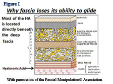
:quoteright_open:Research has shown that the reason fascia is able to glide over and within muscle is due to hyaluronic acid (HA).:quoteright_close: 
Three years ago, I was invited to Italy to meet with Luigi Stecco, PT.  I observed Luigi treat patients for three afternoons, 5 hours each day.  He divides each day into two 5-hour sessions, the morning being devoted to research and reading.
 
Over the course of my career I have had the honor of observing many masters practice, such as Gonstead and Goodheart, among others, but never before have I seen treatments which yield such immediate results. I learned from Luigi’s son Antonio (Antonio Stecco, MD physiatrist who was translating for us) that none of these patients had been to see Luigi for the last 3 to 10 years. Luigi had, indeed, discovered a way to “find it, fix it and leave it alone.”  
 
He had previously sent me one of his two textbooks written in English ¹. At the present time this textbook has been translated into seven languages. I eventually took the first 12 day FM course in Italy that was presented in English and have since introduced it into the US with Antonio Stecco. FM has been taught in Europe for 14 years and is presently being taught around the world. (Go to: www.fascialmanipulationworkshops.com, www.fascialmanipulation.com). The first thing that attracted me about FM is that there is probably more peer review literature on FM than possibly all the soft tissue methods combined.  Go to www.pubmed.com (the national library of medicine) and put in Stecco, C or Stecco, A and you will see about 70 articles, most submitted over the past three years.  I am a consultant for Graston Technique®(GT) and in 2012 a large percentage of the people taking  FM were users of GT.  I told them that FM was the PhD course for GT. Actually it is a PhD course for almost all soft tissue methods.  As stated in the first manual of the first International Fascia Research Congress²  “Hypotheses which accord myofascia a central role in the mechanisms of therapies have been advanced for some time in the fields of acupuncture, massage, structural integration, chiropractic and osteopathy”.  The official definition of fascia from this congress is “Fascia is the soft-tissue component of the connective tissue system that permeates the human body, forming a whole-body continuous three-dimensional matrix of structural support.  It interpenetrates and surrounds all organs, muscles, bones and nerve fibers, creating  a unique environment for body systems functioning.”
 
fascialfigure1So what is this white stuff covering everything in our body?  The infinite wisdom of nature would not just bother to cover our whole body with white stuff that solely functions as a covering.  Probably the most important function of fascia is that it is a sensory organ loaded with mechanoreceptors and proprioceptors. Every single muscle fiber in our body is covered by fascia.  Many muscles originate and insert into fascia, not to mention the intramuscular fascia. Whenever a muscle is stretched or contracts the surrounding fascia must be stretched.  The muscle spindle cell should be called the fascial spindle cell since they are located in the fascia.  As soon as a muscle is activated, spindle cells immediately report back to the CNS the status of the muscle such as its tone, position and movement.  In order for this mechanism to work properly, fascia must have a normal viscosity whereby the spindle cells, Paccini and Ruffini receptors can be normally stretched.  What if fascia is restricted and unable to glide over the muscle or within the muscle during muscle activity?  Alteration of fascial movement due to increased viscosity (density) of the fascia would lessen the normal feedback to the CNS and muscle incoordination would occur.  A recent e-mail exchange with Siegfried Mense, MD a leading expert on muscle pain and neurophysiology was asked if fascial adhesions would have an adverse affect on spindle  cells.  He stated: “Structural disorders of the fascia can surely distort the information sent by the spindles to the CNS and thus can interfere with a proper coordinated movement”.  Antonio Stecco, MD has many ultrasound studies showing the correlation between stiff thickened fascia and diminished sliding being responsible for neck pain³. Helen Langevin, MD did an ultrasound study showing that people with chronic or recurrent low back pain compared to no low back pain had 25% greater perimuscular thickness and echogenicity4. 
 
Research has shown that the reason fascia is able to glide over and within muscle is due to hyaluronic acid (HA).  Due to trauma or stress to soft tissue HA chains begin to entangle and dramatically increase viscoelasticity . Figure I shows HA between the deep fascia and the muscle. HA is also located in the intramuscular fascia. Deep pressure which increases the temperature of tissue three to four degrees untangles the HA and promotes a more fluid medium allowing normal gliding.  Interesting also is that increased alkalinity also helps free up the HA, but I have no particular nutritional information on this subject.
 
fascialfigure2How does FM work to free the fascia and how does it differ from all other soft tissue methods. Over the years Luigi Stecco realized that there were particular areas in the muscles and retinacula around joints that when released solved many musculoskeletal problems. Stecco found that muscles work in groups and that we could consider these groups in terms of myofascial units (MFU).  Luigi thought that if there are muscle fibers contracting together as a group, then for a particular direction some of the fibers will be tensioning their overlying and intra fascia when they contract and somewhere there will be a mathematical point where the vector forces will converge in a given movement pattern.  In other words specific motor units will converge on a point in the deep fascia for a specific direction. He called these points centers of coordination (CCs) which were located in a myofascial unit.  A MFU is defined as particular motor units activating monoarticular and biarticular muscle fibers, their deep fascia and the joint they move in one direction on one plane  (Figure II.)  The CCs are often in the belly of the muscle where most of the fascial spindle cells are located. These areas are often proximal to the painful area and the patient is often surprised that they are tender.  But if these points are involved they will be palpated as densified, tender areas.   Luigi has correlated functional testing (contraction or passive stretching of the area) with these points.  For example anterior forearm pain could be created by palpating and treating anterior points not necessarily on the forearm, see figure II.  Figure II represents 4 MFU making up an anterior sequence.  Fascia is stretched by a muscle due to its fascial expansions and this tension is transmitted during a forward movement of the arm. During forward movement of the whole arm the contraction of the clavicular fibers of the pectoralis major will stretch the anterior region of the brachial fascia due to its myofascical expansion.  The simultaneous contraction of the biceps muscle will stretch the anterior region of the antebrachial fascia by way of the bicipital aponeurosis (lacertus fibrosis) as the palmaris longus pulls on the flexor retinaculum, palmar aponeurosis and thenar fascia. The anatomical fascial/muscle connection for example has been shown in anatomical dissection by the Steccos 6. While this forward movement is occurring the proprioceptors within the fasciae are activated permittingthe perception of the motor direction.
 
Testing for forearm pain along the anterior plane would include testing of the shoulder at 90° (an-hu), testing the biceps area (an-cu), testing of the forearm (an-ca, myofascial of the flexor carpi radialis), and flexion of the thumb (an-di).  The fascia in the sagittal plane is therefore being tested and then palpated for density in the areas where the tests were positive.  Often the forearm pain may be resolved by treating an-hu or even an-di.  In other words, the tension in a particular myofascial plane is evaluated before treatment is applied.  During this examination the horizontal and coronal planes would also be examined.  Examination of function and the most dense and tender points would determine which plane to treat.
 
What is significant about FM is that the fascial planes throughout the body are considered connected, just as acupuncture points as considered in meridians.  Most of Stecco points happen to also be acupuncture points, so often an anterior hip pain could be resolved by treating an anterior lumbar, pelvic, hip, knee or foot area along the plane.  We have all found that working on local points may help a local problem, but unless tension is removed throughout the whole plane the condition will return.  This is true for back and neck problems as well as the whole body. 
 
Carla Stecco, MD is an orthopedic surgeon who has studied the fascia of unembalmed cadavers for years and next year will be the first of its kind ever, a textbook entitled An Atlas of the Human Fascial System.  I have already read a few chapters and the information is amazing.  I guarantee that learning this work will revolutionize what you do as a practitioner.
 
References:
  1. Stecco L, Stecco S. Fascial Manipulation Practical Part, Piccin Nuova Libaria, Padova, It. 2009.
  2. Findley TW, Schleip R. Fascia Research, Basic Science and Imlications for Convential and Complementary Health Care. Elsevier, 2007.
  3. Stecco, A. Evaluation of the role of  ultrasonography in the  diagnosis of myofascial cervical pain. Dept of Medicine & Rehabilition, University of Padua, It. 2012.  
  4. Langevin HM, Stevens-Tuttle D, Fox JR et al. Ultrasound evidence of altered lumbar connective tissue structure in human subjects with chronic low back pain. BMC musculoskeletal disorders, 2009, 10:151.
  5. Matteini P et al; Structural behavior of highly concentrated hyaluronan; Biomacromolecules. 2009 Jun 8;10(6):1516-22.
  6. Stecco A, Macchi V, Stecco C, et al. Anatomical study of myofascial continuity in the anterior region of the upper limb. Journal of Bodywork and Movement Therapies (2009) 13, 53–62.
Warren Hammer DC, MS, has lectured nationally and internationally on soft tissue treatment. He recently completed his third edition of Functional Soft-Tissue Examination and Treatment by Manual Methods available from Jones & Bartlett. He has written over 300 soft tissue articles for Dynamic Chiropractic including articles for the Journal of Manipulative and Physiological Therapeutics and the Journal of Bodywork and Movement Therapies. For more information, visit: www.fascialmanipulationworkshops.com

Evidence-Based Neurological Approach to Multimodal Neurosummation: The Trigenics® Decade Review

:dropcap_open:I:dropcap_close:f you and I are the same, we both became health professionals because we had a deep desire to help people suffering with pain and dysfunction. We are consequently always seeking and learning new ways to do this. I feel blessed to have been able to discover and learn a way to help my patients in an incredibly dramatic and effective way and I would like to share it with you. 
 
neurosummationmultimodalIn January 2006, I had the great honour of being chosen to be featured on the cover of The American Chiropractor Magazine.  The associated article was about a superlatively effective treatment concept which I refer to as multimodal therapeutic “neurosummation®”. This is the fundamental basis of the treatment system, called “Trigenics”, which is designed to neurologically reprogram and correct muscle pull pattern imbalances through sensorimotor, neural re-regulation.  The originality of the Trigenics® method of treatment was that it incorporated the concept of simultaneous, instrument or manually applied, multiple source (multimodal) neural stimulation as an evidence-based / supportive clinical approach for enhanced therapeutic outcome. It was designed as a system of highly advanced neurogenic treatment for patients with musculoskeletal disorders and pain syndromes as well as a method of preventing injuries and augmenting athletic performance.
 
Historically, Trigenics® was the first, in the physical and manual medicine fields, to introduce the concept of simultaneously combining neuro-stimulative soft tissue treatment procedures with resisted exercise movement.   A focused, concentrative, breathing biofeedback component was also added to this bimodal approach to create the “trimodal” interactive treatment, and hence the name “Trigenics”®. 
 
BACKGROUND: Trigenics concepts originated in the early 80’s and were developed fully in the 1990’s. Trigenics, in its current form, was formerly introduced to the world of physical and manual medicine, by way of continuing education courses provided by the Trigenics Institute of Myoneural Medicine,  formed in 1998. Trigenics courses were evaluated for merit, accepted and co-sponsored by National University of Health Sciences in Chicago for CE credits in the early 2000’s as well as by the American Chiropractic Board of Sports Physicians (ACBSP). Trigenics courses are provided in North America by the Trigenics Institute in conjunction with Dr. Christian Guenette in the West, Dr. Stephen Chiu in the East and internationally by Trigenics Directors Dr. Michael Egan in Australia, Lindley Lethard, PT in New Zealand, Martin Toht, PT in Eastern Europe, Dr. Raul Cadagan in South America, Dr. Sashe Ellison in Central America  Dr. Valentin Maltsev, MD in Russia and Dr. Simon Billings in the UK.
 
In 2009, I was asked to teach a sports related manual medicine program to graduate trainers, coaches and physiotherapists in the EU for Tallinn University, Department of Health and Sports Sciences. As a result, in 2010 the Trigenics Institute expanded upon the teaching materials to create specialized Trigenics courses for strength coaches and personal trainers. Elite trainers Tyson Staples and Oliver Thompson are the regional directors for North America and Europe respectively. To date, Trigenics has been officially used by doctors or therapists in 4 Olympics and 3 World games as well as in many other elite and professional sports venues. There are over 2000 Registered Trigenics Practitioners in 25 countries worldwide.
 
TRIGENICS THEORY:  It has been widely accepted that proprioceptive, neurokinesthetic motor and muscle tone deficits with consequent muscle strength and length pull pattern imbalances are known to occur after certain types of stresses or injuries and that these neurogenic  muscle imbalances are the primary cause of neuromusculoskeletal pain and dysfunction conditions.6 In line with Trigenics are the concepts presented by Leon Chaitow, ND, DO, who stresses the importance of core function and motor control. Neuromuscular imbalanced pull patterns of “weak” vs “short” muscles causing global and local movement aberrations have been extensively documented over the last 2 decades by icons such as Vladimir Janda7 and more recent notables such as Karl Lewit and Craig Leibenson. The Trigenics myoneural assessment procedures focus on identifying and graphically mapping (on paper or with the “i3G” iPad program) muscle imbalances through specific strength and length testing procedures. 
 
From a therapeutic corrective perspective, the Trigenics approach has always been to specifically address all muscle imbalances first with neurologically summative lengthening (TL) or strengthening (TS) procedures. If any pain remains following treatment, TL procedures are applied to the muscles which are stressed in the position(s) of pain with distortional pressure applied in the area of pain during contraction for further resolution.What was notably different and unique about the Trigenics neurosummation procedures is that, although various existing and proven neuromuscular and PNF techniques are clearly incorporated which internally generate a neurological response (PIR-Lewit, PFS-Janda, Contract-Hold-Relax, Reciprocal Inhibition, the Inverse Myotatic Reflex etc), these techniques were never before simultaneously combined with additional, externally applied, simulative forces to further magnify their effect to cause a new cumulative combined “super” effect. 
 
This is the real cornerstone of Trigenics, and this is why so many practitioners will watch Trigenics being applied and exclaim that it “looks like” PNF or PIR, etc. Trigenics openly incorporates some well-known therapeutic neuromuscular techniques in its treatment procedures. The difference is that it just makes these techniques far more effective with the simultaneous addition of other new therapeutic procedures which cumulatively stimulate mechanoreceptors and afferent pathways to create a summative effect neurologically and therapeutically. Although some other techniques now claim to create the same effect, it was Trigenics® that first introduced the hypothetical concept of creating an “afferent barrage” to essentially “short-circuit” the brain into resetting and normalizing it’s efferent motor transmission much in the same way that a computer is reset by sending a barrage of electrical impulses to the CPU. Hence the Trigenics phrase “therapeutic neurosummation®” which globally relates to the known neurological term “amplitude summation”. 
 
I believe that one of the main contributions Trigenics has made to the world of manual medicine was, therefore, in simultaneously adding the resistive load exercise component to muscle movement for increased neural firing to the treatmeprotocol.  Another contribution was in changing the soft tissue treatment pressure direction and load for a dynamic neural transduction effect so as to maximally deform muscle fibre length-tension relationship for further neurological amplification of mechanoreceptor activity .
 
In 2003, Dr. Kevin Hooper, a Canadian sports physician and Olympic chiropractor for Team Canada, wrote about the Trigenics phenomenon and published an article on Sports Trigenics in “Dynamic Chiropractic” entitled “Advanced Neuromuscular Medicine for Soft-Tissue Conditions and Athletic Enhancement”. He then collaborated with me that year in using the Trigenics concepts to write a book on Trigenics exercises for the back. In this book, and in the advanced Trigenics courses, resistance tubing, gym balls, weights and various exercise equipment are used to provide the resistance component of the muscle contraction exercise while externally applied soft tissue neuro-stimulation procedures are applied. Trigenics is not only applied using manual methods. Many Trigenics practitioners have also learned to use adjunctive equipment including excellent proprioceptive stimulatory devices such as the arthrostim and vibrocussor, with specialized Trigenics heads, to further stimulate mechanoreceptor activity during Trigenics procedures.  
 
neurosummationmultimodal2Initially, Trigenics® was, considered to be either leading edge or abstract in that it put the initial primary focus of therapy on augmentative correction of aberrant sensorimotor control neurology rather than the aberrant biomechanics found in interosseous dyskinesia and/or soft tissue adhesions with myofascial glide/tightness dysfunction.I can well remember traveling avidly on the lecture circuit for years in the early 2000’s providing presentations for schools and organizations like the OCA, ACA, FCA, American Sports Council, the ACBSP and even Parker Seminar throughout North America, vigorously espousing the need to first address aberrant muscle neurology when the big buzz was then still all about excellent soft tissue myofascial adhesion release techniques like “active release technique” (ART) or “myobrasion”® techniques like “Graston”.
 
Times have changed as many current manual and physical medicine practitioners have become more aware of the critically important neurological connection. As such, Trigenics was formally presented to attendees of the World Federation of Chiropractic symposium in 2011. My colleague and chiropractic college classmate, Dr. Frederick Carrick also played an enormous role in bringing abberant neurology to the forefront for Chiropractors and physical therapists. (One of my favorite quotes is from Dr. Carrick describing Trigenics: “Trigenics is consistent with the principles of neuroplasticity and corticoneural re-organization of the somato-sensory and sensorimotor systems.”) It is plainly apparent that there is a lot more focus today on aberrant neurology and related muscle tone and pull imbalances as the primary cause of musculoskeletal dysfunction. Several studies in the last decade have shown a relation between muscle tightness, injury and muscle strength imbalance6 (Murphy et al., 2003). Certainly many have come before Trigenics and continue to do compelling research in the field of neural information processing with information on such bio-mechanisms as local axon synaptic temporal and spatial neural summation (Coolen, Kuhn, Sollich et al., 2005) and sensorimotor neuronal synaptic “plasticity” as it relates to facilitative or depressive post-synaptic amplitude summation (Ni e, Graen-Nielson et al, 2009). Prior to Trigenics, however, no one had introduced stimulative, mechanically induced, somato-afferent neural summation concepts on a more global musculo-neural (PNS) and afferent converging brain-based (CNS) level for therapeutic outcome relating to pain reduction and gross functional increases in strength and functional mobility. This was most definitely a new and “evolutionary” concept which did, indeed, gain “unprecedented results”!  
 
In the last few years many distinguished notables like Dr. Tom Hyde, and others, who had previously been using and teaching soft tissue techniques, have, much to their credit, now incorporated strikingly similar “afferent barrage” therapeutic neurosummation concepts into their more recent treatment systems like FKTR-PM where ART or Graston are combined with resisted exercise. Even kinesiotaping espouses such “theories”. Of course, as I stated in my book on theoretical Trigenics in 1999, it was only a matter of time before the rest of the therapeutic world would pick up on utilizing the more sensible approach of using the body’s own hardwired neural pathways to enhance treatment outcome. It is nice to see that this is now finally being recognized by so many notables as the new, leading-edge frontier and gold standard in physical therapy and manual medicine. 
 
RESEARCH AND PUBLICATIONS: A groundbreaking peer-reviewed, evidence-based study was published on Trigenics by a group of physiotherapists,  medical doctors and PhDs at Tartu University.5 It was commissioned by the Estonian Olympic Committee and initiated by Trigenics Practitioner and sports physiotherapist, Martin Vahimets, PT, MSc, RTP. The study was ordered after the EOC became aware of the huge value Trigenics was providing to their athletes. The study suggested that Trigenics beneficially alters muscle “tone” as well as creating a state of enhanced tonus and contractibility for injury prevention, rehabilitation, training and performance enhancement. A profound pilot study using sEMG on the soleus muscle was done by Lauri Rannama, MSc, PT at Tartu University, Department of Exercise Biology and Physiology and Institute of Neuroanatomy. In the study entitled “Neurological changes following application of Trigenics Sensorimotor Treatment Protocols”, Rannama proposed that muscular contractile power, strength, speed of movement and structural support all increased while risk of injury decreased following Trigenics.4  In 2011, the Samara Medical Journal in Russia published an informative article on Trigenics® entitled “Trigenics: A new Era in Rehabilitation and Sports Medicine.”3 Further studies are now underway or planned at National Universities in Estonia, Russia and Usbekistan. In the area of rehabilitation, Dr. Ted Forcum published an excellent article in the Sept. 2011 edition of The American Chiropractor entitled “Post Disc Herniation Surgery Rehab: A Case Study using Trigenics ®”.  
 
The principles of Trigenics® were also used to develop the world’s first interactive non-surgical manipulative procedure called “OAT” which has safely restored complete or near-full range of motion in one visit to hundreds of adhesive capsulitis (frozen shoulder)  patients over the last 10 years. A 10 case pilot study was conducted which was published by the author and Dr. Maxim Bakhtadze, MD, PhD in the Russian Journal of Manual Therapy in 20121. Currently an evidence-based peer-reviewed medical research study is being conducted in Canada by Dr. Barham Jam D.P.T. on the OAT which is approved by the Canadian Medical Board’s ethics committee. Publication of this study in a medical journal is expected in 2013. 
 
CONCLUSION:  Although the overwhelming clinical evidence for the tremendously beneficial effects of multimodal therapeutic neurosummation® is strong, much more peer-reviewed medical research would be helpful for unquestionable scientific validation of the outstanding results and to discover what other myoneural and biomechanical changes take place following various applications of Trigenics. 2,3 

References:
  1. “The Oolo-Austin Trigenics Dissection Procedure (OAT) for treatment of adhesive capsulitis using local anesthetic.”  Bakhtadze M., Austin AO,  Journal of Manual Therapy  (Russia) 2012;(1):81-86.
  2. “Effective method of treatment of shoulder adhesive capsulitis (frozen shoulder  syndrome)” Austin AO, . Samara Medical Journal 2012;1-2(65-66):53-58.
  3. “Trigenics: A new era in rehabilitation and sports medicine.” Austin AO.,  Samara Medical Journal 2011; 5-6(63-64):51-53.44.
  4. “Neurological changes following application of Trigenics sensorimotor treatment  protocols.” .  Rannama L. ,  Canadian Chiropractor 2009;Jul. (on-line)
  5. “Influence of Trigenics Myoneural Medicine on lower extremity muscle tone and viscous-elastic properties in young basketball players.” Gapeyeva H, Kaasik P, Ereline J, Paasuke M, Vain A, Vahimets M, Acta Academiae Olympicae Estoniae 2005;14(1-2):49-68. (Indexed in International Databases of sportdata and EBSCO Publishing SPORTDiscus with Fulltext).
  6. “Assessment and treatment of muscle imbalance: The Janda Approach.” Page P, Frank C, Lardner R. Human Kinetics 2010.
  7. “Muscle strength in relation to muscle length, pain and muscle imbalance.”. Janda V, In Harms– Rindahl K, editors.  New York: Churchill Livingston; 1993.
  8. “Theory of Neural Information Processing Systems’, A.C.C. Coolen, R. Kuhn and P. Sollich,(Oxford University Press, 2005).
  9. “Spatial and temporal summation of pain evoked by mechanical pressure stimulation”  Graven-Nielsen PhD,  Lars Arendt-Nielsen PhD.   Journal of Pain, 13 (6): 592–599. July, 2009.
  10. “ A Prospective Study of Overuse Knee Injuries Among Female Athletes With Muscle Imbalances and Structural Abnormalities”, Devan, Pescatello,  Anderson,  J Athl Train. 2004 Jul-Sep; 39(3): 263–267.
  11. “Trigenics Functional Neurology & Myoneural Medicine, Theory”, Oolo-Austin, 1999, (privately published).
Dr. Allan Oolo Austin is a chiropractor and osteopath who, over the last 33 years, attained additional designations in sports medicine, rehabilitation, acupuncture and natural medicine. The developer of Trigenics, Dr. Oolo Austin has been teaching doctors and therapists worldwide for 15 years. As a visiting professor, he developed and taught a manual medicine program at Tallinn University’s Department of Health and Sports Sciences. Dr. Oolo Austin is the founder of Chiropractic in Estonia and the president of the Estonian Manual Medicine and Chiropractic Association. He has worked officially as a sports doctor at many Olympics and World Games. Dr. Oolo Autin is a Rotarian and feels fortunate to be able to still learn from those he teaches, who often share their valuable knowledge with him. He can be reached at +1 416 481 1936 or institute[at]trigenics.com

“PREHABILITATION”: The Gift That Keeps on Giving

:dropcap_open:I:dropcap_close:t would appear that D.D. Palmer was correct when he said that nerves are involved in all diseases; he just didn’t have the research in his age to prove it. As time goes on and research continues, the information coming out clearly substantiates his hypothesis.
 
giftboxThe brain’s ability to change its neural network connections and behavior in response to new information, sensory stimulation, development, damage or dysfunction is called “neuroplasticity.”  However, up until the year 2002, healthcare professionals were taught that we are born with all the brain cells we will ever have, they will die off as we get older, and we will never create any new ones.  It has now been confirmed that we are born and die with millions of unused and unformed stem-cells in our brains that can be used to make new brain cells as we need them throughout our entire life.
 
In the Annual Review of Physiology (1998), B. Kalb and I.Q. Whishaw stated: “The brain monitors its environment continuously and responds accordingly. Many stimuli from the environment will prompt the brain to make changes to the way it is working and re-organize its structure. We call this learning; we also call it brain plasticity. It is possible to craft experiences that will make the brain want to grow and repair itself in the process. The brain can spontaneously create new tools for better functioning with minimal but precise promptings.”
 
We now know that focusing on and practicing new skills or activities causes the brain to change, that is, to grow new cells and new connections between billions of cells. This creates new sensory-motor-learning-thinking-healing maps for each new activity. Moreover, when we think and visualize the practice of the same activities without actually physically performing the activities, we also cause the brain to morph.  These changes can be measured on PET and EEG scans.
 
Skilled Novel Repetitive Motion Exercises Should Be Simple
Pettibon Biomechanics Institute, Inc. has designed an unstable platform named the Neuro-Balance Posture Board. It is patterned after the findings of Vladimir Janda, M.D. He claims a “balance challenge program” causes the brain to re-write and restore the strength to the involved inhibitory muscles.  To use the device, the patient is required to stand on one leg for 30 seconds while wearing a head weight and tossing an object back and forth between hands. Then they change legs and repeat the exercise. The process is repeated three times or more for each leg.  Novel bilateral repetitive exercises engage the prefrontal cortex and the rear of the frontal lobes, particularly the dorso-lateral frontal lobes. These areas are used for problem solving, planning and sequencing new things to learn and do.
 
Learning
The brain wants to learn, and it wants to make things happen. It thrives on learning new skills, like playing musical instruments, learning a new language, or learning how to ski or golf. In fact, any new and challenging activity gives us pleasure. In the beginning, we have to go slowly, but as we learn, we look forward to faster and more challenging things to learn and do. Learning causes the brain’s limbic system, or pleasure center, to be activated. Learning allows us to feel important and gives us a reason for being; we feel good and we cannot wait for the next challenge. The thought of launching a new career or business and making it thrive are what dreams are made of. Dreaming is natural. Every time a person thinks of their dreams, the limbic system in their brain lights up; they feel good just thinking about the possibilities.
 
Brain Cells Are Involved in All Illnesses, Disabilities and Accidents
New brain research has shown us that all illnesses, disabilities and accidents also involve brain cells. Each illness, disability or injury is not contained to just the part that is ill, disabled or injured; the nerve cells that supply the malady with nerve energy are also injured and changed. It has now been confirmed that these nerves have to undergo neuroplasticity normalization before the illness, disability or injury can be rehabilitated and restored. Otherwise, the problem can be treated physically and/or chemically, but until the involved brain cells are rehabilitated, the physical problem can never be cured. Because these normalizing rehabilitation processes have to be performed first, we renamed them “prehabilitation.” After being rehabilitated, the nerves then help with the healing of the injured, disabled or ill organ or part as well as the pain associated with it.
 
The gift that keeps on giving is the body’s ability to restore form and function with precise rehabilitation exercises while experiencing the added benefit of creating new neural pathways. With visualization, this allows for one to change the brain and create the body-mind balance that will make one feel better physically and mentally—anywhere, anytime.
 
The Power of Enthusiasm and Prehabilitation
Patients are exactly like us: slow to change unless we are educated and shown that “new” ideas are better and improved and yield a greater result. We have to keep that in mind when we decide to change how we approach our clinical practices. When something is explained to one in detail and with pictures and then one is shown how to physically do it, the brain shifts to embrace the “difference.” The discussion of the brain and prehabilitation will require that the entire team have their questions answered first so they can become enthusiastic for their personal results and then share it with patients. There is nothing more powerful on the planet than the power of educated enthusiasm.

:dropcap_open:Learning allows us to feel important and gives us a reason for being; we feel good and we cannot wait for the next challenge.:quoteleft_close:
There is work involved on all parts when there is a paradigm shift. It is not like telling a five-year-old child that they no longer can fit on their pull toy and it is time to get on a bicycle. It is getting the mind to embrace a new idea, understanding how and why it works and then asking the body to cooperate in making it happen.
 
Years ago when Dr. Pettibon was experimenting with the first head weight, he had me wear a yellow hard hat with two small five-pound free weights coming off a very impressive rod sticking out in front of the hard hat. Worse yet, he wore one as well as we walked around the island on which we lived. Imagine my paradigm shift—as well as how wonderful we looked—as we walked around the only road on this island. People looked and then looked again and stopped: “What’s that thing on your head?” they asked. Dr. Pettibon never missed a beat and said, “I am rehabbing my spine. Doesn’t everyone wear one of these?”
 
Long story short, as I walked, my misgivings about looking like a yellow walking head disappeared, and I noticed my posture began to improve as I neared the one-mile mark. There was something to this! I knew the research and had read and edited everything Dr. Pettibon had written, so the “why” was there. Now I was experiencing the ”how,” and it brought it all together for me. Patients need the same thing. They will participate if they understand that the outcome is going to be worth it.
 
I have always taught my team to know that you know that you know—if you do not, the day will come when a patient will look you in the eye and ask you point blank: “Do you do this?”  For this very reason, I advocate that the doctor and staff follow the protocol first and tell the patient that something very exciting is coming that will change lives. Get them excited to see what you are going to disclose. Note the changes for yourself and learn how to share those little gains with patients until the day finally arrives when they are ready to jump on board and participate.
 
When one does not grow and learn as new information comes in, one stagnates. Stagnation means the same old patients come in and do the same old thing, and pretty soon the result is obvious and they quit care. A clinic that is energized by education and new information will be one that grows with time, and as it grows the results will reinforce the effort to stay current. There is joy in working in healthcare that does not involve pills, surgery, shots or invasive procedures of any kind and yet gives greater, life-changing results. We have all of the tools to make permanent change that is objectively proven; now is the time to get on board and own it for both yourself and your community.

Sharon Freese-Pettibon, President of The Pettibon System, Inc., has been in the chi­ropractic trenches for 39 years. She ran a successful staff training and practice development consulting com­pany for Pettibon practitioners and has set up 12 chiropractic clinics. Her passion is seeing people succeed. For more information, visit www.PettibonSystem.com.

Burl R. Pettibon, DC, FABCS, FRCCM, PhD. (Hon) has guided The Pettibon Institute’s direction, continuing education offerings, and research since the Institute’s inception as the Pettibon Spinal Bio-Mechanics Institute in 1981. As a teacher, inventor, and researcher, Dr. Pettibon’s influence and contributions to the science of chiropractic are legendary. Dr. Pettibon has been an extension faculty member and lecturer at Palmer College of Chiropractic for more than 35 years. He has also been an extension faculty member at Life University, Logan College of Chiropractic, Parker College of Chiropractic, and Cleveland Chiropractic College—where he received his degree in 1956. Dr. Pettibon has written more than 65 papers and books on chiropractic care and research. Over the course of his career, he has developed 25 clinics. At the present time, the profession is using the more than 50 rehabilitative products that he has invented to make the detection and correction of vertebral displacements both easier and more accurate. He currently holds four patents. Papers and books are also available through www.pettibonsystem.com.


How to Prevent Sports Injuries Using “Functional Muscle Neurology”®

hamstringinjury:dropcap_open:W:dropcap_close:ithout question the main cause of elite athletes not attaining their goals is injuries.  Debilitating injury to muscles or tendons being the culprit. Much has been discovered in recent years relating to the imbalance mechanisms of aberrant and imbalanced pull pattern muscule function. (Janda, Lewit, Leibenson, et al, etc.) Creating an afferent barrage for a neurosummation effect using the Trigenics multimodal procedures provides an advanced neurologically-based  assessment system and a neurologically enhanced treatment system to correct functional motor control imbalances for rapid repair and prevention of injuries as well as augmentation of training outcome.
Injury Development Formula:   
NEUROKINETIC CONTROL  IMBALANCE +  INTENSE MUSCULAR LOAD (TRAINING) =  INJURY,  DAMAGE and COMPETITIVE LOSS
THE PROBLEM:  UNDETECTED AND UNCORRECTED SENSORIMOTOR DEREGULATION AND MOTOR CONTROL IMBALANCE.
Mechanoreceptors in muscles, which monitor changes in muscular length-tension relationships stop functioning properly when muscles are overly stressed or injured. Signals to the lower brain are disrupted and it is thought that the brain responsively sends either too many or too few motor signals downward. Muscles receiving these signals are then either inhibited or over-facilitated causing weakness or shortness which disrupts the balance of pull patterns and movement forces leading to injuries.
This is THE FUNCTIONAL TRAUMA CYCLE.
The Solution: CORRECTION OF ABBERRENT SENSORIMOTOR AND NEUROMOTOR CONTROL AND NORMALIZATION OF FUNCTIONAL MUSCLE NEUROLOGY.
A specialized myoneural assessment for neurological inhibition or over facilitation can first be done by way of mapping and correlating specific orthopedic muscular-break strength testing with specific muscular length assessment procedures. Monitoring and correcting these imbalances is critical to keeping the athlete injury free. It also enables their trainer to be able to properly train their clients maximally for optimum results without fear of injuries occurring.
Once imbalances are noted, the Trigenics® “functional muscle neurology”® assessment, treatment and training system can be used to intercept the trauma cycle early to prevent further functional impairment and optimize neuromotor control. Trigenics treatment is basically a functional sensorimotor correction system which uses augmented muscle neurology to reroute and reorganize efferent motor control from the spinal cord and brain.
Historically, Trigenics was the first treatment to utilize the principle of therapeutic “neurosummation”® to create an afferent barrage by way of direct stimulative treatment being applied to muscles and soft tissues while the subject performs resisted exercise movements.

Thoracic Kyphosis: Part I

In my experience, there is a general lack of appreciation of both the need for radiographic views of the thoracic spine and the influence of the thoracic spine on whole body alignment and health potential. It is for these reasons that I offer this particular series of articles introducing my thoughts on the thoracic kyphosis to readership of the The American Chiropractor. 

thoracicfig1Radiographic Measurements

In the early 1980’s, my father–Dr. Donald Harrison1–developed specific radiographic measurement methods to assess the magnitude and distribution of the thoracic kyphosis. These measurements were termed “the Harrison Posterior Tangents.” Later (2001), we investigated the reliability of these kyphosis measures and identified small standard errors of measurement and good to excellent intra- and inter-examiner reliability.2 Figure 1 shows the Harrison posterior tangent method.

Average Thoracic Kyphosis Angles

Several studies have reported “normal” values of thoracic kyphosis in a wide range of age groups.3-5 The density of the upper ribcage, in the coronal plane, can cause an inability to accurately identify and measure the vertebral segments T1-T4. Because of this, various authors have utilized different vertebral levels when measuring the thoracic kyphosis and a large range of values for kyphosis (20° to 50°) has been reported.3,4

thoracicfig2
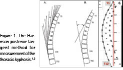
Problematically, some of this normal subject data is contaminated with subjects that should not be considered healthy. For one example, Fon, et al.,5 presented thoracic kyphosis measurement in 316 “normal subjects” aged 2-77 yrs. Their definition of normal was: “…while the general status of some of these patients was not optimal, it was assumed that the patients were sufficiently fit to be ambulatory….”

Beginning in 2001, my colleagues and I proposed a more narrow distribution of thoracic kyphosis values as normal: average values for T3-T10 posterior tangents = 33°-37°.3,4 Part of our reasoning for a narrower range of normal thoracic kyphosis was based on a study done in 2002.6 Here, we identified that translated postures of the ribcage relative to the pelvis in the sagittal plane can have a strong influence on the magnitude of thoracic kyphosis. Specifically, a total change of 26° in thoracic kyphosis was found for maximum posterior translation of the ribcage to maximum anterior translation in normal subjects. (See Figure 2.)

:dropcap_open:Poorer health status, increased disability, and increased pain levels in patients with anterior trunk postures.:quoteleft_close:

Furthermore, biomechanical models have predicted large increases in extensor muscle loads and consequent increased compression and shear loads on the thoraco-lumbar spine discs when sagittal trunk translation is present.7,8 These high compressive and shear loads may produce pain and initiate or contribute to a degenerative remodeling response in the disc. In fact, Glassman, et al.9, identified poorer health status, increased disability, and increased pain levels in patients with anterior trunk postures.

The issues associated with sagittal plane ribcage translation prompted my colleagues and I4 to present thoracic kyphosis data from a group of 50 normal subjects whose sagittal translation was within 1 standard deviation of the mean (neutral alignment). This data is presented in Table 1.

thoracictable1Average & Ideal Thoracic Kyphosis Models

thoracicfig3Harrison and colleagues3 presented average geometric models of the thoracic kyphosis (T1-T12, T2-T11, and T3-T10 segments were modelled) as a segment of an ellipse using pooled data from 80 normal subjects’ lateral thoracic radiographs. Figure 3 shows the average elliptical model of the segments T1-T12.3

Harrison, et al.4 followed this paper with an optimized elliptical model of thoracic kyphosis based in part on data from 50 optimized normal subjects. Since the thoracic vertebral bodies increase in size considerably from T1 to T12, a uniformly increasing model was derived for disc and vertebral body sizes using anatomical literature. We found that the major axis of the ellipse (long axis of an oval) is parallel to the posterior body margin of T12, whereas the minor axis of the ellipse (short axis of the oval) passed through the superior thoracicfig4endplate of T12. The minor axis to major axis ratio was computed to be 0.69.4 Figure 4 shows this optimized elliptical model in a template form that can be used for any height of a patient.

In the more recent literature, investigators have begun to develop individual subject optimized geometric sagittal plane curve models for thoracic kyphosis.10-12 There are certain anatomic variables that have been shown to have a moderate influence on sagittal plane curvature. When these anatomic variables are outside of normal tolerances, a change in sagittal curvature can result. This information will be detailed in Part 2 of this series. 

Summary

Chiropractic has a long history of identifying and attempting to restore normal alignment to the spine, where abnormal alignment is specifically referred to as the mechanical component of vertebral subluxation. The key is to fully understand when the modelling and alignment data presented herein is useful in differentiating a subluxated thoracic spine from a normal spine and how to modify the data and intervene in specific patient situations. Till next time.


Dr. Deed will be presenting a comprehensive, contemporary review of this topic at the upcoming 33rd  CBP Annual Conference on Sept. 23-25th, in Phoenix, AZ.


References

• Harrison DD. Spinal Biomechanics: A Chiropractic Perspective. National Library of Medicine #WE 725 4318C, 1982-97.

• Harrison DE, Cailliet R, Harrison DD, Janik TJ, Holland B. Centroid, Cobb or Harrison Posterior Tangents: Which to Choose for Analysis of Thoracic Kyphosis? Spine 2001; 26(11): E227-E234.

• Harrison DE, Janik TJ, Harrison DD, Cailliet R, Harmon S. Can the thoracic kyphosis be modeled with a simple geometric Shape? The results of circular and elliptical modeling in 80 asymptomatic subjects. J Spinal Disord Tech 2002; 15(3): 213-220.

• Harrison DD, Harrison DE, Janik TJ, Cailliet R, Haas JW. Do alterations in vertebral and disc dimensions affect an elliptical model of the thoracic kyphosis? Spine 2003;463-469.

• Fon GT, Pitt MJ, Thies AC Jr. Thoracic

• kyphosis: range in normal subjects.  AJR 1980;134(5):979-83.

• Harrison DE, et al. How Do Anterior/Posterior Translations of the Thoracic Cage Affect the Sagittal Lumbar Spine, Pelvic Tilt, and Thoracic Kyphosis? Eur Spine J 2002;11:287-293.

• Harrison DE, Colloca CJ, Keller TS, Harrison DD, Janik TJ. Anterior thoracic posture increases thoracolumbar disc loading. Eur Spine J 2005; 14:234-242.

• Kiefer A, Shirazi-Adl A, Parnianpour M. Synergy of the human spine in neutral postures. Eur Spine J 1998; 7:471-479.

• Glassman SD, Bridwell K, Dimar JR, Horton W, Berven S, Schwab F. The impact of positive sagittal balance in adult spinal deformity. Spine 2005;30:2024-2029.

• Berthonnaud E, et al. Analysis of the sagittal balance of the spine and pelvis using shape and orientation parameters. J Spinal Disorders & Techniques 2005;18(1):40-47.

• Vaz G, Roussouly P, Berthonnaud E, Dimnet J. Sagittal morphology and equilibrium of pelvis and spine. Eur Spine J 2002; 11(1):80-87.

• Vialle R, Levassor N, Rillardon L, Templier A, Skalli W, Guigui P. Radiographic analysis of the sagittal alignment and balance of the spine in asymptomatic subjects. J Bone Joint Surgery 2005;87Am:260-267.

Appropriate Use of a Cervical Orthotic for Abnormalities of the Cervical Lordosis

Introduction
More and more, Chiropractors are becoming interested in rehabilitation of the abnormal cervical lordosis due to its potential relationship with a number of patient health disorders.1-3 Chiropractic techniques often recommend the use of both in office equipment as well as supplementation of at home orthotics for rehabilitation of the abnormal cervical lordosis in appropriate cases,4 while other chiropractors use at home orthotics as the primary means of the rehabilitation of the cervical lordosis.  There are a number of home devices to choose from and not enough information on when it is and is not appropriate to use these devices for a given patient presentation.
In the present article, I would like to discuss the appropriate use of two common corrective cervical orthotic devices aimed at rehabilitation of abnormal cervical curvatures by presenting appropriate indications and contra-indications for the use of these devices.

Non-Compression 3-Point Bending
Cervical orthotics provoke a passive 3-point bending force that is generally well tolerated by most patients, as it does not apply compression to the cervical column during patient application. Patient size and the specific abnormality of the cervical lordosis influence the size and choice of what type of cervical orthotic is appropriate for a specific patient. For example, the amount of anterior or posterior head translation dictates the size of the cervical orthotic to be used, however the shape in the cervical lordosis determines the location of the peak of the device (C6-T1, C4-C6, or C2-C4).
There are three primary placements of this cervical orthotic, but only the lower cervical/upper thoracic placement will be discussed. Figure 1

Indications for the Cervical Orthotic Lower Neck Placement:
Upper thoracic/lower cervical placement. This placement of the orthotic will cause significant posterior head translation unless the large device is used. A lower cervical placement will increase the upper thoracic curve and increase the overall cervical lordosis. Specifically, this placement should be used for straightened or kyphotic lower cervical segments with loss of upper thoracic kyphosis and anterior head translation of approximately ≤ 40mm. (Figure 1)
Contra-indications for the Cervical Orthotic:
Quite simply put, the contra-indications for this cervical remodeling orthotic device would be the opposite of the indications listed above and many are the indications for the compression-extension wedge below.

Compression-Extension Remodeling Wedge deed1
The compression extension cervical orthotic device is a common tool available in the chiropractic profession today with several brands on the market. (Figure 2) The device creates the combination of posterior head translation, cervical extension, and compression force down the long-axis of the spine. Patients must be screened for tolerance to this position and loading; canal stenosis would be contra-indicated for this device.
Problematically, the compression extension wedge also creates an anterior translation of the thoracic spine and an extension of the mid-upper thoracic kyphosis. Typically speaking, the wedge only comes in one size (adult) and, thus, patient selection must be considered when using these devices; pediatric cases don’t fit well on this.

Indications for the Compression-Extension Wedge:
The compression-extension wedge will correct-reduce anterior head translation with loss of the cervical lordosis. The patient’s posterior vertebral body margins (black dashed-line) must be well anterior of the ideal cervical lordosis (red-line after Harrison et al.2). Head translation forward ≥ 25mm and up to ≈ 70 mm typically responds well to this device. However, the patient must also have posterior thoracic translation and increased mid- and upper-thoracic kyphosis, as the wedge will cause an opposite effect. Figure 2

deed2Contra-indications for the Compression-Extension Wedge:
Quite simply put, the Contra-Indications for these cervical remodeling orthotic devices would be the opposite of the indications for those listed above and many are the indications for the non compression three-point-bending devices.

SUMMARY
All home cervical corrective orthotic devices, such as the two shown herein, have indications and contra-indications for appropriate patient application. The cervical corrective orthotic should be fit to the following patient presenting conditions: 1) their symptomatology and severity of spinal arthritis/disc disease, 2) their ability to perform specific movements, 3) their presenting posture of the head and thoracic region, 4) their configuration of the cervical curvature, 5) the presence of any unstable segments, and 6) their configuration of the mid-upper thoracic kyphosis. I hope this presentation assists in your delivery of effective at home devices for rehabilitation of the abnormal cervical lordosis.


Dr. Deed will be presenting a comprehensive, contemporary review of this topic at the upcoming 32nd CBP Annual Conference on Sept. 24-26th, in Scottsdale, AZ.
Deed E. Harrison, D.C. is President CBP Seminars, Inc., Vice President CBP® Non-Profit, Inc., Chair PCCRP Guidelines, Editor—AJCC.

References
1.Harrison DE, Betz J, Ferrantelli JF. Sagittal spinal curves and health. JVSR 2009 July 31, pp 1-8.
2.Harrison DD, Harrison DE, Janik TJ, Cailliet R, Haas JW, Ferrantelli J, Holland B. Modeling of the Sagittal Cervical Spine as a Method to Discriminate Hypo-Lordosis: Results of Elliptical and Circular Modeling in 72 Asymptomatic Subjects, 52 Acute Neck Pain Subjects, and 70 Chronic Neck Pain Subjects. Spine 2004; 29:2485-2492.
3.McAviney J, Schulz D, Richard Bock R, Harrison DE, Holland B. Determining a clinical normal value for cervical lordosis. J Manipulative Physiol Ther 2005;28:187-193.
4.Harrison DE, Harrison DD, Haas JW. CBP Structural Rehabilitation of the Cervical Spine. Chapters 2 & 6. CBP Seminars, 2002; pgs:147-151. ISBN 0-9721314-0-X.

Validating the Truth

Validating the Truth

by Robert J. Goodman, D.C.


This year marks the coming of validation for the Upper Cervical Chiropractic techniques. Upper Cervical Chiropractors have appeared on mainstream television programs such as Good Morning America, The Montel Williams Show, and Discovery Health Channel, and our own Dr. Marshall Dickholtz Sr. won the ICA Chiropractor of the Year. Some would say the timing is perfect, including several prominent doctors and researchers in the medical field. The National Upper Cervical Chiropractic Association (NUCCA) has spearheaded this movement with the publishing of a hypertension study in a mainstream medical journal (Journal of Hypertension) and is experiencing a growing awareness in the chiropractic and medical world. Recently, Dr. Bruce Bell, a medical doctor from Chicago, stood before 225 NUCCA chiropractors and chiropractic students and discussed how important correcting the atlas subluxation is to the world and how we should be prepared for a greater interest from the health care field in general and increased awareness from the general public.

Continue reading “Validating the Truth”

Posterior to Anterior Thoracic Spinal Adjusting in the Scoliosis Patient Is Contraindicated by Spinal Biomechanics

Posterior to Anterior Thoracic Spinal Adjusting in the Scoliosis Patient Is Contraindicated by Spinal Biomechanics

by Dennis Woggon, DC, B.Sc.


It would make sense to understand normal spinal biomechanics when putting adjustive forces into the spine, especially in a complicated spine such as a scoliosis. It seems that spinal biomechanical forces are frequently ignored when it comes to spinal adjustments and manipulation.

The sagittal spine should have a lordotic cervical curve, a kyphotic thoracic curve and a lordotic lumbar curve. It is accepted that a loss of cervical lordosis will eventually result in a loss of lumbar lordosis.

The question is what does this loss of cervical and lumbar lever arms have on the thoracic spine? There is a reciprocal influence of the lever arms in the spine. A loss of cervical curve will exert posterior to anterior forces on the thoracic spine. This will cause a dipping of the thoracic spinous processes and a slight elevation of the vertebral body. This will manifest as anterior dorsal saucering or Poettenger’s Saucering.

The thoracic vertebra are somewhat fixed by the rib heads in flexion and extension. Flexion and extension of the thoracic vertebra is not a normal motion, but lateral flexion and rotation, as a coupled motion, is a normal motion. When there is a posterior to anterior leverage force on the thoracic vertebra, Poettenger’s Saucering develops to a point and then the thoracic spine will buckle laterally.

It has been known for a number of years that scoliosis is accompanied by a hypokyphosis.

“Thoracic hypokyphosis with increasing axial rotational instability is claimed to be a primary factor for the initiation of Idiopathic Scoliosis.”1

Rigo states, “. . . thoracic lordosis is the predominant component of the disease.”2

This is further magnified by Winter, who also seems to indicate that the Harrington Rods add to the problem. “The idiopathic cases usually exhibit a flattening of the sagittal curves, which had further deteriorated when the Harrington technique was used.”3

DeJong took a historical perspective stating, “A clinical, cadaveric, biomechanical and radiological investigation of the pathogenesis of idiopathic scoliosis indicates that biplanar asymmetry is the essential lesion. When median plane asymmetry (flattening or, more usually, reversal of the normal thoracic kyphosis at the apex of the scoliosis) is superimposed during growth, a progressive idiopathic scoliosis occurs. Idiopathic kyphoscoliosis cannot and does not exist, from the mildest cases in the community to the most severe cases in pathology museums.”4

Dickson agrees and sees the possibility of reversal in stating, “Idiopathic scoliosis (IS), which is substantially a three-dimensional deformation of a spine, causes not only lateral curvature and axial rotation of vertebral column, but also lordotisation of vertebrae in structural curve extension. In an effect, physiological thoracic kyphosis diminishes or even disappears. Method of asymmetric trunk mobilization in strictly symmetric positions, according to Dobosiewicz, not only deteriorates progression of IS or even reduces lateral curvature, but also significantly rebuilds physiological thoracic kyphosis in cases of IS accompanied by straight back.”5

In comparison groups, Inoue found, “Those patients who had scoliotic deformity with typical vertebral rotation only in thoracic spine (ST group), showed significant decrease compared to normal persons in thoracic kyphosis, but no difference in lumbar lordosis. However those changes in sagittal curvature were not found in FT group patients, who had scoliotic deformity without vertebral rotation. In conclusion, it is not the frontal curvature but the vertebral rotation which influenced the sagittal curvature of spine in patients with idiopathic scoliosis.”6

In a clinical study, a fourteen-year-old patient presented with a descending Cobb angle of 36, 56 and 45 degrees (Figure 1).7

The patient’s posture and X-rays (Figures 2 & 3) revealed a loss of cervical lordosis and forward head posture. The lateral thoracic X-ray demonstrated a hypokyphosis of 18 degrees (Figure 4).

By re-establishing the normal sagittal curves, the scoliosis has been reduced in nine intensive office visits (Figure 5).8

The correct adjustment force for this would be an anterior dorsal adjustment and not a P-A adjustment.

It would appear that a loss of the cervical lordosis can cause an anterior dorsal saucering resulting in a lateral bending motion of the thoracic spine. Based on this, posterior to anterior thoracic adjusting in these areas would appear to be contraindicated. This would also apply to the scoliosis patient or the potential scoliosis patient in regard to P-A thoracic adjusting as well as adjusting on the “high side of the rainbow.”

As Hippocrates said, “First, do no harm.”

For further information, contact Dr. Dennis Woggon at www.clear-institute.org.


References

1. Sagittal configuration of the spine in girls with idiopathic scoliosis: progressing rather than initiating factor. Rigo M, Quera-Salvá G, Villagers M. Elena Salvá Spinal Deformities Rehabilitation Institute, Vía Augusta 185, 08021 Barcelona, Spain. Stud Health Technol Inform. 2006;123:90-4.

2. Excessive thoracic lordosis and loss of pulmonary function in patients with idiopathic scoliosis. Winter RB, Lovell WW, Moe JH. Bone Joint Surg Am. 1975 Oct;57(7):972-7.

3. Sagittal plane correction in idiopathic scoliosis. de Jonge T, Dubousset JF, Illés T. University of Pécs, Faculty of Medicine, Department of Orthopedic Surgery, Pécs, Hungary. Spine. 2002 Apr 1;27(7):761.

4. The pathogenesis of idiopathic scoliosis. Biplanar spinal asymmetry. J Bone Joint Surg Br. 1984 Jan;66(1):8-15. Dickson RA, Lawton JO, Archer IA, Butt WP 1984.

5. Influence of method of asymmetric trunk mobilization on shaping of a physiological thoracic kyphosis in children and youth suffering from progressive idiopathic scoliosis. Stud Health Technol Inform. 2002;91:348-51Dobosiewicz K, et al, Department of Rehabilitation, Medical University of Silesia, Katowice, Poland 40-635 Katowice, ul. Ziolowa 35/37,

6. The sagittal curvature of spine in idiopathic scoliosis—its morphological features and the correlation among sagittal and frontal curvatures and rotation of apical vertebra. Inoue K. Nippon Seikeigeka Gakkai Zasshi. 1985 May;59(5):505-16.

7. Pictures and X-rays used with patient and guardian’s permission. 2008

8. CLEAR Institute and CLEAR Scoliosis Center, St. Cloud, MN.

Protective Postures (Part 2)


Last month we discussed the problems associated with hypolordotic lumbar postures as well as the protective effects of a healthy lumbar lordosis. I want to use this month’s column to cover the same ground as it pertains to the cervical spine. Patients walk into your office every day with complaints related to the cervical spine. Of the many symptomatic presentations we commonly handle as chiropractors, a sizeable percentage can be either directly or indirectly attributed to loss of the normal cervical lordosis. Hypolordotic and/or kyphotic cervical postures are associated with a wide range of problems. Conversely, a healthy cervical lordosis provides a large measure of protection to the neck. Consider the following fun facts.

Fact: Loss of the cervical lordosis predisposes the capsular ligaments to higher risk of injury in the event of trauma.1payne-fig_1

Habitual carriage of the neck in a straightened or kyphotic posture pretensions both the capsular ligaments and the ligamentum flavum. This pre stressing effect can have severe ramifications in the event of sudden neck trauma such as whiplash injuries. A 2005 study in the Journal of Biomechanics, found that trauma to patients with such postures may elongate the capsular ligaments “by up to 70%”, “induce laxity to the facet joint”, and predispose the neck to “accelerated degenerative changes over time.” The authors concluded that “abnormal spinal curvatures enhance the likelihood of whiplash injury and may have long-term clinical and biomechanical implications.”  In the event your patient is involved in an auto accident following care in your office, and statistically the average American is in a car accident about once every six years,  the success or failure of your corrective care efforts to restore the lordosis may well determine how severely they are injured.  
payne-fig_2
Fact:  Hypolordotic/kyphotic neck postures predispose the discs to injury while a healthy lordosis provides a protective effect.
We know that flexed (hypolordotic) postures result in potentially damaging disc mechanics. The anterior disc space is narrowed as the posterior disc space is widened with the result that the nucleus is forced posterior against the tensioned posterior annulus (See Fig. 2). White and Panjabi have stated that “the risk of disc failure is greater with tensile loading as compared to compressive loading.” 2  Restoration of the cervical lordosis provides protection to the posterior annulus by removing/reducing tensile stress to the posterior disc fibers and restoring normal weight bearing so as to discourage posterior migration of the nucleus toward the vital neural elements in the spinal canal and IVF (See Fig.3).
payne-fig_3
Fact: Normal lordosis is vital for the neck to have maximum resilience to compressive loads.
A 2001 study in the Journal of Neurosurgery, looked at the effects of cervical posture on the loadbearing ability of the cervical spine.3  During compressive loading, the straight spines failed through the anterior motor unit (disc and vertebral bodies) while the lordotic spines supported more of the weight on the posterior joints thereby sparing the disc and vertebral bodies. The authors concluded  “that a loss of a lordosis increases the risk of injury to the cervical spine following axial loading.”
In conclusion, the normal cervical lordosis is a highly protective posture for your patients.  Due to the limits of space, I haven’t even touched here on the protective effects of cervical lordosis to the spinal cord and nerve roots, the vertebral bodies, and to the reduction of a wide range of symptomatic complaints. If you aren’t already incorporating postural rehab methods into your treatment plans, you are leaving your patients wide open for future problems. And there’s really no reason for that to happen.  Effective treatment options are easily mastered, require no changes in your adjusting technique, and are affordable to the point of being downright cheap.


payneDr. Mark Payne is president of Matlin Mfg., a manufacturer and distributor of postural rehab products since 1988.  A more detailed report on this subject is available for free, as well as a FREE SUBSCRIPTION to Postural Rehab…electronic newsletter on corrective chiropractic methods. CALL 334-448-1210 or email
[email protected].

References:

  1. Stemper BD, Yoganandan N, Pintar FA.  Effects of abnormal posture on capsular ligament elongations in a computational model subjected to whiplash loading. J Biomech 2005 Jun; 38(6) 1313-23.
  2. White and Panjabi, Clinical Biomechanics of the Spine. J.B. Lippincott Company 1978. Pg. 153.
  3. Oktenoğlu T, Ozer AF, Ferrara LA, Andalkar N, Sarioğlu AC, Benzel EC.J Neurosurg. 2001 Jan;94 (1 Suppl) 108-14 Effects of cervical spine posture on axial load bearing ability: a biomechanical study.

New Brain Science, Old School Thinking- A closer look at the B.E.S.T. Technique


Almost 50 years ago, I entered Logan College with a master’s degree in science education.  Although chiropractic was described as an “art and a science,” I was most interested in the science.  I was taught that the science of this profession evolved and revolved around the concept of encroachment of a nerve in the spine, specifically the intervertebral foramen (IVF).  This encroachment was caused by an apposition between vertebrae, and altered the nerve flow innervating appropriate muscles and organs.  Slowly, this theory has been expanded to include blood and lymphatic vessels surrounding the IVF.  The concept of the chiropractic subluxation began some 100 years ago.  That was then; this is now.

Continue reading “New Brain Science, Old School Thinking- A closer look at the B.E.S.T. Technique”