Posterior to Anterior Thoracic Spinal Adjusting in the Scoliosis Patient Is Contraindicated by Spinal Biomechanics
by Dennis Woggon, DC, B.Sc.
It would make sense to understand normal spinal biomechanics when putting adjustive forces into the spine, especially in a complicated spine such as a scoliosis. It seems that spinal biomechanical forces are frequently ignored when it comes to spinal adjustments and manipulation.
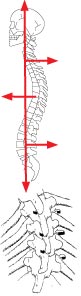
The sagittal spine should have a lordotic cervical curve, a kyphotic thoracic curve and a lordotic lumbar curve. It is accepted that a loss of cervical lordosis will eventually result in a loss of lumbar lordosis.
The question is what does this loss of cervical and lumbar lever arms have on the thoracic spine? There is a reciprocal influence of the lever arms in the spine. A loss of cervical curve will exert posterior to anterior forces on the thoracic spine. This will cause a dipping of the thoracic spinous processes and a slight elevation of the vertebral body. This will manifest as anterior dorsal saucering or Poettenger’s Saucering.

The thoracic vertebra are somewhat fixed by the rib heads in flexion and extension. Flexion and extension of the thoracic vertebra is not a normal motion, but lateral flexion and rotation, as a coupled motion, is a normal motion. When there is a posterior to anterior leverage force on the thoracic vertebra, Poettenger’s Saucering develops to a point and then the thoracic spine will buckle laterally.
It has been known for a number of years that scoliosis is accompanied by a hypokyphosis.
“Thoracic hypokyphosis with increasing axial rotational instability is claimed to be a primary factor for the initiation of Idiopathic Scoliosis.”1
Rigo states, “. . . thoracic lordosis is the predominant component of the disease.”2
This is further magnified by Winter, who also seems to indicate that the Harrington Rods add to the problem. “The idiopathic cases usually exhibit a flattening of the sagittal curves, which had further deteriorated when the Harrington technique was used.”3
DeJong took a historical perspective stating, “A clinical, cadaveric, biomechanical and radiological investigation of the pathogenesis of idiopathic scoliosis indicates that biplanar asymmetry is the essential lesion. When median plane asymmetry (flattening or, more usually, reversal of the normal thoracic kyphosis at the apex of the scoliosis) is superimposed during growth, a progressive idiopathic scoliosis occurs. Idiopathic kyphoscoliosis cannot and does not exist, from the mildest cases in the community to the most severe cases in pathology museums.”4

Dickson agrees and sees the possibility of reversal in stating, “Idiopathic scoliosis (IS), which is substantially a three-dimensional deformation of a spine, causes not only lateral curvature and axial rotation of vertebral column, but also lordotisation of vertebrae in structural curve extension. In an effect, physiological thoracic kyphosis diminishes or even disappears. Method of asymmetric trunk mobilization in strictly symmetric positions, according to Dobosiewicz, not only deteriorates progression of IS or even reduces lateral curvature, but also significantly rebuilds physiological thoracic kyphosis in cases of IS accompanied by straight back.”5
In comparison groups, Inoue found, “Those patients who had scoliotic deformity with typical vertebral rotation only in thoracic spine (ST group), showed significant decrease compared to normal persons in thoracic kyphosis, but no difference in lumbar lordosis. However those changes in sagittal curvature were not found in FT group patients, who had scoliotic deformity without vertebral rotation. In conclusion, it is not the frontal curvature but the vertebral rotation which influenced the sagittal curvature of spine in patients with idiopathic scoliosis.”6
In a clinical study, a fourteen-year-old patient presented with a descending Cobb angle of 36, 56 and 45 degrees (Figure 1).7
The patient’s posture and X-rays (Figures 2 & 3) revealed a loss of cervical lordosis and forward head posture. The lateral thoracic X-ray demonstrated a hypokyphosis of 18 degrees (Figure 4).
By re-establishing the normal sagittal curves, the scoliosis has been reduced in nine intensive office visits (Figure 5).8

The correct adjustment force for this would be an anterior dorsal adjustment and not a P-A adjustment.
It would appear that a loss of the cervical lordosis can cause an anterior dorsal saucering resulting in a lateral bending motion of the thoracic spine. Based on this, posterior to anterior thoracic adjusting in these areas would appear to be contraindicated. This would also apply to the scoliosis patient or the potential scoliosis patient in regard to P-A thoracic adjusting as well as adjusting on the “high side of the rainbow.”
As Hippocrates said, “First, do no harm.”
For further information, contact Dr. Dennis Woggon at www.clear-institute.org.
References
1. Sagittal configuration of the spine in girls with idiopathic scoliosis: progressing rather than initiating factor. Rigo M, Quera-Salvá G, Villagers M. Elena Salvá Spinal Deformities Rehabilitation Institute, Vía Augusta 185, 08021 Barcelona, Spain. Stud Health Technol Inform. 2006;123:90-4.
2. Excessive thoracic lordosis and loss of pulmonary function in patients with idiopathic scoliosis. Winter RB, Lovell WW, Moe JH. Bone Joint Surg Am. 1975 Oct;57(7):972-7.
3. Sagittal plane correction in idiopathic scoliosis. de Jonge T, Dubousset JF, Illés T. University of Pécs, Faculty of Medicine, Department of Orthopedic Surgery, Pécs, Hungary. Spine. 2002 Apr 1;27(7):761.
4. The pathogenesis of idiopathic scoliosis. Biplanar spinal asymmetry. J Bone Joint Surg Br. 1984 Jan;66(1):8-15. Dickson RA, Lawton JO, Archer IA, Butt WP 1984.
5. Influence of method of asymmetric trunk mobilization on shaping of a physiological thoracic kyphosis in children and youth suffering from progressive idiopathic scoliosis. Stud Health Technol Inform. 2002;91:348-51Dobosiewicz K, et al, Department of Rehabilitation, Medical University of Silesia, Katowice, Poland 40-635 Katowice, ul. Ziolowa 35/37,
6. The sagittal curvature of spine in idiopathic scoliosis—its morphological features and the correlation among sagittal and frontal curvatures and rotation of apical vertebra. Inoue K. Nippon Seikeigeka Gakkai Zasshi. 1985 May;59(5):505-16.
7. Pictures and X-rays used with patient and guardian’s permission. 2008
8. CLEAR Institute and CLEAR Scoliosis Center, St. Cloud, MN.