Embryology—Ossification of the first cervical vertebra begins about the seventh fetal week at the lateral masses and proceeds perichondrally in a dorsal direction, creating the posterior arch of the atlas. In the second year of life, a secondary growth center for the posterior tubercle develops between these neural arches. Complete fusion of the posterior arch should be noted between the third and fifth years.1
 |
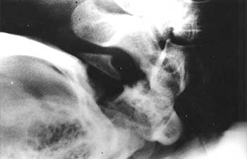
| Figure 1 – Note the congenitally malformed posterior tubercle of C-1 with posterior arch missing. Osteolytic metastatic carcinoma would not leave such a clear posterior tubercle and is very rare to affect the atlas. (Case courtesy of Kelly Jarvis, DC, DABCO; Heber City, Utah) |
Description—The basic defect in agenesis of the posterior arch of the atlas is the lack of a cartilage template on which the ossification process builds. Complete or partial agenesis of the posterior arch is rare, and posterior arch defects, by themselves, should not be the cause of neurologic or biomechanical findings, unless found in association with other anomalies such as Klippel-Feil syndrome.
Radiologic Features—An absent posterior arch can be easily visualized on standard lateral cervical radiographs by the lack of a bony posterior neural arch. A commonly associated finding is enlargement of the superior aspect of the second cervical spinous process, which has been referred to as a mega-spinous process, representing fusion of a rudimentary posterior arch and posterior tubercle of the atlas (not present in this case).2 One may also observe increased size of the anterior arch of C1, which is thought to be stress related and present in this case. This is a helpful radiographic sign and suggests a long-standing congenital origin to the defect.
Medicolegal Implications of Agenesis of the C1 Posterior Arch
The integrity of the transverse ligament may also be compromised in the maldevelopment process; therefore, a cervical flexion radiograph should be performed to evaluate the atlantodental interspace.
References:
1. Gehweiler JA, Daffner RH, Roberts L: Malformations of the atlas simulating the Jefferson fracture. AJR 140: 1083, 1983.
2. Yochum TR, Rowe LJ: Essentials of Skeletal Radiology, 3rd Edition, Lippincott Williams & Wilkins, 2004.
Dr. Terry R. Yochum is a second-generation chiropractor and a cum laude graduate of the National College of Chiropractic, where he subsequently completed his radiology specialty. He is currently Director of the Rocky Mountain Chiropractic Radiological Center, in Denver, CO, an Adjunct Professor of Radiology at the Los Angeles College of Chiropractic, as well as an instructor of Skeletal Radiology at the University of Colorado School of Medicine, Denver, CO. Dr. Yochum is, also, a consultant to Health Care Manufacturing Company that offers a Stored Energy system. For more information, Dr. Yochum can be reached at: 303-940-9400 or by e-mail at [email protected].
Dr. Chad Maola is a 1999 Magna Cum Laude graduate of National College of Chiropractic.