History:
A young 25-year-old male complains of bilateral sacroiliac pain and stiffness.
Diagnosis:
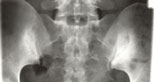
Figure 1. Note the bilateral sclerosis and erosions affecting both sides of the lower two-thirds of the sacroiliac joint. The diagnosis is ankylosing spondylitis.
General Features:
Essentially, the radiographic features of ankylosing spondylitis relate to the inflammatory, erosive, and ankylosing nature of the disease. The basic changes consist of osteoporosis, erosions, and surrounding reactive sclerosis, followed by bony ankylosis. Notably, these changes are bilateral and symmetric in nature.
Target Sites of Involvement:
Sacroiliac Joint. Sacroiliac involvement is the hallmark of ankylosing spondylitis. Roentgen changes in this joint are usually visible on the initial evaluation of the patient, but are often difficult to interpret. In equivocal circumstances radiographic reevaluation in 3 to 6 months should show definitive alterations. Even radionuclide imaging of sacroiliac joint disease is not definitive and emphasizes the role of plain film radiography in early detection. Upright films will not allow optimum visualization of the joint margins due to the sacral base inclination. The most diagnostic projections of the sacroiliac joints are on AP or PA angulated spot views, which visualize the entire joint margin not seen on routine AP views. The use of oblique projections is not usually necessary if this view is obtained.1
Characteristically, the sacroiliac joints are involved bilaterally and symmetrically, although early in the disease this is not invariable. The major radiographic abnormalities are more apparent on the iliac side due to the protective sacral hyaline cartilage, which is three times thicker than the iliac hyaline cartilage. The changes also are more prevalent in the lower two-thirds of the joint corresponding to the synovial portion, where the pathologic process predominates.
Radiographic findings reflect the sequence of inflammation, bone destruction, and ossification. Forestier has classified these sequential changes into three stages.1
Stage 1: Pseudowidening of the joint space. Loss of the articular cortical bone margin simulates widening of the joint cavity. This is due to subchondral osteoporosis and produces hazy joint definition.
Stage 2: Erosive and sclerotic changes. Superimposed on the widened joint space discrete erosive areas will be seen in the subarticular bone, resulting in an irregular joint margin that has been called the “rosary bead” appearance. Reactive sclerosis, particularly in the adjacent ilium, usually accompanies these erosions. This is the most common stage encountered when the diagnosis is first made. CT scan delineates the erosive osteoarticular lesions.
Stage 3: Ankylosis. Narrowing and eventual obliteration of the joint space follows the appearance of the erosions.
|
|
|
Diagnosis: Note the bilateral sclerosis and erosions affecting both sides of the lower two-thirds of the sacroiliac joint. The diagnosis is ankylosing spondylitis.
|

Reactive sclerosis gradually dissipates, to be replaced by generalized osteoporosis. Occasionally, the anterior sacroiliac joint marginal cortex will remain visible through the ankylosis and is referred to as a ghost joint. The upper ligamentous portion of the joint will also demonstrate bridging ossification. When prominent, it will be seen on an AP film as a triangular radiopacity (“star sign”). The time required for ankylosis to occur from the onset of the first radiologic abnormality varies from 7 to 23 years, with a mean of 14 years. At least 50% will develop complete and bilateral sacroiliac fusion, while approximately 40% will progress to stage 2 and resolve. A similar parallel sequence of events and radiographic signs is often observed in the symphysis pubis.

 Dr. Terry R. Yochum is a second generation chiropractor and a Cum Laude Graduate of National College of Chiropractic, where he subsequently completed his radiology residency. He is currently Director of the Rocky Mountain Chiropractic Radiological Center in Denver, Colorado, and Adjunct Professor of Radiology at the Southern California University of Health Sciences, as well as an instructor of skeletal radiology at the University of Colorado School of Medicine, Denver, CO. Dr. Yochum can be reached at 1-303-940-9400 or by e-mail at [email protected].
 Dr. Chad J. Maola is a 1990 Magna Cum Laude Graduate of National College of Chiropractic. Dr. Maola is a Chiropractic Orthopedist and is available for post-graduate seminars. He may be reached at 1-303-690-8503 or e-mail [email protected]