Of all causes, an inherited genetic defect appears to play a significant role with up to 30 percent of patients having another family member with significant scoliosis. A positive family history does not translate into worse curves or progressive curves.
DISCUSSION
The term scoliosis is usually credited to Hippocrates. Its derivation is from the Greek word skolios, meaning twisted or crooked. Within the disciplines of orthopedics and radiology, scoliosis describes any lateral deviation of the spine from the mid-sagittal plane. A structural scoliosis is a lateral curvature that is fixed and that fails to correct on recumbent lateral bending radiographic studies. Many disorders are related to this type of spinal disorder.
Idiopathic Scoliosis
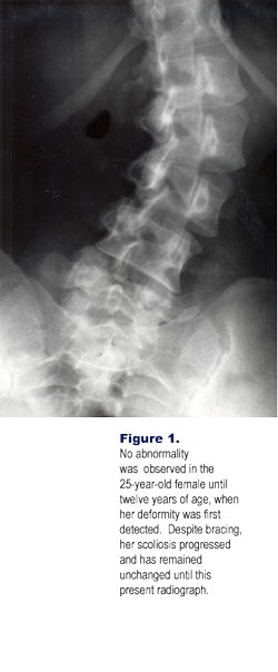
This is the most common form of lateral spinal deviation, accounting for up to 80 percent of scoliosis. The etiology is unknown, although many factors have been implicated. These include connective tissue disease, diet, enzymes, muscular imbalance, vestibular dysfunction, and inheritance. Patients with scoliosis can have associated osteopenia while the intervertebral discs remain immature.

Of all causes, an inherited genetic defect appears to play a significant role with up to 30 percent of patients having another family member with significant scoliosis. A positive family history does not translate into worse curves or progressive curves. The age of onset distinctively occurs within the growth period and allows for an age-based classification—infantile, juvenile, and adolescent. Infantile Idiopathic Scoliosis. This occurs between birth and three years of age. The majority will disappear (resolving infantile idiopathic scoliosis), but some will occasionally progress (progressive infantile idiopathic scoliosis). This progressive form is rare in the United States, is slightly more common in males, and is usually a left convex thoracic curve.Juvenile Idiopathic Scoliosis. The onset is between three and ten years of age, with an average age of seven years. There is female gender predominance of four to one. As many as 30 percent will eventually have corrective surgery.Adolescent Idiopathic Scoliosis. The curvature develops in the period between the age of ten and skeletal maturity. The natural history of scoliosis has not significantly changed over the years. The adolescent form remains the most common type of idiopathic scoliosis. Females are predominantly affected, with a ratio of nine to one over males. There is an increased incidence of coexisting pes cavus deformity. The critical time period for progression, which may be rapid, is between the ages of twelve and sixteen. Once spinal growth has ceased, as indicated by fusion of the iliac apophysis, further progression is unlikely. Later in adult life, superimposed degenerative changes may allow the curvature to increase on an average of 15°, occasionally creating nerve entrapment syndromes.
A frequent finding in a developing scoliosis is a lateral wedged deformity of the vertebral body, which will persist into adult life. This is a result of impaired growth at the discovertebral junction on the concave side of the curvature because of excessive compressive forces (Hueter-Volkmann principle). The most frequent curve pattern is the right convex thoracic type. Three other curves that are frequently present include a right thoracolumbar, a left lumbar, and a combined form of left lumbar and right thoracic. The psoas shadow on frontal radiographs is often absent on the concave side of a lumbar scoliosis.
There is a ten time greater incidence of congenital heart disease than in the general population when the idiopathic curve is more than 20°.
Congenital Scoliosis
Congenital scoliosis is distinguished by anomalies of the vertebrae or ribs. The most frequently observed anomalies include hemivertebrae, block vertebrae, spina bifida, bridging vertebral bars, joint deformities, fusion of ribs and other rib malformations. This curve is typically a short “C” curve and may be rapidly progressive in the growing years. Occasionally, anterior vertebral body defects may cause superimposed kyphosis (kyphoscoliosis). There is a frequent association in congenital scoliosis with anomalies of the genitourinary system.

RADIOLOGICAL ASSESSMENT
The radiologic examination is the most definitive and important diagnostic tool in the assessment and management of the patient with scoliosis. A number of non-radiologic methods, such as moiré contourography and back contour devices, have been employed. The role of the radiograph is multiple: (a) determining etiology; (b) evaluating curvature, including site, magnitude and flexibility; (c) assessing bone maturity; (d) monitoring progression or regression; and (e) aiding in the selection of appropriate treatment. A wide variety of factors are involved in the process of obtaining practical clinical information while avoiding unnecessary radiation exposure.
Standard Radiographic Techniques
Projections
Erect Anteroposterior and Lateral Projections. These projections are the absolute minimum required for accurate assessment of any scoliosis. Preferably, the anteroposterior (AP) projection should be done by a single exposure on a 14″ by 36″ cassette in order to allow for total and continuous curvature evaluation. However, if such equipment is not available, sectional projections can be utilized just as effectively. The disadvantages of the single exposure full spine projection are the irradiation of unnecessary body parts, sacrifice of bone detail for pathology, and the expense of the equipment necessary to do the projections adequately. Wherever possible, a long focal film distance, rare earth screens, density equaling filtration, collimation, optimal kilovoltage, and lead shielding of breasts thyroid, and gonads should be utilized. Obtaining the projection posterior–anterior also significantly reduces the dose to the sensitive tissues and is now the preferred exam of choice.Lateral–Bending Projections. These are primarily projections to evaluate curvature flexibility. The patient laterally flexes as much as possible and the exposure is made. This must be done both to the right and left. Left Hand and Wrist. A spot AP radiograph is taken of the left hand and wrist in patients under twenty years of age. This is compared with the Greulich and Pyle Atlas to ascertain the skeletal age of the patient, which is important in planning the treatment regime. When the growth plate (physis) is closed for the distal radius and ulna, one can assume that the secondary growth centers in the spine have closed and the scoliosis should not show any significant degrees of progression.
Dr. Terry R. Yochum is a second generation chiropractor and a Cum Laude Graduate of National College of Chiropractic, where he subsequently completed his radiology residency. He is currently Director of the Rocky Mountain Chiropractic Radiological Center in Denver, Colorado, and Adjunct Professor of Radiology at the Southern California University of Health Sciences, as well as an instructor of skeletal radiology at the University of Colorado School of Medicine, Denver, CO. Dr. Yochum can be reached at 1-303-940-9400 or by e-mail at [email protected].
Dr. Chad J. Maola is a 1990 Magna Cum Laude Graduate of National College of Chiropractic. Dr. Maola is a Chiropractic Orthopedist and is available for post-graduate seminars. He may be reached at 1-303-690-8503 or e-mail [email protected]
References
1. Yochum TR, Rowe LJ: Essentials of Skeletal Radiology, 3rd ed., Williams & Wilkins, Baltimore, Maryland, p. 513, 2005.