Laser Neurology: Groundbreaking Research

laserneurologyChiropractors and the Nervous System
Chiropractors have always been interested in how subluxation affects the nervous system. DD and BJ Palmer commonly lectured about the wonders of chiropractic and how it could heal the injured nerves. Recently, chiropractors have been seen as leaders in the field of laser therapy and its effects on the central nervous system. This article will explore how this occurs and why it is important for doctors of chiropractic to understand this exciting new research.
 
What is Low-Level Laser Therapy?
Low-level laser therapy (LLLT) is a treatment that uses lasers or LEDs to stimulate or inhibit cellular function.¹  Laboratory studies find that LLLT can stimulate healing, alter cellular metabolism, and stimulate tissue proliferation.² 
 
Laser Has Been Proven
One of the newest trends is the overwhelming number of studies proving the value of laser therapy. There are over 2,000 positive studies and more than 250 randomized double-blind clinical trials that document the value of laser therapy with pain management, reduction of inflammation, and improved rate of healing.³  With this in mind, it is vital that chiropractors understand the latest scientific research regarding the newest and most versatile electrotherapy tool, cold laser, and how it fits into a modern practice. The first question I get from doctors once they are convinced that the use of laser works is, “How does laser compare to other modalities?”
 
Laser Compared to Ultrasound
When comparing laser and light therapy to ultrasound, research indicates that ultrasound has value, but laser has been shown to close wounds more effectively.4  Ultrasound tends to cause a slight retraction of hard and soft tissues, whereas laser causes a proliferation of these tissues.5  Thus, when the goal is wound healing, post-surgical rehab, or tissue strengthening, laser therapy has unique strengths above ultrasound. In summary, to break up old, fibrous scar tissue, I recommend ultrasound as the first phase followed by laser to stimulate healing and blood vessel development.
 
Laser Compared to Electrical Stimulation
When comparing laser therapy to electrical stimulation, research indicates that laser therapy provides as much or more healing stimulation.6  In addition, laser therapy appears to be equal to or more effective than acupuncture with electrical stimulation.7  However, electrical stimulation can provide quick pain relief, so it can be quite effective to use electrical stimulation while painting the area with laser. This appears to accomplish a synergistic effect.8  Thus, laser is unique in that it provides multiple benefits, including improved rates of healing, tissue proliferation, and analgesia, and it can be combined synergistically with other modalities.
 
Lasers in Chiropractic
The primary use of laser therapy in a chiropractic office is for the treatment of pain. The effectiveness of laser at pain management and the lack of serious side effects have been proven.9  We now know that laser can dramatically improve the function of the central nervous system as well.
 
Deep Laser Penetration
There is no question that clinicians who use lasers to treat the spine and extremities treat the spinal cord indirectly. If a doctor places an infrared laser on a patient’s back, six percent of the photons enter the spinal cord; thus, chiropractors are always treating deeper structures.10 For example, if a doctor places a laser over a patient’s sternum or lower ribs when they have costochondral pain, they are indirectly irradiating the heart and liver. There are recent medical studies that demonstrate the beneficial effects of treating the heart and liver with LLLT.11 12
 
Lasers Help the Central Nervous System
Complex regional pain syndrome (CRPS) is a chronic pain syndrome that was originally termed reflex sympathetic dystrophy (RSD) and is characterized by excruciating pain following a very minor injury.13 CRPS is caused by the nervous system getting stuck in a sympathetic mode.14  It has been shown that laser therapy can heal many types of chronic neurological disorders, including CRPS.15
Spinal Cord Injury
Research also documents that laser can simulate spinal cord regeneration. It was found that when the laser was applied to the spine, it significantly improved the average length of axonal re-growth and increased the total number of axons after spinal cord injury. 16

How Does Treating the Brain Affect the Body?
Research documenting that treating the brain could affect the function of the whole body began in the 1970s. Research at UCLA School of Medicine established that descending inhibitory pathways can inhibit pain. This may explain how treating the brain, and even cranial adjusting, can have such powerful, full-body effects.17

Lasers, Depression, and Addiction
LLLT has been shown to be helpful for depression because infrared laser light produces natural opioids and serotonin in the brain.18 The best study on lasers and addiction was done on cigarette smokers. All of the patients who received laser therapy experienced a lessening of withdrawal symptoms and 92 percent of them stopped smoking.19

The New Frontier: Laser Brain Treatment
About five years ago, researchers began to document the value of directly treating the brain with lasers. The first studies in 2006 found that animals that had strokes and were treated with LLLT had a significant improvement in neurological function. 20 21 22

Lasers and Degenerative Brain Disease
Amyotrophic Lateral Sclerosis (ALS) is characterized by progressive loss of motor function and death. Researchers found that by placing the laser directly on the skull, motor function was significantly improved. 23 Another recent study was performed on Parkinson’s disease patients. It was found that laser treatment normalized neurological activity and reduced Parkinson’s symptoms after a single, brief treatment.24

Major Medical Research
:quoteright_open:There is no question that clinicians who use lasers to treat the spine and extremities treat the spinal cord indirectly. If a doctor places an infrared laser on a patient’s back, six percent of the photons enter the spinal cord; thus, chiropractors are always treating deeper structures.:quoteright_close:Harvard Medical School recently studied 10 patients with depression and anxiety. A 250 milliwatt LED was placed on the forehead just a few millimeters above the skin. The researchers noted a significant decrease in depression and anxiety that lasted for four weeks after only one treatment.25 Following this Harvard study, there have been more human studies that demonstrate tremendous benefits from laser therapy on the brain. In the second major human study, 660 patients received laser therapy applied to the skull. Researchers noted a favorable outcome after 90 days and found that the laser was able to penetrate about five inches. The study was performed at some of America’s best medical schools, including UC San Diego, Stanford University, Scripps Hospital, University of Massachusetts, University of Pennsylvania, and Boston University. 26

Wavelength and Dose
Research documents that laser therapy stimulates an increase in ATP, RNA/DNA synthesis, oxygen, and cell metabolism.27Also, it has been found that when treating almost any area of the body, treatment is ineffective if the dose is too low or too high.28 29

It is important that one consider dose because only three percent of the photons delivered to the forehead-scalp surface will reach the cortex.30

Summary
The value of using lasers to directly treat the brain is still in the experimental stage. Yet, it is being validated on human subjects by noted researchers in major medical schools and hospitals with quite dramatic clinical results. Based on the positive findings of these groundbreaking studies, I expect lasers to be an integral part of complementary neurological therapy in the near future. And because chiropractors have been trendsetters in the use of lasers and the nervous system, they should use this information to help them continue to be leaders in this field. Understanding the latest trends in medical research regarding laser and the central nervous system is the first step.

Reference:

  1. Huang, YY, et al. Dose-Response. 2009;7(4):358.
  2. Karu TI, et al. Nonmonotonic behavior of the dose dependence of the radiation effect on cells in vitro exposed to pulsed laser radiation at 820 nm. Lasers Surg Med. 1997;21(5):485-492.
  3. Tuner, J., & Hode, L. (2004). The Laser Therapy Handbook. Grangesberg, Sweden: Prima Books.
  4. Demir, H. (2004). Comparison of the effects of laser and ultrasound treatments on experimental wound healing in rats. J Rehabil Res & Dev, Sept/Oct;41(5).
  5. Lirani-Galvão, AP (2006). Comparative study of how low-level laser therapy and low-intensity pulsed ultrasound affect bone repair in rats. Photomed Laser Surg, Dec;24(6):735-40.
  6. Medlicott, MS. (2006). A systematic review of the effectiveness of exercise, manual therapy, electrotherapy, relaxation training, and biofeedback in the management of temporomandibular disorder. Phys Ther, Jul;86(7):955-73.
  7. Bjordal, JM. (2007). Short-term efficacy of physical interventions in osteoarthritic knee pain. A systematic review and meta-analysis of randomised placebo-controlled trials. BMC Musculoskelet Disord, Jun;22;8:51.
  8. Kato, MT. (2006). TENS and low-level laser therapy in the management of temporomandibular disorders. J Appl Oral Sci, Apr;14(2):130-5.
  9. Bjordal JM, et al. Low-level laser therapy for tendinopathy: Evidence of a dose-response pattern. Phys Ther Rev. 2001;6:91-99.
  10. Byrnes KR, et al. Light promotes regeneration and functional recovery and alters the immune response after spinal cord injury. 2005 Mar;36(3):171-85.
  11. Oron U, et al. Enhanced liver regeneration following acute hepatectomy by low-level laser therapy. 2010 Oct;28(5):675-8.
  12. Yang Z, et al. Low-Level Laser Irradiation Alters Cardiac Cytokine Expression Following Acute Myocardial Infarction: A Potential Mechanism for Laser Therapy. 2011 Feb 24.
  13. Albrecht PJ, et al. Pathologic alterations of cutaneous innervation and vasculature in affected limbs from patients with complex regional pain syndrome. Pain. 2006; 120:244-266.
  14. Wasner G, et al. Vascular abnormalities in acute reflex sympathetic dystrophy (CRPS I): Complete inhibition of sympathetic nerve activity with recovery. Arch Neurol. May 1999;56(5):613-20.
  15. Gibbs GF, et al. Unravelling the pathophysiology of complex regional pain syndrome: Focus on sympathetically maintained pain. Clin Exp Pharmacol P. 2008;35:717-724.
  16. Wu X, et al. 810 nm Wavelength light: an effective therapy for transected or contused rat spinal cord. Lasers Surg Med 2009;41:36-41.
  17. Mayer DJ, et al..Analgesia from electrical stimulation in the brainstem of the rat, Science. 1971 Dec 24;174(16):1351-4.
  18. Hagiwara S, et al. Pre-Irradiation of blood by gallium aluminum arsenide (830 nm) low-level laser enhances peripheral endogenous opioid analgesia in rats. Anesth Analg. 2008 Sep;107(3):1058-63.
  19. Catherine M., et al. Low level laser for the stimulation of acupoints for smoking cessation: a double blind, placebo controlled randomised trial and semi structured interviews. Journal of Chinese Medicine, Number 86, February, 2008.
  20. Oron A, et al.. Low-level laser therapy applied transcranially to rats after induction of stroke significantly reduces long-term neurological deficits. Stroke. 2006 Oct;37(10):2620-4.
  21. Lapchak PA, De Taboada L. Transcranial near infrared laser treatment (NILT) increases cortical adenosine-5=-triphosphate (ATP) content following embolic strokes in rabbits. Brain Res 2009;1306:100-105.
  22. Rochkind S, et al. Increase of neuronal sprouting and migration using 780 nm laser phototherapy as procedure for cell therapy. Lasers Surg Med. 2009 Apr;41(4):277-81.
  23. Moges H, et al. Light therapy and supplementary riboflavin in the SOD1 transgenic mouse model of familial amyotrophic lateral sclerosis (FALS). Lasers Surg Med 2009; 41:52-59.
  24. Trimmer PA, et al.Reduced axonal transport in Parkinson’s disease cybrid neurites is restored by light therapy. Mol Neurodegener. 2009 Jun 17;4:26.
  25. Schiffer F, et al. Psychological benefits 2 and 4 weeks after a single treatment with near infrared light to the forehead: a pilot study of 10 patients with major depression and anxiety. Behav Brain Funct. 2009 Dec 8;5:46.
  26. Zivin J, et al: Effectiveness and safety of transcranial laser therapy for acute ischemic stroke. Stroke 2009, 40:1359-1364.
  27. Lapchak PA, et al.. Transcranial near infrared laser treatment (NILT) increases cortical adenosine-5=-triphosphate (ATP) content following embolic strokes in rabbits. Brain Res 2010;1306:100-105.
  28. Bjordal JM; et al.. A systematic review of low level laser therapy with location-specific doses for pain from chronic joint disorders. Aust J Physiother. 2003;49(2):107–16.
  29. Frigo L, et al. The effect of low-level laser irradiation on melanoma in vitro and in vivo. BMC Cancer. 2009;9:404.
  30. Wan S, et al. Transmittance of nonionizing radiation in human tissues. Photochem Photobiol 1981;34:679-681.
Curtis Turchin, M.A., D.C. is an expert in the field of laser treatment, low force adjusting and therapeutic exercise. He has published 4 books and more than 20 journal articles on chiropractic and laser therapy. He has lectured at many chiropractic colleges and state associations. If you have any questions, please feel free to contact him. Contact him at: [email protected] or 650-224-8789. His teaching website is: http://www.innateadjusting.com

A 21st Century Breakthrough in Ginseng Science: Enzyme Formulation

:dropcap_open:G:dropcap_close:inseng is one of the most well-known of all herbal remedies, and the history of ginseng in natural medicine goes back thousands of years, first mentioned in written medical journals in China over 2,000 years ago. Ginseng is considered the most powerful adaptogenic herb. In fact, ginseng has become the standard by which all other adaptogens are measured. This is why Suma is called Brazilian Ginseng, Maca is called Peruvian Ginseng, Ashwaganda is called Indian Ginseng, and Eleuthero Root is called Siberian Ginseng. None of these common adoptogens are ginseng, yet they are called ginseng. There are 4 types of actual ginsengs which have the plant classification Panax: Korean Ginseng (Panax CA Meyer), Chinese Ginseng (Tianchi Ginseng), American Ginseng (Panax quinquefolius) and Japanese Ginseng (Ginex Japonicus).

ginsengroot3So what is an adaptogen? According to Webster’s Dictionary, “an adaptogen is a nontoxic substance and especially a plant extract that is held to increase the body’s ability to resist the damaging effects of stress and promote or restore normal physiological functioning”. In medical terms, a highly efficacious adaptogen helps to bring the body into homeostatic balance.

How does Ginseng work?
Why has it been the leading herbal remedy for 2,000 years for such a broad range of conditions?

A simple answer is that when the body reaches a homeostatic state its ability to self-heal is optimized. This is no secret for a chiropractic physician. Through making adjustments and removing subluxations we allow the energy in the body to flow naturally and in balance.The result is the body begins to heal itself; this is how it is designed!

So how does Panax Ginseng bring the body into balance?
ginsengchart1In 1963 the unique active ingredient found only in Panax ginseng was discovered, plant triterpene saponins called ginsenosides. The number of different types of ginsenosides determines the range of the adaptogenic capacity of the ginseng. Different ginsenosides have shown in clinical research to have different effects, sometimes opposing effects. For example, some increase blood pressure while others reduce blood pressure, some increase blood glucose while others reduce blood glucose. The ginsenosides in combination with the 32 minerals, 14 amino acids, organic germanium and 7 vitamins and polysaccharides make a remarkable synergy that bring the body into balance.
 
A fully balanced ginseng has tremendous potential. It must be harvested after the optimal length of time, 4 or 5 years, processed as a whole root without peeling, allowed to dry without high heat steam and finally extracted using a low heat vacuum extraction. The result is a remarkably balanced extract with the potential to improve human health.

Ginseng and Chiropractic Care
High Quality Ginseng is an ideal complement to chiropractic treatment. It works synergistically to bring the body into balance and optimize the body’s self-healing potential.

Fermented Ginseng: A 21st century breakthrough in Ginseng Science
“The therapeutic potential of ginseng has been studied extensively, and ginsenosides, the active components of ginseng, are shown to be involved in modulating multiple physiological activities. These molecules exert their functions via interactions with steroidal receptors. The multiple biological actions make ginsenosides important resources for developing new modalities. Yet, low bioavailability of ginsenoside is one of the major hurdles that needs to be overcome to advance its use in clinical settings.” *Pharmacology of ginsenosides: a literature review Kar W Leung* and Alice ST Wong, CMJ Journal, June 2010.

The Challenge: Absorption
There are two main types of ginsenosides: the Protopanaxadiol “Diol Type” and Protopanaxotriol “Triol Type”. The Diol type ginsenosides have been shown in research to have the greatest medicinal efficacy. However, the Diol type ginsenosides have a larger molecular size. A scientific discovery in the late 90s showed that many of the ginsenosides were not being absorbed until they reached the large intestine in the ascending colon. A related problem surfaced in other clinical studies showing that many people were not experiencing full absorption. In one study less than 40% of the subjects were able to have full absorption of all ginsenosides.

ginsengabsorbtion1
The Solution: Create Ginsenoside Metabolites via Fermentation

As we enter 2012, more and more research on the benefits of fermentation as it facilitates digestion and absorption has been made available. This has also been part of the development of ginseng science.

In the 90s, research began in earnest to find a way to improve absorption. The first research was done in the late 90s and utilized human bacteria to create the metabolites of ginsenosides.

In the main area of research the metabolite form of the Diol type ginsenosides became known as “IH 901” or “Compound K”. Ginseng research then expanded.

In 2008 a superior process was developed using enzyme fermentation. The result was overwhelming. In a 2×2 crossover study with 24 healthy male subjects, the enzyme fermented ginseng extract vs. standard ginseng extract showed 15 times increased absorption, 4 times faster absorption and 4 times increased consistency of absorption. *Pharmacokinetic comparison of ginsenoside metabolite IH-901 from fermented and non-fermented ginseng in healthy Korean volunteers. ( Journal of Ethnopharmacology 139 (2012) 664– 667.)

ginsengfermentedextract
Now, high absorption, fully-balanced ginseng is available in the market.

 
Good diet, high quality water, and exercise are all essential components of optimum wellness. We are facing an overwhelming amount of stress and toxicity in our environment. Our bodies need extra help, and the leading natural medicine for several thousand years has been made available using 21st century science.

Now, enzyme fermented ginseng extract can be fully absorbed to provide its balancing effect for bringing everyone’s body into homeostatic balance and optimizing wellness.

Research Opportunities
Today, Doctors can participate in a longitudinal case study with high absorption Enzyme Fermented Ginseng Extract conducted by the Clinic Research Institute.

Each participating physician receives over $450 worth of an enzyme fermented high-absorption ginseng (patent pending) for testing with 3 patients.

To learn more contact CRI at 877-532-8378 or go to www.ClinicResearchInstitute.org

Ronald K. Gilbert, D.C., CCSP, NMD B.S. 1984, Va Commonwealth Univ. 1985, Diplomate, National Board of Chiropractic Examiners D.C 1986, Northwestern College CCSP 1988, Parker College, Sports Medicine 1992 , American Board of Chiropractic Orthopedics NMD 2002, St Luke School of Naturopathic Medicine,, ND 2002, Commission on Certification of Naturopathic Physicians 2002 ,100 hr. Post-Grad Internal Medicine, National University of Health Sciences.

David C. Konn, Double Major Psychology and Kinesiology, Macalester College, St Paul MN, Patent Holder CEO, Neuro Energies Inc. Regional President, Empowered Doctor Managing Director, Ilhwa North America President/CEO, Ilhwa NA Inc.

Increased Injury to Vertebrae Adjacent to Fused Segments

Increased Injury to Vertebrae Adjacent to Fused Segments

Dr. William J. Owens, D.C., D.A.A.M.L.P.


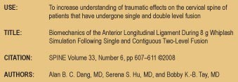
When a clinician is evaluating a traumatically injured victim there are many factors to consider when determining prognosis and diagnosis. The most important are changes in the structure and function of the cervical spine including surgical interventions, specifically fusions. When determining causality in relation to a cervical spine injury in patients with prior surgical fusion, a very recent article published in Spine by Dang et al 2008 may shed some light on this complex scenario. Recognized internationally as the leading journal in its field, Spine is an international, peer-reviewed, bi-weekly periodical that considers for publication original articles in the field of spine care. It is the leading subspecialty journal for the treatment of spinal disorders. Only original papers are considered for publication with the understanding that they are contributed solely to Spine. The Journal does not publish articles reporting material that has been reported at length elsewhere.

This study specifically addressed how cervical fusion alters the peak strain of the Anterior Longitudinal Ligament (ALL) in the adjacent motor segments. Past hypotheses have pointed out that due to decrease in flexibility in the cervical spine, the segments above and below the fused area are more susceptible to injury. The study used cadaveric specimens because “anthropomorphic dummies lack accurate soft tissue representation. Only cadaveric specimens or mathematical models are appropriate to evaluate the ALL.” pp 607. The ALL strain was tested at 8g whiplash.


This study quantified the increase in peak ALL strain during 8g acceleration simulated whiplash in a single-level and 2-level fusion in a healthy spine. “The increase in peak ALL strain in the adjacent motion segments is greater with 2-level fusion than it is with single-level fusion (40.8% vs. 15.5%)” pp610. In conclusion, this study demonstrates “Two-level fusion produced greater increases in peak ligament strain when compared with single level fusion at the adjacent motion segments.” pp 610


Injury to the ligaments surrounding the cervical spine result in permanent injury with long term consequences. When diagnosing traumatically injured patients with a past medical history of surgical fusion, look to the areas adjacent to the fused segments for answers to chronic pain and muscle spasm.


Each issue, a clinical topic will be provided by Dr. William J. Owens of the American Academy of Medical Legal Professionals (AAMLP), which is a national non-profit organization comprised of doctors and lawyers. The purpose of the organization is to provide its members with current research in trauma and spinal-related topics to keep the professional on the cutting edge of healthcare. Members may also sit for a Diplomate examination and be conferred a DAAMLP. The organization also offers support to the individual member’s practice. To learn more, go to www.aamlp.org
or call 1-716-228-3847.


The above review is provided for educational purposes only. It is not designed or intended to reproduce or replace the authors work. Readers are encouraged to obtain full licensed versions of the article as determined by Copyright Law. For information on how to obtain a licensed copy please contact the Academy directly. 2008.

Whiplash Injury and Surgically Treated Cervical Disc Disease


http://theamericanchiropractor.com/images/murphy.jpg

Key Points from Dan Murphy, D.C.

 1) The purpose of this study was to determine if whiplash injury predisposes to surgically proven cervical disc degeneration.

2) 11% of the whiplash-injured patients required cervical disc surgery, while only 5% of the control population required cervical disc surgery. Therefore, the cervical disc surgery rate was more than doubled in the whiplash-injured group.

3) Although both the whiplash injury group and the control population were age matched, the mean age for surgery in the whiplash-injured group was 45 years and for the control non-injured group it was 55 years. This 10-year difference was “significantly less.”

4) “This study demonstrates an increased association between whiplash injury and cervical disc disease.” These authors reference a number of studies that make these points:

A) Acute post-traumatic cervical angular kyphosis developed into frank cervical spine degenerative change within 5 to 7 years. (1974)

B) The prevalence of degenerative changes in the cervical spine in patients with whiplash in their 4th and 5th decade were equivalent to those found in the control population that were 10 and 20 years older. (1991)

C) MRI findings of cervical hyperextension injuries showed separation of the cervical disc from the endplate, anterior annular tears, occult anterior vertebral endplate fractures, and anterior longitudinal ligament injuries at multiple levels. (1991)

D) A study was “unable to demonstrate that psychosocial stress played any role in the outcome of whiplash injury.” (1991)

E) Four studies “demonstrated that patients continued to experience symptoms after settlement of litigation, suggesting that financial gain played little part.” (1964, 1965, 1975, 1990)

5) “These data suggest that the symptoms and signs of whiplash injury cannot be attributed solely to psychological factors and the organic pathology is a more constant explanation.”

6) “This study provides further evidence that whiplash injury causes structural changes predisposing to premature degenerative disc disease.”


Dr. Dan Murphy graduated magna cum laude from Western States Chiropractic College in 1978. He received Diplomat status in Chiropractic Orthopedics in 1986. Since 1982, Dr. Murphy has served part-time as undergraduate faculty at Life Chiropractic College West, currently teaching classes to seniors in the management of spinal disorders. He has taught more than 2000 postgraduate continuing education seminars. Dr. Murphy is a contributing author to both editions of the book Motor Vehicle Collision Injuries and to the book Pediatric Chiropractic. Hundreds of detailed Article Reviews, pertinent to chiropractors and their patients, are available at Dr. Murphy’s web page, www.danmurphydc.com.

Cell Phones and Brain Tumors

TITLE: Cell phones and brain tumors: A review including the long-term epidemiologic data

CITATION: Surgical Neurology, September 2009; 72; pp. 205–215

AUTHORS: Vini G. Khurana, Ph.D., F.R.A.C.S., Charles Teo, M.B.B.S., F.R.A.C.S., Michael Kundi, Ph.D.,

Lennart Hardell, M.D., Ph.D., Michael Carlberg, M.S.

 Key Points from Dr. Dan Murphy

 1. This article has 68 references. An editorial comment ascribed to this study indicates that it is “the most comprehensive study and analysis to date of this topic.”

2. These authors found 11 long-term studies in the PubMed database of participants using cell phones for ≥10 years.

3. “The results indicate that using a cell phone for ≥10 years approximately doubles the risk of being diagnosed with a brain tumor on the same (“ipsilateral”) side of the head as that preferred for cell phone use.”

4. “The authors conclude that there is adequate epidemiologic evidence to suggest a link between prolonged cell phone usage and the development of an ipsilateral brain tumor.”

5. The power [and danger] generated by a cell phone will vary according to the amount of interference with the signal. Higher power is required when using a cell phone in a moving vehicle, within a building, or in an elevator.

6. “The output power [and danger] of the phone is generally set to the highest level during ‘handovers’ between networked base stations as a user moves from one geographic area to another or when signal interference is greatest.”

7. Evidence presented suggests that cordless phones are also not safe.

8. Cell phones emit electromagnetic radiation only during calls.

9. Cell phone systems have been presumed to be safe because their longer wavelengths are nonionizing, lacking sufficient energy to break intermolecular bonds. Therefore, their increased cancer risk is not as a consequence of ionization.

10. “Science Magazine has recently acknowledged that there are several peer-reviewed studies from laboratories in at least 7 countries, including the United States, showing that cell phone or similar low-intensity electromagnetic fields can (contrary to expectations of non-ionizing sources) break DNA or modulate it structurally.”

11.”Irrespective of the type of phone, exposure is highest on the side of the head against which the cell phone is held and appears to be even higher in children owing to thinner scalps and skulls, increased water content of their brain, and lower brain volume.”

12.”Many independent laboratory investigations have suggested adverse biologic effects of cell phone radiation.” (12 references)

13.The authors present evidence that cell phones can be DNA-damaging as a consequence of “non-thermal interaction between incoming microwaves and exquisitely sensitive oscillatory electrical processes found in living tissues.” “This is akin to the reception of a clock radio being susceptible to interference from a nearby cell phone.” This “oscillatory similitude may lead to genetic or epigenetic damage through increased local production of reactive oxygen species or free radicals.” [Wow!]

14.”There are several hundred studies that support the existence of low-intensity, non-thermal effects of cell phone radiation on biological systems. The consequences are mostly adverse: DNA single- and double-strand damage, changes in gene transcription, changes in protein folding, heat shock protein generation, production of free radicals, and effects on the immune system.”

15.”Taken together, the long-term epidemiologic data suggest an increased risk of being diagnosed with an ipsilateral brain tumor related to cell phone usage of 10 years or more.”

16.There are also “significantly elevated odds for the development of ipsilateral parotid gland tumors among heavy cell phone users.”

17.The Central Brain Tumor Registry of the United States maintains comprehensive tumor incidence rates in the USA, and their data shows an increase in incidence of brain tumors of about 36% in less than a decade. This increase is not explained by an aging population (because these figures were age-adjusted) or by better detection.

18.”The authors believe that the aforementioned epidemiologic and laboratory findings underscore the need for reassessment by governments worldwide of cell phone and also most radiation exposure standards and the usage and deployment of this technology. If the epidemiologic data continue to be confirmed then, in the absence of appropriate and timely intervention and given the increasing global dependence on cell phone technology, especially among the young generation, it is likely that neurosurgeons will see increasing numbers of primary brain tumors, both benign and malignant.”


Dr. Dan Murphy graduated magna cum laude from Western States Chiropractic College in 1978. He received Diplomat status in Chiropractic Orthopedics in 1986. Since 1982, Dr. Murphy has served part-time as undergraduate faculty at Life Chiropractic College West, currently teaching classes to seniors in the management of spinal disorders. He has taught more than 2000 postgraduate continuing education seminars. Dr. Murphy is a contributing author to both editions of the book Motor Vehicle Collision Injuries and to the book Pediatric Chiropractic. Hundreds of detailed Article Reviews, pertinent to chiropractors and their patients, are available at Dr. Murphy’s web page, www.danmurphydc.com.

A National Survey of Physician-Industry Relationships

FROM ABSTRACT


BACKGROUND

Relationships between physicians and pharmaceutical, medical device, and other medically related industries have received considerable attention in recent years.

We surveyed physicians to collect information about their financial associations with [the drug] industry and the factors that predict those associations.


 METHODS

We conducted a national survey of 3167 physicians in six specialties (anesthesiology, cardiology, family practice, general surgery, internal medicine, and pediatrics).


 RESULTS

Most physicians (94%) reported some type of relationship with the pharmaceutical industry, and most of these relationships involved receiving food in the workplace (83%) or receiving drug samples (78%).

More than one third of the respondents (35%) received reimbursement for costs associated with professional meetings or continuing medical education, and more than one quarter (28%) received payments for consulting, giving lectures, or enrolling patients in trials.

Cardiologists were more than twice as likely as family practitioners to receive payments.

Family practitioners met more frequently with [drug] industry representatives than did physicians in other specialties, and physicians in solo, two-person, or group practices met more frequently with industry representatives than did physicians practicing in hospitals and clinics.


CONCLUSIONS

The results of this national survey indicate that relationships between physicians and [the drug] industry are common and underscore the variation among such relationships according to specialty, practice type, and professional activities.


KEY POINTS FROM DR. DAN MURPHY

1) This study shows that most physicians (94%) have some type of relationship with the drug industry. Because “respondents may have underreported their associations with [drug] industry,” the relationship between physicians and drug companies is probably higher than the 94% documented in this study.

2) Physician benefits from drug companies include receiving free drug samples, free meals, free tickets to events, free travel from drug companies, financial benefits from drug companies, reimbursement for costs associated with professional meetings or continuing medical education, payments for consulting, payments for giving lectures, and payments for enrolling patients in drug clinical trials.

3) “Family practitioners reported the highest average number of meetings with [drug] industry representatives (16 meetings per month), followed by internists (10 per month), cardiologists (9 per month), pediatricians (8 per month), surgeons (4 per month), and anesthesiologists (2 per month).”

4) The drug industry may focus marketing efforts on “physicians who are perceived as influencing the prescribing behaviors of other physicians.”

5) There is a “higher frequency of [drug] industry payments to physicians who have developed clinical practice guidelines.”

6) In 2000, the average number of meetings between physicians and drug industry representatives was 4.4 per month. This study showed an elevated average to 16 meetings per month. The reason for this 400% increase in meeting rates “may reflect an intensification of [drug] industry marketing since the 1990’s.”


Dr. Dan Murphy graduated magna cum laude from Western States Chiropractic College in 1978. He received Diplomat status in Chiropractic Orthopedics in 1986. Since 1982, Dr. Murphy has served part-time as undergraduate faculty at Life Chiropractic College West, currently teaching classes to seniors in the management of spinal disorders. He has taught more than 2000 postgraduate continuing education seminars. Dr. Murphy is a contributing author to both editions of the book Motor Vehicle Collision Injuries and to the book Pediatric Chiropractic. 

Making a Case for Chiropractic Care

KEY POINTS FROM DR. DAN MURPHY

1) In chronic low back pain, there is an integration between connective tissue fibrosis and the nervous system perception of pain.

2) Adverse connective tissue fibrosis can be remodeled by applying mechanical forces to soft tissues, including chiropractic spinal adjusting. [Note, chiropractic was included as the applying of a mechanical force to reverse adverse connective tissue fibrosis and its influence on the nervous system.]

3) The “association between symptoms and imaging results (X-ray, CT, MRI) has been consistently weak, and up to 85 percent of patients with low back pain cannot be given a precise pathoanatomical diagnosis using these methods.”

4) “Ongoing pain is associated with widespread neuroplastic changes at multiple levels within the nervous system and including primary afferent neurons, spinal cord, brainstem, thalamus, limbic system and cortex.”

5) Neuroimaging has shown that there are distinct “brain networks” involved in acute vs. chronic pain. Chronic pain is specifically related to regions for cognition and emotions.

6) Chronic back pain results in neuronal or glial loss in the pre-frontal and thalamic gray matter. [Brain atrophy]

7) “Increased connective tissue stiffness due to fibrosis is an important link in the pathogenic mechanism leading to chronicity of pain.” [Very Important: The Fibrosis of Repair]

8) “Abnormal movement patterns can have important influences on the connective tissues that surround and infiltrate muscles.” [Very important because the subluxation complex includes abnormal movement patterns.]

9) “A hallmark of connective tissue is its plasticity or ‘remodeling’ in response to varying levels of mechanical stress.” [This is important because it implies that spinal adjusting can initiate remodeling of abnormal connective tissues.]

10) “Both increased stress due to overuse, repetitive movement and/or hypermobility, and decreased stress due to immobilization or hypomobility can cause changes in connective tissue.” [Both increased and decreased motion are deleterious.]

11) A chronic local increase in stress leads to micro-injury and inflammation. [Subluxation can cause micro-injury and inflammation.]

12) “A consistent absence of stress leads to connective tissue atrophy, architectural disorganization, fibrosis, adhesions and contractures.” [Fibrosis]

13) “Fibrosis can be the direct result of hypomobility or the indirect result of hypermobility via injury and inflammation.” [Very Important]

14) During the early phase of immobilization, loss of muscle length is primarily due to shortening of muscle-associated connective tissue, which is later followed by actual shortening of muscle fibers.

15) Muscle connective tissue fibrosis promotes hypomobility. “Connective tissue fibrosis is detrimental, as it leads to increased tissue stiffness and further movement impairment.” [Important: fibrosis]

16) “Tissue microinjury, inflammation and fibrosis not only can change the biomechanics of soft tissue (e.g., increased stiffness) but also can profoundly alter the sensory input arising from the affected tissues.” [Very Important: Many contend that the tissue changes associated with the subluxation alter the afferent input into the central nervous system, which is the nerve interference of the subluxation.]

17) “Connective tissue is richly innervated with mechanosensory and nociceptive neurons.” [Very Important]

18) Activation of nociceptors can contribute to the development or worsening of fibrosis and inflammation, causing even more tissue stiffness and movement impairment. [Important]

19) Chronic low back pain may be caused by pathological connective tissue fibrosis, which causes adverse changes in movement. This is well documented in ligaments and joint capsules. [Very Important] This pathological connective tissue fibrosis is plastic and can, therefore, be remodeled. However, the remodeling must take place over time.

20) “In fibrosed connective tissue and muscle, blood and lymphatic flow may be chronically compromised by the disorganized tissue architecture and, thus, vulnerable to unusual muscle activity (e.g., beginning a new work activity or sport), or to conditions causing further decrease in perfusion such as prolonged sitting.”

21) Pain leads to reduced motion, and movement restriction increases fibrosis, “setting the patient up for more painful episodes.” [Very Important: fibrosis]

22) “In addition to its role in the pathological consequences of immobility and injury, the dynamic and potentially reversible nature of connective tissue plasticity may be key to the beneficial effects of widely used physical therapy techniques as well as ‘alternative’ treatments involving external application of mechanical forces (e.g., massage, chiropractic manipulation, acupuncture), changes in specific movement patterns (e.g., movement therapies, tai chi, yoga) or more general changes in activity levels (e.g., increased recreational exercise).”

23) “Manual or movement-based treatments have the advantage of not causing drug-induced side effects (e.g., gastritis, sedation),” but excessive motion may lead to inflammation.

24) A “carefully applied direct tissue stretch may be necessary in cases of long standing hypomobility with pronounced fibrosis and stiffness.” [Very Important, as an adjustment may be considered to be a “carefully applied direct tissue stretch.”]

Dr. Dan Murphy graduated magna cum laude from Western States Chiropractic College in 1978. He received Diplomat status in Chiropractic Orthopedics in 1986. Since 1982, Dr. Murphy has served part-time as undergraduate faculty at Life Chiropractic College West, currently teaching classes to seniors in the management of spinal disorders. He has taught more than 2000 postgraduate continuing education seminars. Dr. Murphy is a contributing author to both editions of the book Motor Vehicle Collision Injuries and to the book Pediatric Chiropractic.

The Importance of Antioxidants with Fish Oil Dietary omega-3 fatty acids for women

KEY POINTS FROM Dr. DAN MURPHY

ALA: Alpha-linolenic acid, plant derived 18-carbon long omega-3 fatty acid. Primarily from flaxseed (linseed), walnut, and hemp oils.

EPA: Eicosapentaenoic acid, animal derived 20-carbon long omega-3 fatty acid. Primarily from cold-water fatty fish.

DHA: Docosahexaenoic acid, 22-carbon long omega-3 fatty acid. Primarily from cold-water fatty fish. There are vegetarian sources (algae) for DHA.

1) Adequate maternal omega-3 fatty acid intake “ensures the optimal cerebral and cognitive development of the infant.”

2) Human milk contains both ALA [flaxseed oil, etc.] and DHA, unlike that of other mammals. [This is one of the reasons that cow’s milk is not a substitute for human milk for infants.]

3) Vegetarian and vegan mother’s milk have altered fatty acid profiles which impair the cerebral and cognitive development of their infants. [Very Important]

4) ALA [flaxseed oil, etc.], DHA and EPA are important for preventing ischemic cardiovascular disease in women of all ages.

5) Omega-3 fatty acids can help to prevent the development of certain cancers, particularly those of the breast and colon, and possibly of the uterus and the skin, and are likely to reduce the risk of postpartum depression, manic-depressive psychosis, dementias (Alzheimer’s disease and others), hypertension, toxemia, diabetes and, to a certain extent, age-related macular degeneration.

6) Omega-3 fatty acids play a positive role in the prevention of menstrual syndrome and ostmenopausal hot flushes.

7) The normal Western diet contains little ALA [flaxseed oil, etc.], providing less than 50% of the RDA.

8) The best sources for EPA and DHA are fish, seafood and “omega-3” eggs.

9) Both the omega-6 fatty acid linoleic (LA, 18:2(n-6)) and the omega-3 fatty acid alpha-linolenic acid (ALA) [flaxseed oil, etc.], 18:3(n-3)) are “physiologically essential.” [Important, ALA is physiologically essential]

10) The intake of ALA [flaxseed oil, etc.] is far too low.

11)Pregnant women that consume more fish oil improve the pregnancy for both the mother and the baby, reducing prematurity and low birth weight in the infant, and reducing hypertension and pre-eclampsia in the mother.

12)Vegetarians are more prone to premature births and Caesarean sections.

13)Vegetarian mothers are more likely to have premature babies with low birth weight.

14)Daily maternal supplementation of fish oil containing a DHA/EPA mixture is good for fetal development.

15)“Omega-3 fatty acids are most important as structural elements in the developing nervous systems of the fetus and newborn, and this is linked to the mother’s food.”

16)ALA [flaxseed oil, etc.], influences vision, behavior and brain structure and function.

17)“Adding omega-3 fatty acids to baby formula, to make it more like mother’s milk, influences the visual, cerebral and intellectual capacities of newborn babies.”

18)“The fetus uses most of the portion of dietary omega-3 fatty acids supplied to it for its developing brain.” [Very Important]

19)Maternal DHA status decreases during pregnancy.

20)“The cerebral and overall DHA status of breast-fed babies is better than that of infants fed formula lacking DHA.”

21)Human milk contains considerable concentrations of both DHA and ALA [flaxseed oil, etc.].

22)Eating fatty fish, taking fish oil capsules and eating omega-3 eggs increases the DHA in maternal milk.

23)A diet containing flaxseed oil, which has a high ALA content, increases the ALA and EPA in the milk and erythrocytes of lactating women, but not DHA. [Very Important for vegan mothers]

24)“Although the diets of vegans and vegetarians contain reasonable amounts of ALA [flaxseed oil, etc.], it is unlikely that enough is converted to DHA to satisfy the needs of pregnancy and lactation.” [Very Important]

25)Dietary fish, seafood or omega-3 supplements are advisable and prudent for pregnant and lactating women. [Very Important]

26)Omega-3 fatty acids prevent age-related macular degeneration.

27)“There is no doubt that supplements of omega-3 fatty acids, generally taken as fish oil, improve infant visual acuity.” [Important]

28)A lack of omega-3 fatty acids damages hearing and leads to premature aging of the auditory nervous system. “Omega-3 fatty acids are important dietary components for preserving hearing throughout life.”

29)Children given fish oil during the first year of life are less likely to develop type I diabetes, perhaps because of the anti-inflammatory action of very long chain omega-3 fatty acids.

30)Fish consumption reduces the risk of breast cancer.

31)“Excess omega-6 fatty acids seems to increase the risk of breast cancer metastasis, while omega-3 fatty acids have the opposite action.”

32)“There should always be a good intake of antioxidants to restrict the peroxidation of fatty acids, as these peroxide derivatives are genotoxic and cytotoxic.” [Very Important]

33)“Omega-3 fatty acids, if adequately preserved from oxidation,” benefit atherosclerosis, chronic hepatitis, inflammatory bowel diseases, psoriasis, and rheumatoid arthritis. [Important, again: this is why everyone should take antioxidants with each gram of fish oil.]

34)Eating fish and omega-3 fatty acids reduces the risk of suicide attempts, reduces the frequency of bipolar disorder (manic-depressive patients), and reduces the risk of dementia, particularly Alzheimer’s disease.

35)Studies show that ALA [flaxseed oil, etc.] significantly protects against cardiovascular disease.

36)Assuming ALA [flaxseed oil, etc.] intake is sufficient, newborn babies can only in small quantities convert ALA to DHA and, therefore, DHA is considered an essential nutrient for babies. [Important for strict vegetarians.]

37)Both ALA [flaxseed oil, etc.] and DHA are essential nutrients.

38)People who eat no animal lipids are very deficient in DHA. [Important for strict vegetarians.]

39)Omega-3 fatty acids help to prevent menstrual syndromes,  particularly dysmenorrhea and menopausal hot flushes and reduce the risk of osteoporosis.

40)“Only seafood provides adequate EPA and DHA.”

41)“There is practically no toxicological risk from eating too much omega-3 fatty acid.” [Important]

42)“Women, therefore, have specific requirements for omega-3 fatty acids that should be recognized and fulfilled, either by the diet or with capsules.”

COMMENTS FROM DR. DAN MURPHY

A central issue from this article is that alpha-linolenic acid (ALA, from fl axseed oil and other sources) is essential for health and must be included in the diet. Many individuals taking fish oil (omega-3 supplements) are unaware that fish oil capsules usually do not contain ALA. Again, this article supports the importance of using antioxidants when consuming omega-3 fatty acids.

Dr. Dan Murphy graduated magna cum laude from Western States Chiropractic College in 1978. He received Diplomat status in Chiropractic Orthopedics in 1986. Since 1982, Dr. Murphy has served part-time as undergraduate faculty at Life Chiropractic College West, currently teaching classes to seniors in the management of spinal disorders. He has taught more than 2000 postgraduate continuing education seminars.

Dr. Murphy is a contributing author to both editions of the book Motor Vehicle Collision Injuries and to the book Pediatric Chiropractic.


Chiropractic Works

OBJECTIVE:

To study the immediate sensorimotor neurophysiological effects of cervical spine manipulation using somatosensory evoked potentials (SEP’s).


METHODS:

Twelve subjects with a history of reoccurring neck stiffness and/or neck pain, but no acute symptoms at the time of the study were invited to participate in the study.

An additional twelve subjects participated in a passive head movement control experiment.

Spinal brainstem and cortical SEP’s to median nerve stimulation were recorded before and for thirty minutes after a single session of cervical spine manipulation, or passive head movement.


RESULTS:


There was a significant decrease in the amplitude of parietal and frontal SEP components following the single session of cervical spine manipulation compared to pre-manipulation baseline values.

These changes lasted on average twenty minutes following the manipulation intervention.

No changes were observed in the passive head movement control condition.


CONCLUSIONS:


Spinal manipulation of dysfunctional cervical joints can lead to transient cortical plastic changes, as demonstrated by attenuation of cortical somatosensory evoked responses.


SIGNIFICANCE:


This study suggests that cervical spine manipulation may alter cortical somatosensory processing and sensorimotor integration.

These findings may help to elucidate the mechanisms responsible for the effective relief of pain and restoration of functional ability documented following spinal manipulation treatment.


Key Points from Dan Murphy

1. Spinal manipulation is a commonly used conservative treatment for neck, back, and pelvic pain.

2. The effectiveness of spinal manipulation in the treatment of acute and chronic low back and neck pain has been well established by outcome-based research.

3. Spinal dysfunction will alter afferent input to the central nervous system.

4. Altered afferent input to the central nervous system leads to plastic changes in the central nervous system. (Very Important)

5. Neural plastic changes take place both following increased and decreased afferent input. (Extremely Important)

6. Both painful and painless joint dysfunction will inhibit surrounding muscles.

7. Joint dysfunction causes afferent driven increases in neural excitability (facilitation) to muscles that can persist even after the initiating afferent abnormality is corrected. (This suggests that a muscle afferent problem can persist even after the joint component of the subluxation is corrected. The chronic component of the subluxation may be plastic changes that cause long-term alteration of muscle afferentation.) This article clearly supports that the joint component, the muscle component, and the neurological component of the subluxation complex are influenced by traditional joint-cavitation spinal adjusting.

8. The altered neural processing that occurs as a consequence of joint dysfunction provides a rationale for the effects of spinal manipulation on neural processing that have been described in the literature. (Very Important)

9. Spinal dysfunction alters the balance of afferent input to the central nervous system and this altered afferent input may lead to maladaptive neural plastic changes in the central nervous system, and spinal manipulation can effect this. (Very Important)

10.The clinical evidence for joint dysfunction that requires manipulation includes:

A. Tenderness on joint palpation,

B. Restricted intersegmental range of motion,

C. Palpable asymmetry of intervertebral muscle
tension,

D. Abnormal or blocked joint play and end-feel,

E. Sensorimotor changes in the upper extremity.

[I recall in the teachings of Richard Stonebrink, DC, in the orthopedic diplomate program twenty-five years ago, the importance of always documenting (in our daily records) the evidence that the patient had a manipulatable spinal lesion (subluxation). His evidence was identical to these. Dr. Stonebrink would stress that such documentation would always make the case unique to chiropractic and consequently make the chiropractor the only expert in the case.]

11.The most reliable spinal-dysfunction-indicators are tenderness with palpation of the dysfunctional joint, and alterations of segmental range of motion.

12.High velocity, low amplitude thrust spinal manipulation with the head held in lateral flexion, with slight rotation and slight extension is a standard manipulative technique used by manipulative physicians, physiotherapists and chiropractors.

13.High velocity manipulation alters reflex EMG activity and alters afferent input to the central nervous system. (Important)

14.High-velocity manipulation causes significant cortical SEP amplitude attenuation in at least the frontal and parietal cortexes.

15.Passive head movements do not cause changes in cortical firing.

16.A single session of spinal manipulation of dysfunctional joints resulted in attenuated cortical (parietal and frontal) evoked responses. These changes most likely reflect central changes. (Very Important)

17.The cortical function of different individuals responded differently to spinal adjusting. [This indicates that variables other than the adjustment, itself, can influence the cortical responses in a given individual]

18.The significantly decreased somatosensory cortical SEP occurred in all post-manipulation measurements, indicating enhanced active inhibition because the cervical manipulations could have altered the afferent information originating from the cervical spine (from joints, muscles, etc.).

19.The passive head movement SEP experiment demonstrated that no significant changes occurred following a simple movement of the subject’s head. Our results are, therefore, not simply due to altered input form vestibular, muscle or cutaneous afferents as a result of the chiropractors touch or due to the actual movement of the subject’s head. This, thus, suggests that the results in this study are specific to the delivery of the high-velocity, low-amplitude thrust to dysfunctional joints. [Extremely Important]

20.Displacement of vertebrae is signaled to the central nervous system by afferent nerves arising from deep intervertebral muscles, and this is improved with adjusting the adjacent dysfunctional joint.

21.Joint dysfunction leads to bombardment of the central nervous system with Ia afferent signaling from surrounding intervertebral muscles. Spinal manipulation reduces excessive afferent signals from adjacent intervertebral muscles, which improves altered afferent input to the central nervous system. This changes the way the central nervous system responds to any subsequent input.

22.Episodes of acute pain following injury induce plastic changes in the sensorimotor system, prolonging the episode of pain and playing a roll in establishing chronic neck pain conditions. (Very Important) The reduced cortical SEP amplitudes observed in this study following spinal manipulation may reflect a normalization of such injury/pain-induced central plastic changes, which may reflect one mechanism for the improvement of functional ability reported following spinal manipulation. (Extremely Important)

23.Spinal manipulation of dysfunctional joints may modify transmission of neuronal circuitries, not only at a spinal level, but at a cortical level, and possibly also deeper brain structures such as the basal ganglia. (Very Important)

24.Cervical spine manipulation alters cortical [brain] somatosensory processing and sensorimotor integration.

25.These findings may help to elucidate the mechanisms responsible for the effective relief of pain and restoration of functional ability documented following spinal manipulation treatment.


Comment by Dan Murphy

One of the central themes of the neurology diplomate program taught by Ted Carrick, DC, is that chiropractic spinal adjusting influences the cortical brain, creating plastic changes. This article very much supports that perspective.


Dr. Dan Murphy graduated magna cum laude from Western States Chiropractic College in 1978. He received Diplomat status in Chiropractic Orthopedics in 1986. Since 1982, Dr. Murphy has served part-time as undergraduate faculty at Life Chiropractic College West, currently teaching classes to seniors in the management of spinal disorders. He has taught more than 2000 postgraduate continuing education seminars. Dr. Murphy is a contributing author to both editions of the book Motor Vehicle Collision Injuries and to the book Pediatric Chiropractic.


What you and your patients need to know about Fish and Mercury

Many patients are familiar with at least some of the health benefits of fish oil supplementation and dietary consumption of fatty fish, especially species higher in the n-3 fatty acids eicosapentaenoic acid (EPA) and docosahexaenoic acid (DHA). There are a multitude of benefits, including improved cardiovascular health, better functioning immune system, help with arthritis, atherosclerosis, depression, adult-onset diabetes mellitus and some cancers. There is reduced risk for stroke, asthma, dementia and kidney disease. In recent research, it was noted that modest consumption of fish (e.g., 1-2 servings/wk), especially species higher in the n-3 fatty acids EPA and DHA, reduces risk of coronary death by 36 percent (95 percent confidence interval, 20-50 percent; P<.001) and total mortality by 17 percent (95 percent confidence interval, 0-32 percent; P=.046) and may favorably affect other clinical outcomes.1

Many of the benefits are attributed to the anti-inflammatory effects of these fatty acids. There are thousands of research articles touting the benefits of polyunsaturated fatty acids or omega-3’s obtained from our diet as well as supplementation. (A search of Medline with the key word “omega-3” results in just under 10,000 articles.) Many well-known organizations, such as the American Heart Association, recommend eating fish as a regular part of a healthy diet. However, we are being made increasingly aware that, along with these benefits, come risks. As a result, consumers are rightly concerned about health risks of contaminants from eating. Many Americans have dangerous levels of methylmercury in their bodies, including 5- 8 percent of American women of childbearing age.

In 2004, the Food and Drug Administration and the Environmental Protection Agency issued a joint consumer advisory about mercury in fish and shellfish. The significance of this becomes apparent when one considers that this is the first time these two organizations have combined their advice to form a single uniform advisory.2 Their advice was directed toward women who might become pregnant, women who are pregnant, nursing mothers, and young children. In July of 2003, the Food Standards Agency in the United Kingdom issued a similar precautionary warning, however they also included children under sixteen years of age.3

In a 2004 article published in the Annals Of Internal Medicine, Wilson states that “studies have linked over consumption of certain fish (particularly popular ones such as swordfish, tuna steaks, Chilean sea bass, and some kinds of salmon) to neurologic deficits, cancer, autoimmune and endocrine disorders, and even some heart disorders. The risks stem mainly from two toxins: mercury, which accumulates over the lifetime of larger, longer-living fish, and polychlorinated biphenyls (PCB’s), which are found in fish living in polluted waters and in some farmed fish.”4

In adults, methylmercury poisoning causes tremor, difficulty with concentration, vision deficits, and numbness and tingling. In children, numerous health problems may result, including brain damage, mental retardation, blindness, and seizures. With lower levels of methylmercury exposure in the womb, there may be subtle but irreversible deficits in learning ability as well as altering the male reproductive organs and increased risk for cancer.4

Mercury is an element that is naturally found in the environment. It is also released into the air through industrial pollution, including the burning of fossil fuels and solid wastes. This mercury accumulates in streams and oceans where bacteria cause chemical changes that transform mercury into methylmercury. Fish and shellfish absorb the methylmercury as they feed in these waters and tend to build up in the larger predatory species. Of these, shark, tilefish, tuna and bottom-feeders, such as certain crab, tend to accumulate the highest levels. Essentially, the higher up the food chain, the higher the bioaccumulation of toxins. According to Wilson, exposure to methylmercury comes almost solely from eating fish. She also notes that “methylmercury is absorbed from the gastrointestinal tract and binds readily with proteins; the highest levels in the body are found in the kidneys.”

Methylmercury crosses the blood-brain barrier, affecting the brain. According to some researchers, the health effects of low-level methylmercury in adults are not clearly established.1 However, we suggest it is best to err on the side of caution and limit our exposure to toxic elements.

In addition to the dangers posed by methylmercury, fish are also the main source of concentrated polychlorinated biphenyls (PCB’s) with the highest dietary levels found in farmed salmon. PCB exposure is associated with liver and breast cancer, neurologic and endocrine problems, and possibly even increased risk for heart disease. “In children, PCB exposure in utero and from breast milk consumption has been linked with neurodevelopmental delays, impaired cognition, immune problems, and alterations in male reproductive organs…. Generally, freshwater fish that live in inland lakes, such as bluefish, lake trout, and smelt, are more likely to be contaminated.”4

Farmed salmon sounds innocent enough until you know a little more about it. Wilson states that “any more than a single eight-ounce portion of farmed salmon a month posed an ‘unacceptable cancer risk’ to consumers.”

Foran, et al., analyzed farmed vs. wild salmon and reported that organic arsenic was significantly higher in farmed than in wild salmon, whereas cobalt, copper, and cadmium were significantly higher in wild salmon. In addition, he stated that none of the contaminants exceeded federal standards or guidance levels.5 In a follow-up article, he stated that “health risks (based on a quantitative cancer risk assessment) associated with consumption of farmed salmon contaminated with PCB’s, toxaphene, and dieldrin were higher than risks associated with exposure to the same contaminants in wild salmon.”6

In the journal Science, Hites, et al., note, “The annual global production of farmed salmon has increased by a factor of forty during the past two decades.” He and fellow researchers analyzed over two metric tons of farmed and wild salmon for organochlorine contaminants and discovered that concentrations of these contaminants are significantly higher in farmed salmon than in wild.7

This data is frightening, considering over 90 percent of the salmon sold in the United States is farmed and that farmed salmon is sometimes labeled as “wild” in grocery stores.

Many researchers recommend purified fish oils that have been tested and found to be free of contaminants. We noted previously, “Untainted fish oils containing abundant levels of omega-3 fatty acids should be a routine supplement during pregnancy and lactation.”8 Our suggestion is that you consider for your patients a high quality purified fish oil as a daily supplement to help reduce exposure to environmental toxins and yet reap the benefits of a diet high in polyunsaturated long chain fatty acids.

After reviewing a number of articles and studies including information from the EPA, FDA and the Food Standards Agency in the United Kingdom, we have produced a guide of fish and shellfish to avoid and those that are lower in mercury. In addition, we list some commonly eaten fish with their mercury levels. Note that this list does not include PCB and other contaminants.

Our recommendation is, “Do Not Eat: shark, swordfish, king mackerel, tilefish, farmed salmon and marlin.” (The FDA recommends that pregnant and breast-feeding women, and women who intend to become pregnant limit their consumption of tuna to no more than two medium size cans or one fresh tuna steak per week.)

Five of the most commonly eaten fish that are low in mercury are shrimp, canned light tuna, salmon, pollock, and catfish. Others include flounder, rainbow trout, sole, anchovies and clams. Lower levels of mercury are also found in lobster (spiny), oysters and sardines.

For those of your patients that are fishermen, it is recommended that they check local advisories about the safety of locally caught fish. If no advice is available, eat one average meal per week of fish from local waters, but don’t consume any other fish during that week.

“Lean ocean fish, such as cod, flounder, and haddock, are the least likely to be contaminated with PCB’s.”4

Earlier this year in the Journal Environmental Science and Technology, Oregon State University and the EPA published the results of a survey of over 2700 fish involving 600 rivers and streams in the western United States. The fish were analyzed and found to contain alarmingly high levels of mercury. According to Associate Professor Alan Herlihy, mercury was literally in every single fish sampled. Based on this and other information presented, it would be wise to exercise caution in our dietary selection of fish. In addition, our suggestion for you and your patients is to consider a high quality purified fish oil as a daily supplement to help reduce exposure to environmental toxins and yet reap the benefits of a diet high in polyunsaturated long chain fatty acids.


Dr. Dan Murphy graduated magna cum laude from Western States Chiropractic College in 1978. He received Diplomat status in Chiropractic Orthopedics in 1986. Since 1982, Dr. Murphy has served part-time as undergraduate faculty at Life Chiropractic College West, currently teaching classes to seniors in the management of spinal disorders. He has taught more than 2000 postgraduate continuing education seminars. Dr. Murphy is a contributing author to both editions of the book Motor Vehicle Collision Injuries and to the book Pediatric Chiropractic.  


Michael L. Underhill, D.C., C.C.S.P., C.C.S.T., is a 1981 graduate of Western States Chiropractic College. He is certified in Chiropractic BioPhysics® as well as being certified as a sports chiropractor and in spinal trauma. He holds a diplomate in thermography. Dr. Underhill is also a contributing author to both editions of the book Motor Vehicle Collision Injuries: Mechanisms, Diagnosis, and Management. He has been in private practice in Beaverton, Oregon, for the past twenty-five years and has taught both chiropractic postgraduate and undergraduate classes. Dr. Underhill can be reached by email at [email protected].


References

1. Mozaffarian D, Rimm EB. Fish intake, contaminants, and human health: evaluating the risks and the benefits. JAMA. 2006 Oct 18;296(15):1885-99

2. US Food and Drug Agency website: http://www.fda.gov/fdac/features/2004/304_fish.html

3. United Kingdom Food Standards Agency, www.food.gov.uk/multimedia/faq/mercuryfish/

4. Wilson JF. Balancing the Risks and Benefits of Fish Consumption. Annals of Internal Medicine 2004 Dec;141:12, pp977-980

5. Foran JA, Hites RA. A Survey of Metals in Tissues of Farmed Atlantic and Wild Pacific Salmon. Environmental Toxicology and Chemistry: 2004 Sep;23, pp. 2108–2110.

6. Foran JA, Carpenter DO, Hamilton MC, Knuth BA, Schwager SJ. Risk-based consumption advice for farmed Atlantic and wild Pacific salmon contaminated with dioxins and dioxin-like compounds. Environ Helath Perspec. 2005 Oct;113(10):p655-6

7. Hites RA, Foran FA, Carpenter, Hamilton M, Knuth B, Schwager SJ. Global Assessment of Organic Contaminants in Farmed Salmon. Science 92004 January: Vol. 303. no. 5655, pp. 226-229.

8. Murphy DJ, Underhill ML. Omega-3 Fatty Acids in Pregnant Women and Infants. The American Chiropractor. Jan 2007:1, pp16-20.

9. US Food and Drug Administration website: http://www.cfsan.fda.gov/~frf/sea-mehg.html


Daniel J. Murphy, DC and Michael L. Underhill, DC