History and Presenting Symptoms
A 45-year-old male presents with occasional, moderate pain in his left hip and groin. He has been evaluated for hernia and arthritis, with no definitive diagnosis and no successful treatment. Physical therapy with various modalities and exercises felt good, but didn’t resolve his problem. He says that he has noticed these symptoms for at least the past six years, and possibly longer. He recalls no injury to his hip or groin, and reports that he has never been active in sports. On a 100mm Visual Analog Scale, he rates his left groin pain as about 40mm. He has not identified any specific activities that consistently worsen or improve the symptoms.

Exam Findings
Vitals. This middle-aged male weighs 187 lbs, which at 5’11’’ results in a BMI of 27–he is slightly overweight, but has not established a regular exercise program because of his concern about his groin pain. He tries to eat a low-fat diet, but travels for business and is frequently unable to eat healthily. He was a pack-a-day cigarette smoker, but quit successfully four years ago. His blood pressure is 124/84 mmHg and his pulse rate is 80 bpm.
Posture and gait. Standing postural evaluation finds a lower iliac crest on the left, and a low left greater trochanter. The right shoulder is somewhat lower than the left, with no history of fracture or surgery. His knees are well-aligned, but there is medial bowing of the left Achilles tendon, associated with calcaneal eversion. Dynamic examination of walking revealed excessive pronation of the left foot.
Chiropractic evaluation. Motion palpation identifies several mild limitations in spinal motion: the left SI joint, the lumbosacral junction, T11/12, and the cervicothoracic junction. There is no localized tenderness in these regions, and all spinal and hip ranges of motion are full and pain-free. Provocative regional orthopedic and neurological tests are negative.
Lower extremities. Closer examination finds that the left medial arch of the foot is lower than the right when standing. When he is seated and non-weightbearing, the left arch appears equal to the right, and when he performs a toe-raise while standing, the left arch returns. Manual testing finds no significant muscle weakness in the hip abductor or adductor muscles, although the right adductor muscles are “sore” when stressed isometrically.
Imaging
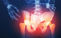
AP and lateral lumbopelvic X-rays in the upright, standing position are taken while weightbearing. An obvious pelvic tilt and leg length discrepancy are noted, with the left femur head 7 mm lower. A moderate lumbar curvature (4°) is noted, convex to the left side, and both the sacral base and the iliac crest are lower on the left. The sacral base angle and measured lumbar lordosis are increased, but within normal limits. No loss of joint spacing or osteophyte formation is seen in the hip joints.
Clinical Impression
Moderate functional leg length discrepancy (left short leg) when standing, with associated pelvic tilt and slight lumbar curvature.
Treatment Plan
Adjustments. Specific, corrective adjustments for the SI joints and the lumbar, thoracic, and cervical regions, as well as soft tissue manipulation, were provided as needed. Manipulation of the left navicular, cuboid, and calcaneal bones was also performed.
Support. Flexible, custom-made stabilizing orthotics were supplied, with a pronation correction added to the left side. He had no difficulty in adapting to the orthotics.
Rehabilitation. He was shown a series of upright strengthening exercises for all hip ranges of motion, in order to speed the process of adaptation to the new alignment. After two weeks, he began a daily brisk walking program, progressing from 20 minutes to 40 minutes.
Response to Care
The spinal, pelvic, and foot adjustments were well tolerated, and the orthotics made a noticeable improvement in his postural alignment, at the feet and in the lumbopelvic region. After four weeks of adjustments (seven visits) and daily home exercises, including walking with orthotic support, he was released to a self-directed maintenance program.
Discussion
Chronic biomechanical stress can cause low-grade nociception that is difficult to track down. Had this patient been more athletically involved, it is likely that his lower extremity asymmetry would have become more obviously symptomatic. With chiropractic care and foot stabilization, he could safely initiate a walking and exercise program for health and weight control.
Dr. John J. Danchik, the seventh inductee to the ACA Sports Hall of Fame, is a clinical professor at Tufts University Medical School and formerly chaired the U.S. Olympic Committee’s Chiropractic Selection Program. Dr. Danchik lectures on current trends in sports chiropractic and rehabilitation.
