Sacral and Coccygeal Fractures

coccygealfracturesCASE HISTORY

This young male patient fell off a horse and landed on his buttocks.

SACRAL FRACTURES

Sacral fractures usually occur as the result of a fall upon the buttocks, direct trauma or in association with pelvic fractures.  Isolated fractures of the sacrum are uncommon and a diligent search for an associated fracture of the pelvic ring or symphysis pubis is often beneficial (as seen in this case).  Two types are horizontal and vertical.

Horizontal (transverse) Fractures.

These are the most common types of sacral fractures.  The most common location is at the level of the third and fourth sacral tubercle, which is near the lower end of the sacroiliac joints.  The fracture line is frequently difficult to identify due to overlying intestinal contents, which may require reexamination or enema.  Careful identification of the cortex outlining each sacral foramen (“foraminal lines”) should be scrutinized for disruption or distortion.    The lateral radiograph occasionally demonstrates the fracture with disruption of the anterior cortex.  Often, the lower segment of the sacrum may be displaced or angled forward.  (1)

A horizontal fracture of the upper sacrum, affecting the first or second sacral segments, may occur from high falls such as attempts at suicide (“suicidal jumpers” fracture). (1)

Vertical Fractures.

These usually occur as a result of indirect trauma to the pelvis with more than 50% suffering pelvic organ damage.  They are visible on the frontal radiograph, but not the lateral view.  The cephalic tilt up view and/or CT may be necessary in order to demonstrate the vertical fracture line, which usually runs nearly the entire length of the sacrum.  The normally symmetrical transverse sacral foraminal lines should be carefully scrutinized for detection of the fracture line.

Coccygeal Fractures

Most fractures of the coccyx are transversely oriented, similar to those of the sacrum.  Seldom are they seen on the frontal radiograph; the lateral film best demonstrates this type of fracture.  The fracture line is usually oblique in presentation, and slight anterior displacement of the distal coccyx is quite common.  Developmental variation in the position of the distal coccygeal segment may provide some concern to the inexperienced observer.


Dr. Terry R. Yochum is a second generation chiropractor and a Cum Laude Graduate of National College of Chiropractic, where he subsequently completed his radiology residency. He is currently Director of the Rocky Mountain Chiropractic Radiological Center in Denver, CO, and Adjunct Professor of Radiology at the Southern California University of Health Sciences, as well as an instructor of skeletal radiology at the University of Colorado School of Medicine, Denver, CO. Dr. Yochum can be reached at 1-303-940-9400 or by e-mail at [email protected].

Dr. Chad Maola is a 1990 Magna Cum Laude Graduate of National College of Chiropractic. Dr. Maola is available for post-graduate seminars. He may be reached at 1-727-433-0153 or by email at [email protected].


REFERENCES

1. Yochum TR, Rowe LJ:  Essentials of Skeletal Radiology, 3rd ed., Williams & Wilkins, Baltimore, Maryland, 2005.

100% of My Patients Get Custom Orthotics What about Yours?


footbones:dropcap_open:W:dropcap_close:e all are victims, in many ways, of the existing healthcare crisis.  The auto industry is addressing their crisis through new laws for improved mileage per gallon and stricter pollution controls.  The energy crisis continues to search for more efficient and alternative means to produce renewable energy.  The healthcare crisis, unlike the above two, is merely shifting responsibilities with regard to who will pay for it.  This “system” will continue until it can no longer sustain itself.

Chiropractic’s Role

As chiropractors, we are interested in the neuro-musculo-skeletal system (NMS).  The current healthcare system has 3 major flaws in addressing NMS disorders: 1) We wait until someone breaks, 2) we only look at the sight of the break, and 3) our goal is only to remove the pain or symptoms.  This approach costs exponentially more, as we all know it is less expensive to “pay now” vs. “pay later,” especially when the proactive approach will produce slower aging, delayed degeneration and a much greater quality of life.  Joint replacement surgery is a growth industry.  It doubled between 2000 and 2006.  Osteoarthritis, the leading arthritis, better known as the wear and tear arthritis, is predicted to have costs and numbers rise by 40% by 2030.

As chiropractors, we know that a joint that has lost mobility is predisposed to a more rapid degeneration.  We know that joints will fixate when under greater stress (abnormal mechanical loading), such as with traumas or biomechanical imbalances.  These fixations prevent people from moving or exercising, which is the breeding ground for obesity, elevated blood pressure, increased anxieties, elevated cholesterol and much more.  We know that we, as a profession, are the most equipped to both detect and improve biomechanical fixations and imbalances.  This approach will make a dramatic impact on the “fixing” of this healthcare crisis.

Altered Biomechanics and Bone Marrow Edema?

According to an article published in the journal Radiology 1996 by Mark E. Schweitzer, M.D., and Lawrence M. White, M.D., from Thomas Jefferson University Hospital in Philadelphia, PA, a very unique study was performed to evaluate the effects of altered biomechanical stress on the human skeleton.  The details of this study are as follows:

» Twelve asymptomatic volunteers (6 women and 6 men) ranging between the ages of 19 and 41 were chosen to be evaluated in this biomechanical study.  All 12 of these asymptomatic volunteers had MR images of their hips, knees, ankles and feet performed at the initial time of commencement of the study.  No evidence of pathology or bone marrow edema was seen affecting any of these 12 volunteers and the MR imaging was done bilaterally.  Three sets of images were obtained before, baseline and 2 weeks after altered weight bearing and then 3 volunteers two weeks after removal of a pad, which had forced the patient into altered biomechanical weight bearing.   The alteration in weight bearing was accomplished by placing an extra large 9/16” (1.4 cm) longitudinal metatarsal arch pad underneath the lateral aspect of one foot to increase pronation. This orthotic was placed in the shoe, but the volunteers did not undergo casting with the foot in this position. Therefore, movement was altered somewhat voluntarily.  The volunteers were instructed not to alter their daily or recreational activities in any way other than that caused by the pronation.   The volunteers were given an adequate number of pads for all pairs of their shoes.  The pads were placed unilaterally to minimize discomfort.1

:dropcap_open:We know that joints will fixate when under greater stress (abnormal mechanical loading), such as with traumas or biomechanical imbalances.:quoteleft_close:

» After 2 weeks of altered weight bearing, MR images of both the lower extremities were obtained in the STIR (short tau inversion recovery) or fluid sensitive or fat suppression images) imaging sequence.   All three sets were done with STIR sequences.1

» The results of these MR images was that 11 of the 12 volunteers demonstrated bone marrow edema on the over pronated side in 10 of the volunteers.  One of the volunteers with medial involvement had the findings only on the non over pronated side.   These changes were seen most frequently in the foot, four metatarsals and calcaneus.  Changes were predominately lateral in 6 volunteers.  The tibia was affected in 3 volunteers, two proximally and one distally and in an additional 3 volunteers, the femur was involved, one affecting the proximal femur and two affecting the distal femur.  Eleven of these 12 volunteers had pain directly over the areas where bone marrow edema was identified.  At MR follow-up, after the pad was removed in two of the three volunteers, the MR images returned completely to normal but, in the third volunteer, MR images demonstrated minimal persistent edema with approximately 50% having been resolved.  All of the volunteers were completely asymptomatic immediately after the pad removal and at clinical follow-up.  (1 week, 1 month, 1 year)1 

Bone is dynamic, undergoing hypertrophy in response to stress.  Alternatively, after immobilization from casting or paralysis or in a gravity free environment, bone atrophy occurs.  What was most interesting about this study is that bone marrow edema and symptoms directly over the area of edema were created with only 2 weeks of altered biomechanical weight bearing with over pronation of one foot.  One wonders if there would be altered biomechanics (subluxation) of the lower extremity and/or the lumbar spine for an extended period of time, what kind of stress this would place on the human skeleton and what long-standing effects it could have on premature degenerative changes within the freely moveable joints of the spine and/or pelvis and lower extremities.   The results of this study clearly shows that increased signal intensity on fluid sensitive images or STIR images (fat suppression images) can occur and may represent a bone contusion or bone bruise.  The results of this study indicates that the increased signal intensity is the result of a bony response to the stress created upon it without actual fracture occurring.   On the basis of Schweitzer’s study, I believe that the altered biomechanics should be added to the list of causes of increased intramedullary signal intensity on T2 and/or STIR weighted images.1,2

It is of interest to note that I (Dr. Yochum) personally interviewed Dr. Mark Schweitzer and asked him if any of these 12 volunteers had lower back pain and/or sacroiliac pain. He told me that those questions were never asked of these volunteers.  From my chiropractic perspective, I would have to believe that a large number of these patients would have had sacroiliac and/or lower lumbar pain.  It would have been interesting to perform pre and post MRI images of the bones adjacent to the sacroiliac joint and/or the lumbar facets to determine whether bone marrow edema could have been identified there as a result of the altered biomechanical stress from the disturbance of the lower kinetic chain.1,2 

Imaging Bone Marrow Edema

Imaging stress to the human skeleton may be done by means of plain films, bone scan, CT or MRI scan.  The most sensitive imaging modality to detect stress to the human skeleton reflected as bone marrow edema is magnetic resonance imaging.  While bone scans can certainly reflect an increase in turnover of bone, they are not as sensitive as the 1% sensitivity of marrow change occurring with MRI scans.  Understanding this concept becomes extremely important to evaluate the highly motivated athlete who may or may not have the presence of a spondylolysis and/or spondylolisthesis on plain film radiographs and may only be seen by means of magnetic resonance imaging scans.   It is possible that a patient may have normal plain film radiographs, but yet have pain on extension and have, in fact, the early fatigue fracture (stress fracture) of spondylolysis and be hidden or “PENDING.” Since the plain film radiographs may not be sensitive enough to detect the “PENDING” spondylolysis or certainly not see edema adjacent to existing pars defects (spondylolysis), specialized physiologic imaging such as magnetic resonance imaging should be given clinical consideration.2

Since on standard MR imaging with standard T1- and T2-weighted images, it is quite possible that bone marrow edema may be missed on the T2-weighted image in a patient who may be “PENDING” without defect or in a patient with an existing pars defect, who may have bone marrow edema adjacent to the pars defect.   With that being the case, it is important that an additional imaging series referred to a short tau inversion recovery (STIR) images, otherwise known as fluid sensitive images or fat suppression images, should be performed routinely in patients where there is a high suspicion of the possibility of a hidden or pending pars defect or bone marrow edema adjacent to an existing pars defect.  The imaging sequence of choice, which should be added to the standard routine MRI scan, is a sagittal STIR imaging sequence, which will unequivocally rule in or out the possibility of bone marrow edema in the region of the pars interarticulares, with or without a defect.2

For further discussion in patient management and evaluation of the problematic cases of spondylolysis and/or spondylolisthesis in the lower lumbar spine and how it relates to the highly motivated athlete, please see chapter 5 of Dr. Yochum’s textbook,  “Essentials of Skeletal Radiology” which is the 3rd edition published in 2005.2

Evidence Based

figure1orthoticsThe industry is pushing for all care to be evidence based.  The irony is that under this heading, less imaging is encouraged.  Less treatment is encouraged.  And, in the end, less correction will have been done.  The chiropractic profession would do well to redefine our identity, and associate itself closely with the detection and correction of biomechanical faults, an identity our forefathers fought hard to protect.  We would then be the only profession whose goal would be to correct these structural imbalances (even in the absence of symptoms), and not just provide symptomatic care.  This identity would enhance the public’s perception of our profession, as the public is begging for someone to help them achieve structural preservation, especially when this approach would improve their long-term quality of life.

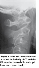
In order to achieve this result, we must look at all people from a biomechanical perspective, as everyone has biomechanical faults and imbalances.  Just as the orthodontist improves the alignment of the teeth long before problems occur, it’s easy to understand the benefit of doing this to the NMS system as well.  As seen in Fig. 1, all people have biomechanical imbalances, and these imbalances always originate in the feet.

If we ignore the imbalances in the feet, we would be ignoring the importance of a balanced foundation.  Introductory Architecture teaches the importance of a balanced foundation.  This is the very reason our office puts every patient into custom orthotics at the beginning of all correction programs.

Biomechanics of Feet

footprintsThere are 3 arches in each foot, with each being critically important for providing foundational balance.  Upon scanning of the feet (Fig. 2), it is easily detectable if any or all of these arches have fallen.  Most patients have multiple fallen arches, when scanned in the standing position.  In addition, aging, gravity and stress over time will encourage further falling of these arches, which will alter centers of gravity in each and every joint of the body.  Abnormal centers of gravity, combined with aging, will further accelerate the degenerative process in joints.

Custom Orthotics

The simple solution to current foot/structural imbalances as well as future structural weaknesses is to put the patient into flexible custom orthotics at the start of their corrective program.  Regardless of whether the patient appears to have pronated, supinated or even normal arches, the digital foot scan will demonstrate that most people will have some degree of fallen arches, as well as imbalances with body weight distribution.  Secondly, although we don’t test for this in our office, a significant percentage of people will over-pronate during the gait cycle, and this over-pronation is blocked with flexible custom orthotics.  Many injuries, especially sports injuries, occur or are aggravated during this over-pronation phase.

After we digitally scan each patient, along with our explanation of Fig. 1 (Crooked Man), along with the explanation of the potential for acceleration of degeneration if left imbalanced, most people will excitedly agree to the inclusion of custom orthotics as soon as possible.  All people have a similar lifetime goal: to have a higher quality of life combined with greater activity.

More patients wearing custom orthotics in your office will convert to improved clinical results, improved patient satisfaction and greater patient compliance.  And, if an office can “manage” patients properly, patients will require new orthotics every two years, keeping more people engaged in active care.  This truly is the beginning of making your practice more successful and fixing the healthcare crisis.


Dr. Terry R. Yochum is a second generation chiropractor and a Cum Laude Graduate of National College of Chiropractic, where he subsequently completed his radiology residency. He is currently Director of the Rocky Mountain Chiropractic Radiological Center in Denver, CO, and Adjunct Professor of Radiology at the Southern California University of Health Sciences, as well as an instructor of skeletal radiology at the University of Colorado School of Medicine, Denver, CO. Dr. Yochum can be reached at 1-303-940-9400 or by e-mail at [email protected].

Dr. Maggs currently practices full time, while also lecturing for Foot Levelers.  He is the developer of The Structural Management® Program, as well as the 10 Week Webinar Series, “How to Build Your High School Athlete Practice.”  He can be reached at 1-518-393-6566 or [email protected]. His website is www.StructuralManagement.com.


References:

1. Schweitzer, Mark E; White, Lawrence M., Radiology; 198:851-853, 1996.

2. Yochum, TR, Rowe LJ:  Essentials of Skeletal Radiology, ed. 3, 2005, Chapter 5.
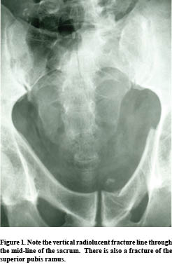
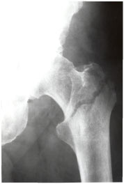
OS ODONTOIDEUM

odontoidfig1
CASE HISTORY

This young adult patient presents with neck pain following an MVA.  You note an abnormality in the upper cervical spine at C1 & C2.  Is this a fracture?


DISCUSSION

Anomalies of the odontoid process are considered uncommon (1) and are usually discovered by the principle “traumatic determinism.”  This means that the underlying condition predated the injury and is not caused by this current trauma.  These anomalies may be associated with Down’s syndrome, Klippel-Feil syndrome, Morquio’s syndrome, and spondyloepiphyseal dysplasia.


odontoidfig2Clinical Features:

Any symptoms the patient may manifest are usually the result of atlantoaxial instability with resultant cord compression; however, if there is compression of the vertebral artery resulting from stretching of the artery during C1 subluxation on C2, then the symptoms may be considerably greater.  Increased deep-tendon reflexes, proprioceptive loss, or sphincter incompetence may be encountered.  Additionally, compression of the vertebral arteries may result in local thrombosis and vascular occlusion.  The thrombus may also serve as a source for emboli to the brain.

Clearly, the combination of os odontoideum with high-velocity injury can produce central cord syndrome or even fatal injury.


Radiologic Features:

The X-ray diagnosis of os odontoideum in a child below the age of 5 years can be made, if there is demonstration of hypermobility of the odontoid process on the body of C2 during flexion and/or extension.  In the adult, an X-ray diagnosis is certain, if a smooth, wide, lucent defect is seen to separate the odontoid process from the C2 body at the level of the superior articular processes, and there is an associated stress hypertrophy (enlargement) of the anterior tubercle of the atlas.  This finding will not be present in the child, as the biomechanical stresses on the anterior arch of the atlas will not have been present for a long enough period to allow the hypertrophy to develop.  Os odontoideum must be differentiated from an acute fracture of the odontoid process.  A helpful radiographic sign that may be present and that confirms a developmental defect of the odontoid process is a “molding: of the anterior arch of C1 into the ventral aspect of the odontoid process.

Magnetic resonance imaging is useful in evaluating the spinal cord for angulation, compression, and intramedullary injury (contusion).


Dr. Terry R. Yochum is a second generation chiropractor and a Cum Laude Graduate of National College of Chiropractic, where he subsequently completed his radiology residency. He is currently Director of the Rocky Mountain Chiropractic Radiological Center in Denver, CO, and Adjunct Professor of Radiology at the Southern California University of Health Sciences, as well as an instructor of skeletal radiology at the University of Colorado School of Medicine, Denver, CO. Dr. Yochum can be reached at 1-303-940-9400 or by e-mail at [email protected].

Dr. Chad Maola is a 1990 Magna Cum Laude Graduate of National College of Chiropractic. Dr. Maola is available for post-graduate seminars. He may be reached at 1-727-433-0153 or by email at [email protected].


Reference

1.Yochum, T. R., Rowe, L.J.:  Essentials of Skeletal Radiology, 3rd ed., Williams & Wilkins, Baltimore, Maryland, 2005.

Digital X-Ray

:dropcap_open:T:dropcap_close:he technology used in conventional X-ray film is 100 years old. There are not many technologies that do not evolve and improve in 100 years, and the X-ray field is no different, although many doctors have not yet made the technology jump in this area. In the last 10 years alone, we have seen the development of affordable technology in the field.

The world of digital technology has ushered in incredible savings in cost and time across the board in our daily lives. We have seen vivid changes in the way that we take our family snap shots and the trickledown effect has been the acceptance of digital technology—in all aspects of life. The societal push for less waste and more efficiency should make most doctors at least explore the options afforded to their practices by going digital when it comes to X-rays.

digitalxray
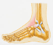
What are the dollars and cents of going with digital X-ray opposed to conventional film? There is a front-loaded cost to digital radiography but, as an investment, digital radiography not only offers cost savings (no film, no chemicals, no processors), it can actually become a profit center. Going digital, X-ray moves from a cost per film, to a fixed cost—meaning that, dependent upon the number of X-rays that you take, it will make the practice a profit. As the doctor sees more patients, the cost of taking X-rays, per patient, actually decreases.

When upgrading from film to a digital system, the DC should consider that there may need to be additional equipment that they must upgrade as well, depending on which system they choose. In most instances this is the case but, with some systems, you can use the existing generators and bucky stand, so the doctor only has to purchase the digital plate, making the transition more affordable but easier to install. The doctor needs to determine which system requires new equipment and which ones can use existing modules, as this element does factor into over all cost and complexity—or lack thereof—of the switch to digital.

Radiation Level Comparison

I often get asked about the comparison of radiation levels between the conventional X-ray to digital. The newest text books in radiology now indicate that you can increase the kVp to higher levels with digital imaging because the software now controls contrast. So, you can limit the patient dosage by increasing the penetration of the beam—thereby reducing the absorbed dose. An increase in kVp does have to be within reason but, by increasing kVp, you can decrease mAs. Of course, with heavier patients you must increase the kVp in order to penetrate the excess mass and fluids.

There are also other safety benefits by going digital. There is no doubt that—as the office no longer uses processing chemicals—the air becomes much more clean and fresh; no one is exposed to toxic chemicals in film development or in having to dispose of the chemicals.

:quoteright_open:There is no doubt that—as the office no longer uses processing chemicals—the air becomes much more clean and fresh:quoteright_close:

What about maintenance?

Every company and system has different requirements for continuing maintenance. Most companies charge a monthly fee for technical support and also charge for all computer software upgrades. But not every company charges for technical support or computer software upgrades. It would be prudent to ask each vendor about their policies in these areas, as there could be hidden costs a doctor doesn’t realize unless he looks into maintenance and upgrade charges with each system.

Required Office Space and Training

Once a practice goes true digital X-ray, it eliminates the need for a processor, dark room and all of the required space to store film. CR still requires the use of a processor.

As far as training required to go digital, a doctor does need to traverse and understand the difference between digital images compared to film. The doctor needs to consistently evaluate diagnostic images and will require some training to do so. Some vendors have radiographers on staff who work with their clients to develop a technique chart that enables the doctor to obtain the desired images, leading to accurate diagnoses.

Image Quality Comparisons

Digital allows for further manipulation of the X-ray. In your general terms, it is the ability to window and level, or in layman’s terms, the ability to manipulate contrast and density, that enables the doctor to see different qualities in the image. Using digital X-ray allows the DC to get a consistent, quality image, because of the software manipulation ability, as compared to using film.

:dropcap_open:Using digital X-ray allows the DC to get a consistent, quality image, because of the software manipulation ability, as compared to using film.:quoteleft_close:

HIPPA Compliant Issues

As with many things these days in a chiropractor’s office, you must consider HIPPA compliancy when making decisions on new technologies. With digital systems, the software allows password protection of the data via internal processes and procedures set for onsite security, thereby complying with HIPAA. (For doctors that want off-site HIPAA compliant storage, we have partnered with Central Data Storage, which stores and manages electronic patient imagining and documents.)

Summary

The standard for images must remain constant throughout the health care field. In order for chiropractors’ roles to become more vital today, they must take advantage of improved—and evolved—technology.  That isn’t to say all facets of chiropractic care have to evolve, as many key elements of spinal adjustments may remain constant. But using conventional film X-ray is a technology that is 100 years old. As is the case with your family photos—now taking advantage of digital technology—it makes sense for the doctor of chiropractic to afford some of those same, related advantages by no longer using film in the process.

Fracture of the Wrist Affecting the Young and Old

yochumCASE HISTORY:
This 8-year-old fell and hyperextended his wrist.

Diagnosis: Torus fracture–distal radius.

FRACTURES OF THE WRIST

Distal Radius Fractures.
The distal radius is one of the most common sites of fracture in the wrist. Care must be taken to search all projections for these fractures, many of which are often subtle and obscure. In the absence of an obvious fracture, close observation on the lateral projection for displacement of the pronator quadratus fat line is a useful indicator for the presence or absence of fracture. Normally, this fat line appears as a well-defined linear lucency, oriented parallel with the plane of the radius, 2 to 5 mm from its anterior surface. In almost all cases of distal radius fractures, the pronator quadratus fat line will be altered.1 These alterations include anterior displacement, blurring, irregularity and obliteration of the line. The most common fracture of the wrist in the elderly is Colles’ fracture and in a child is a torus fracture of the distal radius.

Colles’ Fractures.
In 1814, Abraham Colles wrote the definitive description of this fracture which, consequently, bears his name. The injury is defined as a fracture of the distal radius, approximately 20 to 35 mm proximal to the articular surface, with posterior angulation of the distal fragment. More than 60% will have an accompanying fracture of the ulnar styloid process. The usual mechanism is a fall on an outstretched, extended hand. The physical appearance of the fractured distal forearm and wrist has led to its being called the dinner fork deformity. The incidence of the fracture increases with age, and this increase is so rapid in women that, by age 65, the lesion is six times more common in women than in men. Osteoporosis appears to be the underlying influencing factor. Complications are common and may be severe.

Torus Fracture.
This is the most common fracture of the wrist between 6 and 10 years of age. Typically, the fracture is located 2 to 4 cm from the distal growth plate. A torus fracture can occur in any long bone and is the term applied to a buckled cortex following trauma. As such, the key radiologic sign is a localized cortical bulge, bump or offset.1

Dr. Terry R. Yochum is a second generation chiropractor and a Cum Laude Graduate of National College of Chiropractic, where he subsequently completed his radiology residency. He is currently Director of the Rocky Mountain Chiropractic Radiological Center in Denver, CO, and Adjunct Professor of Radiology at the Southern California University of Health Sciences, as well as an instructor of skeletal radiology at the University of Colorado School of Medicine, Denver, CO. Dr. Yochum can be reached at 1-303-940-9400 or by e-mail at [email protected].

Dr. Chad Maola is a 1990 Magna Cum Laude Graduate of National College of Chiropractic. Dr. Maola is available for post-graduate seminars. He may be reached at 1-727-433-0153 or by email at [email protected].

References
1.Yochum TR, Rowe LJ, Essentials of Skeletal Radiology, 3rd ed. Lippincott, Williams and Wilkins, Baltimore, 2005.

Chiropractic Clinician and the Radiologist: A Partnership with a Caveat

radiologist:dropcap_open:I:dropcap_close:t is critical for every chiropractor to have a support team of health care specialists in order to render the highest level of care for our patients. The radiologist is the first specialist that you should add to your team. In the triaging of our patients, the progression is to create an accurate diagnosis, prognosis and treatment plan and then to treat. Imaging, both basic X-rays and advanced imaging such as MRI’s, CAT scans and bone scans are often a critical step in getting to “first base,” with the diagnosis. Without any diagnosis, we cannot treat our patients and, without an accurate diagnosis, we can go from spinal experts rendering quality care to well meaning practitioners who hurt their patients.

It was reported by Lurie, Doman, Spratt, Tosteson, and Weinstein (2009) that 42.2% of the cases reported by radiologists were not clear in their description of the morphology of the lesion seen in MRI. While this does not conclusively represent that the radiologist made an error (although, in many cases, gross errors are seen nationally), it does create a huge issue for the clinician who relies on the radiologist to paint a “verbal picture” of the tissue and lesion structure, in order to create an accurate diagnosis, prognosis and treatment plan.

The research study was done with general radiologists and not neuroradiologists. As a note, neuroradiology is not a level of licensed practitioner, but a level of academic degree. A neuroradiologist goes into a fellow program and studies only brain and spine for an additional 18-24 months. A general radiologist does rotations in brain and spine in residency, called a “neuro” rotation, and the spine rotation is usually five weeks. The rest of the education for a radiologist is general radiology, studying joints and other organs. Many MRI companies hire general radiologists and not neuroradiologists for economic reasons, and some of these general radiologists have not seen spine MRI for five, ten or twenty years since their residency. It is always suggested that, if you have a choice, request that a neuroradiologist review your MRI’s.

:dropcap_open:It was reported by Lurie, Doman, Spratt, Tosteson, and Weinstein (2009) that 42.2% of the cases reported by radiologists were not clear in their description of the morphology of the lesion seen in MRI.:quoteleft_close:

According to Magdy Shady, M.D., a neurosurgeon and fellow in neuro-trauma, he disagrees with the general radiologist over 80% of the time in the description of the morphology of the lesion, and will not make a surgical decision unless he has personally reviewed the films. This begs the question for the chiropractic profession, “At what level should we accept the radiologist’s interpretation of the film, and at what level of reading expertise should the individual chiropractic practitioners involve themselves in the process of interpreting the images rendering an accurate diagnosis?”

From a clinical perspective, some of the more common errors seen by radiologists are referencing the wrong side and calling cauda equina compressions “cord compressions.” Just recently, a radiologist in Washington state reported an enlarged artery on the intervertebral foramen erroneously. Each of these miscues alters the diagnosis, prognosis and treatment plan and creates havoc for both the patient and the practitioner.

In each of the above scenarios, the errors were realized by a chiropractor who was trained in MRI spine interpretation. Although chiropractors can return to school and get a diplomate in radiology (D.A.C.B.R.) which is highly recommended for those who want to focus primarily on interpreting film, the vast majority of us want to stay in our offices. For those practitioners, it is highly suggested that, as a profession, every D.C. take post-doctoral training in MRI spine interpretation, so that we all understand and can interpret the films to create an agreement with the radiologists’ reports.

Call it an ability to do an over read on the radiologist and it must be done. The 2009 Spine article mandates that we, as a profession, take responsibility for an accurate diagnosis of our patients. With a 42.2% unclear (error rate) reporting of the morphology of the lesion, circumstances require that we, at least, know the basics to say, “Something isn’t right and we need another opinion.” When there is a discrepancy in the interpretation, the team approach comes into play and we confer with the radiologist and ask what they see vs. what you see. If there is not an agreement, then a third party, hopefully a neuroradiologist, will be the final arbiter, so that an accurate diagnosis can be rendered. In some instances, another MRI sequence might be necessary. Either way, from a posture of clinical excellence, unless an accurate diagnosis is rendered, the clinician cannot create a treatment plan and render care. How can you treat what you do not know?


Reference
1.  Lurie, J. D., Doman, D. M., Spratt, K. F., Tosteson, A. N., & Weinstein, J. N. (2009). Magnetic resonance imaging interpretation in patients with symptomatic lumbar spine disc herniations: Comparison of clinician and radiologist readings. Spine, 34(7), 701–705

Intertrochanteric Fracture Of The Proximal Femur

Intertrochanteric Fracture Of The Proximal Femur

by Dr. Terry R. Yochum, D.C.; D.A.C.B.R.; Fellow, A.C.C.R. and Dr. Chad J. Maola, D.C.


CASE HISTORY

This elderly female patient slipped getting out of the bath tub. She heard a crack and felt immediate pain.

DIAGNOSIS

Fractures around the proximal femur are relatively uncommon in young to middle-aged patients with a sharp increase in the geriatric patient.1 Severe forces are necessary to fracture the proximal femur in the young and middle years, while only moderate to minimal trauma may induce a fracture in the osteoporotic bone of the elderly. Certain predisposing factors may allow fractures to occur, such as the presence of Paget’s disease, fibrous dysplasia, benign or malignant bone tumors, osteoporosis, osteomalacia and radiation-induced osteonecrosis.1

The overall incidence of all types of fractures of the proximal femur shows a two to one female-to-male ratio. A five to one female predominance exists with intracapsular fractures. The average age is approximately seventy years. It has been estimated that 10 percent of white females and 5 percent of white males will sustain fracture of the proximal femur by the age of eighty years. The incidence by the age of ninety years increases to 20 percent for women and 10 percent for men. Many elderly patients with fractures of the proximal femur die within six months of the original injury. This occurs secondary to pulmonary or cardiac complications. Therefore, fracture of the proximal femur and their attendant sinister1 complications are of such proportions that they represent a major health hazard to the elderly and constitute a significant public health issue because of their frequency, morbidity, and cost.


The standard radiographic examination of the hip joint includes an anteroposterior (AP) full pelvis, AP hip spot (involved side) and an oblique or frog-leg projection.1


Types of Hip Fractures

The types of hip fractures are divided into intracapsular and extracapsular, as determined by the relationship of the fracture line to the joint capsule. In general, intracapsular fractures have a high incidence of nonunion and avascular necrosis due to probable disruption of the tenuous blood supply. 1

Intracapsular Fracture. Any fracture involving the femoral head or neck proximal to the trochanters is classified as being intracapsular. These are then named according to the fracture location:

a) subcapital (involving the junction of the femoral head and neck;

b) midcervical (through the midportion of the fermoral neck);

c) basicervical (traversing ) the base of the femoral neck and its junction with the trochanters.

Most femoral neck fractures are subcapital; midcervical and basicervical fractures are uncommon.

Extracapsular Fracture. This type of fracture occurs outside of the joint capsule and includes intertrochanteric, subtrochanteric and avulsion fractures of the greater or lesser trochanters. Avascular necrosis and nonunion are uncommon complications in extracapsular fractures.

The intertrochanteric fractures are usually comminuted, with the greater or lesser trochanter, or both, forming separate fragments. The oblique fracture line usually splits the trochanters, separating the femur into two components. The proximal component consists of the head and neck, and the distal component includes the shaft and the remainder of the trochanter.

The subtrochanteric fracture is found in the area two inches below the lesser trochanter. This is an uncommon type of fracture of the proximal femur. Middiaphyseal fractures follow severe trauma and are prone to malalignment unless treated appropriately. Pathologic fractures of the proximal femur often occur in the subtrochanteric region. Paget’s disease and metastatic lesions in the proximal femur may be predisposing factors to the development of a subrochanteric fracture; thus the presence of a subtrochanteric fracture should be a signal to the observer to look closely for roentgen signs of adjacent bone disease.

Dr. Terry R. Yochum is a second generation chiropractor and a Cum Laude Graduate of National College of Chiropractic, where he subsequently completed his radiology residency. He is currently Director of the Rocky Mountain Chiropractic Radiological Center in Denver, Colorado, and Adjunct Professor of Radiology at the Southern California University of Health Sciences, as well as an instructor of skeletal radiology at the University of Colorado School of Medicine, Denver, CO. Dr. Yochum can be reached at 1-303-940-9400 or by e-mail at [email protected].

Dr. Chad J. Maola is a 1990 Magna Cum Laude Graduate of National College of Chiropractic. Dr. Maola is a Chiropractic Orthopedist and is available for post-graduate seminars. He may be reached at 1-303-690-8503 or e-mail [email protected].


References

1. Yochum TR, Rowe,LJ: Essentials of Skeletal Radiology, 3rd ed., Williams & Wilkins, Baltimore, Maryland., 2005

Osteolytic Metastatic Carcinoma

Osteolytic Metastatic Carcinoma

by Dr. Terry R. Yochum, D.C.; D.A.C.B.R.; Fellow, A.C.C.R. and Dr. Chad J. Maola, D.C.


Osteolytic Metastatic Carci


CASE HISTORY

 This adult male patient complains of back pain, especially at night, and it awakens him from sleeping.

Diagnosis: Osteolytic metastatic carcinoma of the left L-3 pedicle and vertebral body. This is the “one-eyed pedicle” sign or the “winking owl sign” of lytic metastatic disease.
(Figure 1)


Table 1.  Radiologic Features
of Spinal Metastasis

LOCATION:v   Lumbar/thoracic spine v   Vertebral body, pedicles 

SIGNS: Altered bone densityv   Decreased: moth-eaten, permeative diffusev   Increased: localized, ivory vertebra   Cortical destruction   Disc space unaffected     Pathologic collapse      v   Decreased posterior vertebral body heightv   Endplate disruption (malignant Schmorl’s node)   Pedicle destructionv   One-eyed pedicle sign (winking owl sign)

v   Blind vertebra (both pedicles destroyed


DISCUSSION: Pedicle. The pedicle is an important radiologically detectable site for osteolytic metastatic carcinoma. Any component of the neural arch can be involved, although the pedicle is by far the most common location. Destruction of the posterior vertebral body with contiguous involvement of the pedicle attachment results in loss of the cortical outline of the pedicle.1 This has been referred to as the one-eyed pedicle sign or the winking owl sign and is most commonly found in the lower thoracic and lumbar spine. It is most easily visualized on the AP radiograph. Most cases of pedicle destruction involve a single vertebra; however, multiple levels can be affected. Occasionally, bilateral pedicular destruction may occur and is referred to as the blind vertebra. (Table 1)

The most common cause for a missing pedicle is osteolytic metastatic carcinoma; however, agenesis of a pedicle may also occur. The key to radiologic differentiation is to search for a stress-related reactive sclerosis and enlargement of the contralateral pedicle. If this sign is present, it represents a firm assurance that osteolytic metastatic carcinoma is not present. Those cases of agenesis of the pedicle that create no stress hypertrophy of the opposite pedicle must be considered metastatic tumors until proven otherwise. Previous radiographs in this circumstance will be very helpful. Destruction of a pedicle in a patient under the age of thirty years is most commonly due to aneurysmal bone cyst (ABC), osteoblastoma, neurofibroma, or other cord tumors.

Dr. Terry R. Yochum is a second generation chiropractor and a Cum Laude Graduate of National College of Chiropractic, where he subsequently completed his radiology residency. He is currently Director of the Rocky Mountain Chiropractic Radiological Center in Denver, Colorado, and Adjunct Professor of Radiology at the Southern California University of Health Sciences, as well as an instructor of skeletal radiology at the University of Colorado School of Medicine, Denver, CO. Dr. Yochum can be reached at 1-303-940-9400 or by e-mail at [email protected].

Dr. Chad J. Maola is a 1990 Magna Cum Laude Graduate of National College of Chiropractic. Dr. Maola is a Chiropractic Orthopedist and is available for post-graduate seminars. He may be reached at 1-303-690-8503 or e-mail [email protected]


Reference

1. Yochum TR, Rowe LJ: The Essentials of Skeletal Radiology, 3rd ed.,

Baltimore, Williams & Wilkins, 2005.

Tourette Syndrome and Chiropractic Care

Tourette Syndrome and Chiropractic Care

by Lisa Brittain, D.C., B.S.


History and Presenting Symptoms The patient is an 11-year-old male who was diagnosed at the age of four with Tourette syndrome. He has complex vocal and motor tics, which are made worse by stress. The tics affect his daily life; school, sports and his self esteem. The patient also notices he has fatigue and irritability. The patient has been on many different medications—clonidine, inversine, depakote, orap, clarinex, and singular—several causing side effects but never alleviating his tics. Exam Findings The patient is a very active young man; he plays golf, basketball, football, and baseball, as well as excelling at school. He has a good diet. Postural examination revealed anterior head carriage. Motion Palpation found a decrease in range of motion of the cervical spine along with tender spots at C1 and C2. An increase in muscle spasm throughout the right side of the thoracic spine, due to a side bend tic was noted. Surface EMG along with a Rolling Thermal Scan were performed. The sEMG revealed large amounts of nervous system disturbances at C1, C3, C5, C7, T1, T2, T4, T6, T8, T10, T12, L1 and L3. The Rolling Thermal Scan only showed one area of disturbance at T1. Imaging AP and Lateral cervical spine X-rays were performed and revealed a slight decrease in the cervical lordotic curve. Severe rotation and lateral fl exion of C2, along with lateral fl exion of the atlas, were also noted. Clinical Impression Cervical and thoracic segmental joint dysfunctions with associated myospasm in thoracic spine were present. Treatment Plan Diversifi ed adjustments were used to correct the subluxations. Bilateral pisiform adjustments were used to address the thoracic spine; side posture with specifi c contact was used to correct the lumbar spine. For the cervical spine, master cervical was used and a seated occiput adjustment was used to correct the occipital subluxation. The patient was adjusted three times a week for four weeks then re-evaluated. Response to Care The patient tolerated the adjustments very well. During the course of treatment, the patient stopped all of his medications. At the beginning of care, the patient rated his tics, both motor and vocal, an eight out of ten in severity; after twelve treatments, the patient rated his vocal tics a two out of ten and a three out of ten for motor tics. Also the patient noticed he had less fatigue and less irritability, since starting his chiropractic treatments Discussion The chiropractic adjustment relieved pressure that was being put on the nerves in the cervical spine which has direct relation to the brain and nervous system. In this case, the chiropractic treatments were the only thing that relieved the patient from his symptoms, allowing him to be medication free and back to his normal self.


Dr. Lisa Brittain graduated from Cleveland Chiropractic College in August 2006. That is where she met her husband, Jacob, who is also a Chiropractor. They opened their own practice in Overland Park. Dr. Brittain can be contacted at offi ce # 913-825-3900, cell # 913-269-1625. Offi ce address 10841 W. 87th Suite 200, Overland Park, KS 66214. Home 950 E. 126th Terr Olathe, KS 66061

Rasterstereography: Radiation – Free Technology for the Analysis of the Spine and Pelvis

Rasterstereography: Radiation – Free Technology for the Analysis of the Spine and Pelvis

by Jean Pierre Gibeault, P: eng.


RECENTLY IN THE LITERATURE: In a recent study, researchers demonstrated an excellent correlation between X-ray measurements and measurements made with rasterstereography (Schulte 2008). Schulte, et al., produced rasterstereographs and compared them to digitized A-P radiographs of the same 16 patients with idiopathic scoliosis. In this longitudinal study design, with a mean follow-up period of 8 years (range 3 to 10 yrs.), lateral vertebral deviation and vertebral rotation were measured between C7 and L4. This study design mimics actual clinical environments and, therefore, provides invaluable data to the clinician. During the 10 year time frame, Cobb angle measurements increased on average by 13 degrees. The study showed an excellent correlation between rasterstereographic and radiographic progression. The mean difference between rasterstereographs and radiographs was 3.21 mm for lateral vertebral deviation and 2.45 degrees for vertebral rotation. In conclusion, using the parameters of lateral vertebral deviation and vertebral rotation, rasterstereography accurately refl ects the radiograph measured progression of idiopathic scoliosis during the long-term follow-up. The authors do comment that the parameters used are not directly comparable with the Cobb angle. However, they recommend a rasterstereographic examination every 3 to 6 months and a radiographic examination every 12 to 18 months, provided that rasterstereography does not show rapid deterioration of the scoliosis. If this result is reproducible, patients and practitioners alike will want to change the way conditions, such as scoliosis and other global postural distortions are managed. Further studies confi rming the high accuracy of this technology can be found on both normal populations (Hackenberg 2003), as well as those with rotational scoliosis (Drerup 1997). With respect to these studies, it is reasonable to say, rasterstereographs can signifi – cantly reduce the amount of X-rays needed, without sacrifi cing clinical data. In fact, the sophistication of rasterstereography today affords the practitioner a cornucopia of postural information, well beyond what is possible with plain fi lm projections alone. Of course, X-rays do provide unique information about bone and soft-tissue that cannot be gleaned from any other source and, because of this, I believe that both will be a necessary part of chiropractic practice in the future. The Diers Formetric produces the rasterstereographs using an optical light scanner and computer software which automatically fi nds anatomical landmarks without markers or user placement. Thousands of images are taken over an adjustable time frame, which provides information for the quantifi cation of sway patterns, breathing tendencies, weight distribution, and a 3-D computer generated representation of the spine and pelvis. Although not as breakthrough, rasterstereography equipment available in the United States also offers range of motion analysis, cervical spine imaging, and a dynamic forceplate which can invert, dorsifl ex, plantarfl ex and elevate one foot at a time. This is particularly useful when assessing the effects of an orthotic or shoe lift on pelvic and spinal alignment. Rasterstereography is not new; Stokes and Moreland utilized the same technology twenty years ago (Stokes, et al., 1987) when they evaluated for changes in rib hump in seated, standing and forward bending postures.