Statins and All-Cause Mortality in High-Risk Primary Prevention: A Meta-analysis of 11 Randomized Controlled Trials Involving 65,229 Participants

Archives of Internal Medicine

June 29, 2010, Vol. 170, No. 12, pp. 1024-1031


Kausik K. Ray, MD; Sreenivasa Rao Kondapally Seshasai, MD; Sebhat Erqou, MD, PhD; Peter Sever, PhD; J. Wouter Jukema, MD, PhD; Ian Ford, PhD; Naveed Sattar:

The authors are from University of Cambridge


BACKGROUND FROM DAN MURPHY:

LDL-C means low density lipoprotein cholesterol. This is the “bad” cholesterol because it can plaque on the arterial wall. Ideally, it should measure less then 100 mg/dL. High is over 130 mg/dL.

In 2007, the New England Journal of Medicine published the JUPITER study. This study claimed that individuals with low cholesterol but high levels of inflammation [high sensitivity C-Reactive protein {hs-CRP}] could “significantly reduce all-cause mortality by 20%” by taking statin drugs. However, other studies have “questioned these findings as a chance or exaggerated observation.”

[Ridker PM, Danielson E, Fonseca FA, et al; JUPITER Study Group. Rosuvastatin to prevent vascular events in men and women with elevated C-reactive protein. N Engl J Med. 2008;359(21):2195-2207].

Therefore, these authors analyzed 11 randomized controlled trials involving a total of 65,229 participants to provide the most robust information to assess whether statins reduce all-cause mortality. All of the participants had high cholesterol, but none of them had cardiovascular disease. The study represented about 244,000 person-years of follow-up.

Statin drugs are commonly prescribed for two groups of patients:


Group I

Group II

Those that have established cardiovascular disease

Those that do not have cardiovascular disease, but do have high cholesterol levels

It is thought that statins for this group will prevent atherosclerosis and subsequent heart attack

It is thought that statins for this group will prevent cardiovascular disease and heart attack


KEY POINTS FROM DAN MURPHY

1) Some researchers have questioned the benefits of statins among individuals without cardiovascular disease, noting there is little evidence for reductions of all-cause mortality, and the potential to cause “serious unrecognized harm.”

2) These authors assessed the effect of statin therapy (compared with placebo) on all-cause mortality in individuals who did not have cardiovascular disease. They used 65,229 subjects and a follow-up period of 3.7 years:


Placebo Group

Statin Group

# of participants: 32,606

# of participants: 32,623

# of deaths: 1,447

# of deaths: 1,346

LDL Cholesterol level: 134 mg/dL

LDL Cholesterol level with statins:

94 mg/dL

Death rate: 4.44%

Death rate: 4.13%


3) Taking statin drugs lowered LDL-C by 40 mg/dL compared to the placebo group. This lowered the death rate by .31% (4.44% – 4.13%). The authors considered such a small reduction in death to be nonsignificant.

4) “This literature-based meta-analysis of 11 clinical trials involving 65,229 participants with approximately 244,000 person-years of follow-up and 2,793 deaths provides more reliable evidence than previously available on the impact of statin therapy on all-cause mortality among high-risk individuals without prior CVD. These data indicate that, over an average treatment period of 3.7 years, the use of statin therapy did not result in reduction in all-cause mortality.”

5) “There were, on average, an estimated 7 fewer deaths for every 10,000 person-years of treatment” with statin drugs.

6) “Our meta-analysis was based on data from only those individuals without clinically manifest CVD, including previously unpublished data, thus providing the most reliable effect estimates about the effect of statins in this population.”

7) “The present data suggest that the all-cause mortality reduction of 20% reported in JUPITER is likely to be an extreme and exaggerated finding as often occurs when trials are stopped early, hence, indicating that more liberal use of potent statin regimens, particularly in the setting of lower risk primary prevention subjects, is unlikely, at least in the short term, to have a major impact on all-cause mortality reduction.”

8) Fibrates are drugs that primarily lower triglyceride levels. A 2007 meta-analysis of randomized controlled trials published in the American Heart Journal “showed that, despite a significant reduction in nonfatal myocardial infarction, all-cause mortality was approximately 7% higher among individuals randomized to a fibrate.”

9) “In conclusion, based on aggregate data on 65,229 men and women from 11 studies, yielding approximately 244,000 person-years of follow-up and 2,793 deaths, we observed that statin therapy for an average period of 3.7 years had no benefit on all-cause mortality in a high-risk primary prevention population.”

10) “There is no evidence that prescribing cholesterol-lowering drugs known as statins to patients at risk of heart disease reduces their chances of premature death in the short term.”

11) “There is little evidence that statins reduce the risk of dying from any cause in individuals without heart disease.”

12) People taking statin drugs may have higher risks of liver dysfunction, kidney failure, muscle weakness and cataracts.

13) “While low-density lipoprotein (LDL), or ‘bad’ cholesterol levels, were higher among those taking placebo than those taking statins (134 milligrams per deciliter versus 94 milligrams per deciliter), this had no effect on the risk of premature death.”


COMMENTS FROM DAN MURPHY

The number needed to treat (NNT) is an epidemiological measure used to assess the effectiveness of a health-care intervention. The NNT is the number of patients who need to be treated in order to prevent one additional bad outcome. The ideal NNT is 1, where everyone improves with treatment and no one improves with placebo or in the control group. The higher the NNT, the less effective is the treatment.

NNT values are time-specific. A study’s NNT would be multiplied by the number of years of the study. For example, if a study ran for 3.7 years and it was found that the NNT was 321 during this 3.7-year period, in one year the NNT would have to be multiplied by 3.7 to correctly assume the right NNT for only the one-year period (in the example, the one year NNT would be 1,188).

Even though NNT is an important measure in a clinical trial, it is infrequently included in medical journal articles reporting the results of clinical trials.

In this study, the Number Needed to Treat (NNT) was 321 over a period of 3.7 years. The one-year NNT was 1,188:

1) This means for a period of 3.7 years for every 321 people taking a statin drug, only one is benefited, and 320 are not benefited, although they are spending about $1000/year on the drugs, and often experiencing numerous side effects.

2) This means, for a period of 1 year for every 1,188 people taking a statin drug, only one is benefited, and 1,187 are not benefited.

One can calculate the NNT using a calculator, or there are web pages that will do it for you by plugging in the numbers, such as www.graphpad.com, or just Googling “Number Needed to Treat” or “NNT.”

In the end, the consumer is paying for all of this, either through taxes (the government pays), or health insurance premiums, or cash.

Omega-3 Fatty Acids in Pregnant Women and Infant

by Daniel J. Murphy, DC and Michael L. Underhill, DC


There is mounting evidence implicating omega-3 deficiency as a determinant of various maternal and pediatric afflictions.  As a consequence, physicians should consider recommending a purified fish oil supplement during pregnancy and lactation.


Continue reading “Omega-3 Fatty Acids in Pregnant Women and Infant”

The Omega-6/Omega-3 Fatty Acid Ratio

The Omega-6/Omega-3 Fatty Acid Ratio

by Dr. Daniel J. Murphy D.C., D.A.B.C.O.


KEY POINTS FROM DR. DAN MURPHY

1. “Human beings evolved on a diet with a ratio of omega-6 to omega-3 essential fatty acids of approximately 1/1, whereas in Western diets the ratio is 15/1–20/1.”

2 “Western diets are deficient in omega-3 fatty acids, and have excessive amounts of omega-6 fatty acids compared with the diet on which human beings evolved and their genetic patterns were established.”

3. “Excessive amounts of omega-6 polyunsaturated fatty acids (PUFA) and a very high omega-6/omega-3 ratio, as is found in today’s Western diets, promote the pathogenesis of many diseases, including cardiovascular disease, cancer, and inflammatory and autoimmune diseases, whereas increased levels of omega-3 PUFA (a lower omega-6/omega-3 ratio) exert suppressive effects.”


4. The ratio of omega-6/omega-3 should be below 4/1. A ratio of 2.5/1 reduced rectal cell proliferation in colorectal cancer patients, whereas a ratio of 4/1 with the same amount of omega-3 PUFA had no effect. [Important]

5. A lower ratio of omega-6/omega-3 fatty acids is more desirable in reducing the risk of many of the chronic diseases of high prevalence in Western societies.

6. “Genetic factors determine susceptibility to disease and environmental factors determine which genetically susceptible individuals will be affected.” “Nutrition is an environmental factor of major importance.”

7. The spontaneous mutation rate for nuclear DNA is estimated at 0.5% per million years, which means our genes today are very similar to the genes of our ancestors during the Paleolithic period 40,000 years ago.


8. Humans today live in a nutritional environment that differs from that for which our genetic constitution was selected, particularly in the type and amount of essential fatty acids and in the antioxidant content of foods. Today’s industrialized societies diets are characterized by

• An increase in calories

• An increase in cereal grains

• An increase in saturated fat

• An increase in trans fatty acids

• A decrease in omega-3 fatty acid intake

• A decrease in complex carbohydrates and fiber

• A decrease in energy expenditure

• A decrease in fruits and vegetables

• A decrease in protein

• A decrease in antioxidants

• A decrease in calcium

9. “The increase in trans fatty acids is detrimental to health.”

10. The beneficial health effects of omega-3 fatty acids, eicosapentaenoic acid (EPA) and docosahexaenoic acid (DHA) are low rates of coronary heart disease, asthma, type 1 diabetes, multiple sclerosis, cancer, inflammatory bowel disease, rheumatoid arthritis, and psoriasis.

11. “The rapid changes in our diet, particularly in the last 150 years, are potent promoters of chronic diseases such as atherosclerosis, essential hypertension, obesity, diabetes, arthritis and other autoimmune diseases, and many cancers, especially cancer of the breast, colon, and prostate.”

12. The present Western diet is deficient in omega-3 fatty acids with a ratio of omega-6 to omega-3 of 15-20/1, instead of 1/1 as is the case with wild animals and Paleolithic humans.

13. “An absolute and relative change of omega-6/omega-3 in the food supply of Western societies has occurred over the last 150 years. A balance existed between omega-6 and omega-3 for millions of years during the long evolutionary history of the genus Homo, and genetic changes occurred partly in response to these dietary influences. During evolution, omega-3 fatty acids were found in all foods consumed: meat, wild plants, eggs, fish, nuts and berries.”

14. “Omega-6 and omega-3 fatty acids are essential because humans, like all mammals, cannot make them and must obtain them in their diet.”

15. Omega-6 fatty acids are represented by linoleic acid (LA) and are plentiful in seeds and grains.

16. Omega-3 fatty acids are represented by alpha-linolenic acid (ALA), and are found in the chloroplasts of green leafy vegetables, and in the seeds of flax, rape, chia, perilla and in walnuts.

17. EPA and DHA are found in the oils of fatty fish.

18. (AA) is found predominantly in the phospholipids of grain-fed animals and eggs.

19. DHA is one of the most abundant components of the brain’s structural lipids.

20. When humans ingest fish or fish oil, the EPA and DHA from the diet replace the omega-6 fatty acids, especially AA, in the membranes of all cells.

21. “Because of the increased amounts of omega-6 fatty acids in the Western diet, the eicosanoid metabolic products from AA, specifically prostaglandins, thromboxanes, leukotrienes, hydroxy fatty acids, and lipoxins, are formed in larger quantities.” These eicosanoids from AA contribute to the formation of thrombus and atheromas, to allergic and inflammatory disorders, shifting the physiological state to prothrombotic and increase in blood viscosity, vasospasm, and vasocontriction.

22. An omega-6/omega-3 Ratio of 1/1 decreases C-reactive protein.

23. “The higher the ratio of omega-6/omega-3 fatty acids in platelet phospholipids, the higher the death rate from cardiovascular disease.”

24. “Excessive amounts of omega-6 PUFA and a very high omega-6/omega-3 ratio, as is found in today’s Western diets, promote the pathogenesis of many diseases, including cardiovascular disease, cancer, and inflammatory and autoimmune diseases, whereas increased levels of omega-3 PUFA (a lower omega-6/omega-3 ratio), exert suppressive effects.”

25. The total omega-3 fatty acids are associated with lower levels of pro-inflammatory markers and higher anti-inflammatory markers.

26. Increased dietary AA significantly enhances atherosclerosis, whereas increased dietary intake of omega-3 fatty acids EPA and DHA blunt this effect.

27. Dietary omega-6 fatty acids promote, whereas omega-3 fatty acids EPA and DHA inhibit, leukotriene-mediated inflammation that leads to atherosclerosis.

28. Olive oil increases the incorporation of omega-3 fatty acids into membranes.

29. Reducing the ratio to 4/1 of LA/ALA for two years decreases total mortality by 70 percent.

30. Supplementing with 850–882 mg of omega-3 fatty acids at a ratio of 2/1 EPA to DHA decreases sudden cardiac death by 45 percent.

31. EPA is a promising treatment for prevention of major coronary events, especially nonfatal coronary events.

32. Studies show that reducing the levels of omega-6 and increasing the levels of omega-3 fatty acids:

• Is antiinflammatory

• Is cardioprotective

• Reduces the risk for heart disease

• Reduces the prevalence of non-insulin diabetes mellitus

• Is beneficial for patients with rheumatoid arthritis

• Is beneficial for patients with asthma

• Reduces colorectal cancer risk

• Reduces breast cancer risk

• Reduces the incidence of osteoporosis by helping adolescents establish a better bone mineral base early in life and in preserving skeletal integrity in old age

• Reduces depressive illness

• Reduces the incidence of dry eye syndrome

• Reduces age-related macular degeneration

33. “Asthma is a mediator driven inflammatory process in the lungs and the most common chronic condition in childhood. The leukotrienes and prostaglandins are implicated in the inflammatory cascade that occurs in asthmatic airways.” The inflammatory mediators eicosanoids are the products of AA metabolism, and are important mediators in the underlying inflammatory mechanisms of asthma.

34. “Leukotrienes and prostaglandins appear to have the greatest relevance to the pathogenesis of asthma. The leukotrienes are potent inducers of bronchospasm, airway edema, mucus secretion, and inflammatory cell migration, all of which are important to the asthmatic symptomatology.”

35. “Fatty acid levels in breast adipose tissue (which reflect dietary intake) suggest a protective effect of omega-3 fatty acids on breast cancer risk and support the hypothesis that the balance between omega-3 and omega-6 fatty acids plays a role in breast cancer.”

36. Omega-3 fatty acids are beneficial to bone health. Omega-3 fatty acids may attenuate postmenopausal bone loss.

37. “Psychologic stress in humans induces the production of proinflammatory cytokines. An imbalance of omega-6 and omega-3 PUFA in the peripheral blood causes an overproduction of proinflammatory cytokines. There is evidence that changes in fatty acid composition are involved in the pathophysiology of major depression.”

38. “Diets with a high omega-6/omega-3 ratio may enhance the risk for both depression and inflammatory diseases.”

39. Inflammation of the lacrimal gland, the meibomian gland, and the ocular surface plays a significant role in dry eye syndrome. A higher ratio of omega-6/omega-3 consumption is associated with a significantly increased risk of dry eye syndrome.

40. “Age-related macular degeneration (AMD) is the leading cause of vision loss among people sixty-five and older,” and ingestion of omega-3 fatty acids reduce the risk of AMD.

41. “Western diets are characterized by high omega-6 and low omega-3 fatty acid intake, whereas, during the Paleolithic period when human’s genetic profile was established, there was a balance between omega-6 and omega-3 fatty acids.”

42. Increased dietary intake of LA [omega-6 fats from grains and seeds] leads to oxidation of LDL and platelet aggregation. [Very Important]

43. “Inflammation is at the base of many chronic diseases, including coronary heart disease, diabetes, arthritis, cancer, osteoporosis, mental health, dry eye disease and age-related macular degeneration. Dietary intake of omega-3 fatty acids may prevent the development of disease, particularly in persons with genetic variation.”

44.”It is essential to increase the omega-3 and decrease the omega-6 fatty acid intake in order to have a balanced omega-6 and omega-3 intake in the background diet.”


Dr. Dan Murphy graduated magna cum laude from Western States Chiropractic College in 1978. He received Diplomat status in Chiropractic Orthopedics in 1986. Since 1982, Dr. Murphy has served part-time as undergraduate faculty at Life Chiropractic College West, currently teaching classes to seniors in the management of spinal disorders. He has taught more than 2000 postgraduate continuing education seminars. Dr. Murphy is a contributing author to both editions of the book Motor Vehicle Collision Injuries and to the book Pediatric Chiropractic. Hundreds of detailed Article Reviews, pertinent to chiropractors and their patients, are available at Dr. Murphy’s web page, www.danmurphydc.com

Spinal Cord Injury without Radiographic Abdnormalities SCIWORA

Spinal Cord Injury without Radiographic Abdnormalities SCIWORA

by William J. Owens, D.C., D.A.A.M.L.P. & Mark E. Studin, D.C., F.A.S.B.E.(C), D.A.A.P.M., D.A.A.M.L.P.


When evaluating the traumatically injured patient, correlation of all aspects of the diagnostic process is paramount to proper care. The diagnostic process starts with eliciting a complete and thorough history of the trauma and the resulting symptoms. “Eliciting” a history is different than “taking” a history. Eliciting a history involves the clinician asking pointed questions about each and every part of the patient’s body, to ensure subtle complaints are not overlooked. In many cases, these subjective complaints help to direct objective testing. Correlating the patient’s history with physical examination, lab results and imaging studies helps to complete the diagnostic picture and provide the doctor with an accurate causal relationship, prognosis and treatment plan.

What if the history, objective and radiological testing do not correlate? Did the patient have an accident? Go to the ER for evaluation only to be released with radiographs and CT being normal? Is it your policy to evaluate the patient or do you rely on the testing of others to diagnose your patients?


This very recent paper, published in The Journal of Trauma in 2008, by Kasimatis, et al., discusses in depth the presentation of patients with cervical spine injury that had a “radiologic-clinical mismatch.” The authors state, “This retrospective review provides information on adult patients with cervical spinal cord injuries whose radiographs and computed tomography studies were normal.” (Kasimatis, et al., 2008, p. 86) By definition, SCIWORA consisted of negative radiologic investigation with plain radiographs and CT scans, but not MRI. It is important to note that the authors reported, “No patient with an upper cervical spine injury did have any radiological and clinical mismatch.” (Kasimatis, et al., 2008, p. 90) It was shown that, in the cases presented in this paper, the highest level of involvement was the C4-5 level. In all patients reviewed that had SCIWORA, it was the non-boney components of the spinal cord, intervertebral disc or ligamentum flavum, that caused injury to the spinal cord, or it was an epidural hematoma. The epidural hematoma was particularly observed in the patients with a fused spinal column, such as ankylosing spondylitis or cervical spondylosis. The authors go on to say, “SCIWORA was first described by Pange, et al. (1982), and is a well known entity in pediatric populations, but its incidence in adults varies significantly among studies.” (Kasimatis, et al., 2008, p. 90). When imaging studies are reviewed, they are often read as having, or not having, radiological evidence of trauma. This is an important point of discussion since all structures in the spine must be evaluated. The physical examination plays a very important role, as does MRI. Since SCIWORA is an entity that evolved because of the lack of soft tissue imaging in the trauma patient, MRI becomes a very important diagnostic tool prior to the chiropractic adjustment.

The mechanism of injury in Spinal Cord Injury (SCI) is also important to understand, since two phases have been identified. In the first phase, it is the primary injury or mechanical insult that results in the damage to spinal cord tissue. This is particularly important in patients with bone spurs, as the cord can be stretched across these structures. Secondly, there is a physiological and histochemical response to the initial trauma that causes further damage to the spinal cord, “…among which free radical formation, vascular disturbances and apoptosis are the principal ones.” (Kasimatis, et al., 2008, p. 91).


This study reports some interesting final points:

1. “A rate of around 5% may be considered as representative of the true incidence of cervical SWIWORA.” (Kasimatis, et al., 2008, p. 90).

2. “Children below 8 years and the elderly above 60 are mostly affected, but the two populations have different characteristics.” (Kasimatis, et al., 2008, p. 90). In children, the disproportionate size of the head related to the neck causes excessive injury, while in the elderly it has more to do with pre-existing degenerative changes and loss of disc height. Since the pre-existing changes in the spine are the issue, consideration of this phenomenon in any patient that has pre-existing changes in the spine, regardless of age, is appropriate.

3. “….the physician dealing with a possible SCIWORA patient should be aware that the correlation of physical examination with the radiological studies is mandatory, and he should perform MRI in all cases.” (Kasimatis, et al., 2008, p. 92)

When evaluating patients that are traumatically injured, correlation of all clinical findings is mandatory, especially prior to chiropractic adjustment. In the case of spinal cord compromise, direct and speedy neurosurgical referral is critical.


Each issue, a clinical topic will be provided by Drs. Mark Studin & William J. Owens of the American Academy of Medical Legal Professionals (AAMLP), which is a national non-profit organization comprised of doctors and lawyers. The purpose of the organization is to provide its members with current research in trauma and spinal-related topics to keep the professional on the cutting edge of healthcare. Members may also sit for a Diplomate examination and be conferred a DAAMLP. The organization also offers support to the individual member’s practice. To learn more, go to www.aamlp.org or call 1-716-228-3847.

Chiropractic VS Allopathic Care for Kids

Chiropractic VS Allopathic Care for Kids

by Dr. Daniel J. Murphy D.C., D.A.B.C.O.


KEY POINTS FROM DR. DAN MURPHY

1. There is a “definite correlation between chiropractic care and superior health.”

2. 94% of medical children are vaccinated while only 25% of chiropractic children are vaccinated (from abstract).

3. 69% of the chiropractic children have never had otitis media [middle ear infection], while only 20% of the medical children never had otitis media.

4. Chiropractors contend that chiropractic care improves the quality of life, stimulates the immune system, and confers superior health.

5. Allopathic health care is primarily disease treatment. Chiropractic healthcare is primarily preventative.


6. 91% of the chiropractic children started chiropractic care by age 1.

7. “95% of the chiropractors considered that chiropractic had a positive effect on their children’s health.”

8. “This study has shown that children raised under chiropractic care are less prone to infectious processes such as otitis medial and tonsillitis, and that their immune systems are better able to cope with allergens such as pollen, weeds, grasses, etc., as compared to children raised under allopathic care.”

9. There is a significant decreased history of antibiotic use among the chiropractic children, “indicating a lower susceptibility to bacterial infections as a result of greater immune system response.”

10. The chiropractors responded that the “recovery period from any trauma or illness by their children was more rapid than that of their children’s peers.”

11. Non-immunized chiropractic children who came down with those childhood diseases seemed to have minor diseases that were not incapacitating. “The hypothesis that chiropractic care maintains the immune system is supported by the observation that, even though the children did have the disease, their lifestyles were not affected and the recovery periods were short.”


12. Comments from medical pediatricians indicate that a number of them believe that, essentially, all children suffer from otitis media and that all children have at least one course of prescribed antibiotics. “These are fallacies propagated within allopathic medicine, as demonstrated by this study.”

13. “This study has shown that there is validity in the premise that chiropractic has a positive effect on the health status of individuals.”

14. The results of this study confirm the benefits of the chiropractic model of health care on the health status of children.

Dr. Dan Murphy graduated magna cum laude from Western States Chiropractic College in 1978. He received Diplomat status in Chiropractic Orthopedics in 1986. Since 1982, Dr. Murphy has served part-time as undergraduate faculty at Life Chiropractic College West, currently teaching classes to seniors in the management of spinal disorders. He has taught more than 2000 postgraduate continuing education seminars. Dr. Murphy is a contributing author to both editions of the book Motor Vehicle Collision Injuries and to the book Pediatric Chiropractic. Hundreds of detailed Article Reviews, pertinent to chiropractors and their patients, are available at Dr. Murphy’s web page, www.danmurphydc.com

Vestibular Deficits after Whiplash Injuries

Vestibular Deficits after Whiplash Injuries

by William J. Owens, D.C., D.A.A.M.L.P. & Mark E. Studin, D.C., F.A.S.B.E.(C), D.A.A.P.M., D.A.A.M.L.P.


This manuscript addresses one of many important issues to keep in mind when evaluating patients that present with neck or head trauma. In the case of whiplash type injuries, it is the inertia of the trauma that causes the injury. The context of this paper was relative to whiplash injuries without direct head trauma; however, any type of acceleration/deceleration syndrome can produce symptoms of tinnitus, dizziness or visual field deficits (falls, fights, contact sports). It is reported that “dizziness and vertigo are reported in 25 percent-50 percent of [whiplash] cases.”

When evaluating a patient that complains of the above symptoms, in what ways do you strive to objectify those subjective complaints? Determining the proper diagnostic test will assist you in a proper and timely medical specialty referral.

In this paper, the authors reviewed three individual cases where the patients had experienced acute peripheral vestibular dysfunction following car accidents. The following cases were discussed and, as you can see, the speed and collision type varies.


1: Case #1: “Fifty-seven-year-old male, frontal collision, speed approx 100km/hr.”

2: Case #2: “Twenty-two-year-old female, rear-end collision, speed approx 60km/hr.”

3: Case #3: “Fifty-six-year-old woman, lateral collision, speed approx 50km/hr.”


Most importantly, “Lesions of the vestibular organs, particularly the otolithic organs after whiplash injuries, are probably underestimated by attributing dizziness and vertigo symptoms mainly to cervical damage and lesions of the central nervous system.” They also stated in the article that “otolithic dysfunction seems not to be directly correlated to the severity of the whiplash injury”.

It was determined that these types of symptoms can be found in all five categories of whiplash injury; therefore, it is most important to evaluate the patient properly to obtain an accurate diagnosis.

Two tests that are specific to these injuries and subjective complaints include:

1. Brainstem Auditory Evoked Potential (BAEP): This test, as the name implies, targets the brainstem area and is a recording of the electrical activity coming from the brain stem.

2: Electronystagmography (ENG): Electronystagmography is a test to look at voluntary and involuntary eye movements.

It evaluates the acoustic nerve, which aids with hearing and balance.

The paper also references that Oosterveld, et al., 1991, “demonstrated that of 262 patients investigated six months to five years after a whiplash injury, 85 percent complained of persistent dizziness such as rotary vertigo (50 percent of cases), and 35 percent complained of erroneous body sensations (floating sensations). Tinnitus was present in 14 percent of patients and unilateral or bilateral hearing loss was reported in 5 percent of cases.


Finally, it was stated that, “Lesions of the vestibular organs, particularly the otolithic organs after whiplash injuries, are probably underestimated by attributing dizziness and vertigo symptoms mainly to cervical damage and lesions of the central nervous system. A complete otoneurological examination should be undertaken as soon as possible after the accident, that is, within the first days to weeks.


Each issue, a clinical topic will be provided by Drs. Mark Studin & William J. Owens of the American Academy of Medical Legal Professionals (AAMLP), which is a national non-profit organization comprised of doctors and lawyers. The purpose of the organization is to provide its members with current research in trauma and spinal-related topics to keep the professional on the cutting edge of healthcare. Members may also sit for a Diplomate examination and be conferred a DAAMLP. The organization also offers support to the individual member’s practice. To learn more, go to www.aamlp.org or call 1-716-228-3847.

Thirty Percent of Intervertebral Disc Herniations Are Missed on MRI

Thirty Percent of Intervertebral Disc Herniations Are Missed on MRI

by Dr. William J. Owens, D.C., D.A.A.M.L.P.


Diagnostic: attentiveness has always been a hallmark of accurate trauma assessment. TheMedi Visual Inc. 
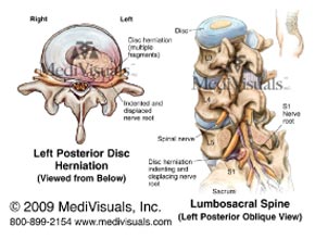
diagnostic dilemma relates to finding the traumatic lesion, and clinically correlating it to causality and, when appropriate, to persistent functional loss. There may be a single, straight forward answer to subjective complaints, or there may be a more subtle presentation that may involve several pain generating structures concurrently. The clinical examination is the most important step in determining the traumatic lesion, the patient’s diagnosis, treatment plan and subsequent prognosis.

The article that is being reviewed was published in SPINE. This study was a collaborative effort between the Department of Orthopedic Surgery, The First Affiliated Hospital of Soochow University, China, and the Department of Orthopedic Surgery, UCLA, Los Angeles, CA. This manuscript was designed to review the accuracy of traditional MRI studies in the diagnosis of lumbar intervertebral disc herniation. The authors cite a study that was published in the journal Radiology in 1995 showing that, in cases of acute lumbar radiculopathy, only 70% of the patients that were diagnosed with a lumbar disc herniation based on clinical examination had a lumbar disc herniation confirmed by MRI. That obviously means, 30% of patients with a disc herniation do not show that herniation on traditional MRI studies.

That is where the concept of the functional lesion comes into being. Once they are out of the initial acute inflammatory phase, many trauma patients will describe the pain varying throughout the day and with specific activities. These differences are generally based on the position and biomechanics of the spine, such as walking up or downstairs, standing or sitting. They can also vary based on the time of day, which indicates that water content of the disc is an influencing factor in the generation of pain. Structural continuity of the spine is an important factor, as different positions will have different effects on spinal anatomy.

The following list may assist you in connecting the dots in relation to the lumbar spine and functional positioning:

1. Lumbar extension – loads the posterior elements of the spine (facet joints) and decreases the size of the intervertebral foramen.

2. Lumbar flexion – increases intradiscal pressure and is associated with sitting and  standing.

3. Lateral flexion – take either of the above positions and induce a lateral flexion component and you will also compress neural elements on ipsilateral side. This will also increase intradiscal pressure at the lateral aspect of the disc and compress the facet joint complex on that side.

These important concepts are the basis for this entire article, since cases where there is limited association between diagnostic testing and clinical symptoms have perplexed clinicians for a long time. The authors stated, “Flexion and extension radiographs and computed tomography myelography were the standard methods of obtaining positional images of the spine. However, because MRI yields an image that is superior to radiographs and less invasive than myelography, physicians have been experimenting with ways of using MRI to obtain positional images of the spine.”1

This brings us to the concept of functional MRI studies. The article utilized an MRI with vertical orientation of the opposing magnet donuts, allowing scanning of the patient in an upright axially loaded position. That is an interesting concept and set up; however, many areas of the country do not have that luxury, so we may need to work these concepts into studies, using traditional recumbent MRI. The fascinating part of this paper was that, not only were more intervertebral disc lesions visualized and, therefore, diagnosed, but also those that were present, increased in size when provocative positions were implemented. Imaging patients in the position of most pain has been a helpful addition into solving the diagnostic dilemma. The authors wrote, “A significant increase in the degree of lumbar disc herniation was found by examining flexion and extension views when compared with neutral views alone.”2

The results of the study showed:3

1. For patients with normal or 3mm bulge in extension and 15.29% demonstrated an increase to >3mm in flexion.

2. Patients in the neutral view that had baseline disc pathology of 3 to 5mm, 13.28% had increased herniations to >5mm in extension and 8.47% had increased herniation to >5mm in flexion.

3. For patients with baseline disc pathology of 5 to 7mm in neutral, 10.58% increased in extension and 5.78% increased in flexion. In addition, for patients with a baseline disc pathology of 7 to 9mm in neutral, 9.09% increased in extension and 4.55% increased in flexion.


Orthopedic testing in the office is nothing more than provocative testing of injured structures. This approach should be implemented into our advanced imaging procedures in trauma to insure the traumatic lesion is identified in a timely manner. Looking to functional positioning during MRI evaluations, especially in the lumbar spine, should be a part of every clinician’s diagnostic protocols.


WDEIn each issue, a clinical topic is covered by William J. Owens of the American Academy of Medical Legal Professionals (AAMLP), which is a national, non-profit organization, comprised of doctors and lawyers. The purpose of the organization is to provide its members with current research in trauma and spinal related topics, to keep the profession on the cutting edge of healthcare. Members may also sit for a Diplomate examination and be conferred a DAAMLP. The organization also offers support to the individual member’s practice.
To learn more or join, go to www.aamlp.org or call 1-716-228-3847.

References:

1) Zou, et al., 2008, ppE142

2) Zou, et al., 2008, ppE142

3) Zou, et al., 2008, ppE140

Hearing Loss following Whiplash

Hearing Loss following Whiplash

by Dr. Daniel J. Murphy D.C., D.A.B.C.O.


TITLE:

A Review of the Otological Aspects of Whiplash Injury

CITATION:

Journal of Forensic and Legal Medicine, Volume 16, Issue2, February 2009, Pages 53-55
AUTHORS: R.M.D. Tranter and J.R. Graham

From abstract:

Approximately 10% of patients who have suffered with whiplash injury will develop otological symptoms such as tinnitus, deafness and vertigo. Some of these are purely subjective symptoms; nevertheless, for the majority, there are specifi c tests that can be undertaken. These tests can quantify the extent and severity of the symptoms, as well as provide guidance as to the correct rehabilitation pathway.


KEY POINTS FROM DR. DAN MURPHY

1.    Approximately 10% of patients who have suffered whiplash injury will develop otological symptoms such as tinnitus, deafness and vertigo.

2.    “Significant [whiplash-related] injuries can occur following even low speed car collisions.”

3.    “Simulated accidents have shown that a 5-mile an hour rear end car collision can result in a positive acceleration of 8.2 G and 4.7 G of the head and chest, respectively. These forces explain the damage that can occur to an unsupported neck.”

4.    “Balance and hearing problems occur in 5-50% of whiplash injuries.”

5.    15–20% of whiplash patients develop “persistent complaints including headache, vertigo, instability, nausea, tinnitus and hearing loss.”

6.    “High frequency hearing loss is the most common form of hearing loss associated with whiplash injury and is easily demonstrated with a pure tone audiogram. This type of hearing loss produces difficulties hearing the high frequency consonant sounds and makes it difficult for the patient to discriminate speech, especially in the presence of background noise or when several people are talking.”

7.    These authors consider hearing loss for whiplash injury to be a permanent injury.

8.    The majority of tinnitus related to whiplash is purely subjective, with no objective measurement testing available, and there is no effective medical treatment. [See Comments Below]. Consequently, the prognosis for the resolution of tinnitus is “very guarded.”

9.    The sensation of balance relies on the input of three systems:

a. The inner ear vestibular apparatus, the labyrinth and semi-circular canals;

b. Proprioception sensors [very important for chiropractors];c. Vision.

10. Post-whiplash unsteadiness may be due to altered posture to protect the injured neck.

11. Following whiplash, the most common type of vertigo seen is benign paroxysmal positional     vertigo (BPPV), “which is characterized by a short duration of vertigo, associated with movement of the head.”

12. “Following trauma, the crystals of calcium carbonate in the utricle become displaced and lie within the labyrinthine fluid and, in certain positions, will stimulate the balance nerve endings in the semi-circular canals, causing brief sensations of spinning.” The appropriate treatment is a “series of movements of the head which move the loose particles of crystal into the utricle, where they will not cause stimulation of the sensitive nerve endings in the semi-circular canals.”

13. Therefore, BPPV is usually curable, but other forms of labyrinthine damage are not so easily managed and may not be curable.

14. Legally, “the expert medical witness simply needs to be satisfied that there is at least a 51% chance (the balance of probability) that the claimant’s symptoms are attributable to whiplash injury rather than any other cause.”


COMMENTS FROM DAN MURPHY

Although this article notes that there is no effective medical treatment for tinnitus, below are three recent interesting non-medical approaches:

1.    Burkhard Franz and Colin Anderson. The Potential Role of Joint Injury and Eustachian Tube Dysfunction in the Genesis of Secondary Meniere’s Disease. International Tinnitus Journal; 2007, Vol. 13, No. 2, pp. 132-137. (This article suggests that tinnitus can be treated by managing dysfunctions of the upper cervical spine joints or TMJ.)

2.    Tullberg M, Ernberg M. Long-term effect on tinnitus by treatment of temporomandibular disorders: a two-year follow-up by questionnaire. Acta Odontologica Scandinavica; 2006 Apr;64(2):89-96. (The results of this study showed that TMD symptoms and signs are frequent in patients with tinnitus and that TMD treatment has a good effect on tinnitus in a long-term perspective.)

3.  Gungor A, Dogru S, Cincik H, Erkul E, Poyrazoglu E. Effectiveness of transmeatal low      power laser irradiation for chronic tinnitus. The Journal of Laryngology & Otology; May 2008 (This was a prospective, randomised, double-blind study using a 5 mW laser with a wavelength of 650 nm, or placebo laser, applied transmeatally for 15 minutes, once daily, for a week. Loudness improved 49%; duration of annoyance improved 58%; degree of annoyance improved 56%. The authors concluded: “transmeatal, low power (5 mW) laser irradiation was found to be useful for the treatment of chronic tinnitus.”


Dr. Dan MurphyDr. Dan Murphy graduated magna cum laude from Western States Chiropractic College in 1978. He received Diplomat status in Chiropractic Orthopedics in 1986. Since 1982, Dr. Murphy has served part-time as undergraduate faculty at Life Chiropractic College West, currently teaching classes to seniors in the management of spinal disorders. He has taught more than 2000 postgraduate continuing education seminars. Dr. Murphy is a contributing author to both editions of the book Motor Vehicle Collision Injuries and to the book Pediatric Chiropractic. Hundreds of detailed Article Reviews, pertinent to chiropractors and their patients, are available at Dr. Murphy’s web page, www.danmurphydc.com TAC.

Permanency Issues and Ligament/Tendon Injuries The biology of soft tissue injury and permanent functional impairment

Permanency Issues and Ligament/Tendon Injuries The biology of soft tissue injury and permanent functional impairment

by Dr. William J. Owens, D.C., D.A.A.M.L.P.


The human body, as we are well aware, is a complex system of anatomical and physiological mechanics that continues to amaze and frustrate clinicians. I would venture to say the latter is more common when triaging and caring for the traumatically injured patient. Diagnostic dilemmas abound in trauma cases.

Continue reading “Permanency Issues and Ligament/Tendon Injuries The biology of soft tissue injury and permanent functional impairment”

Low Velocity Impacts Cause Whiplash

Low Velocity Impacts Cause Whiplash

by Dr. William J. Owens, D.C., D.A.A.M.L.P.


Have you ever had a patient present to your office for evaluation after a low-velocity rear-end collision?

Conversations in the medical legal community abound regarding injuries sustained as a result of a motor vehicle accident and the force of impact. Many clinicians and legal professionals understand each patient must be evaluated as an individual, being worked up as they present. There are many instances where the case has the potential of being dismissed because of a low impact and low property damage situation.

What is important to point out is that the objective evidence of injury and its correlation to causation and persistent functional loss must be the primary objective.

Continue reading “Low Velocity Impacts Cause Whiplash”