Herniated Discs & Advanced Imaging

iStock_000002390175SmallThe patient enters your office and has significant pain radiating into an extremity. Being responsible, you adopt the policy of the insurers and treat the patient conservatively for 4-8 weeks and the pain doesn’t diminish; you then refer the patient to the imaging company for an MRI.

In the report, there are minimal findings, so you continue to treat with marginal results. After a few weeks, you refer to an orthopedist who, in turn, refers to a neurosurgeon, who eventually orders another MRI prior to surgery. At first, you get angry because you are out of the loop; secondly, you feel, over time, the patient could have responded to your care; and, finally, you lose sleep worrying about a malpractice suit over something you did or didn’t do.

This is the litany of emotions that thousands of us have gone through because we were not in control as a result of our ignorance of anything other than clinical evaluations, spinal X-rays, some electrodiagnostics and adjustments.

Being the best-of-the-best in chiropractic includes your ability to understand the innate component of the adjustment and/or the joints’ ability to function normally once put in the proper position. These basics make chiropractic unique and are the foundation for patients getting well in a way no other healing discipline can compete.

However, this is 2009 and, with advanced technology, we can see more, understand more and do more in triaging the severe, mild and sub-acute patients. The tool is advanced imaging, specifically MRI and, when contraindicated, 3-D CAT scans. Regarding technology, a 1-Tesla MRI machine will soon be released and will be capable of scanning patients with pacemakers

Not every condition responds to chiropractic care, and knowing when to refer will affect the outcome of your patients’ care positively, protect your license, and give you many peaceful nights of sleep. In addition, it will grow your practice over time, better than any marketing scheme you can devise.

The reason your practice will grow is twofold. First, your patients will recognize that you have found the cause of their problem, especially if surgery is necessary; you will be the reason that they got better and will tell everyone they know. Secondly, the medical community will recognize you as being the best, based upon your referrals, documentation, triaging and your clinical conclusions and will engage professionally with you, resulting in dialogue and cross referrals.

You will become part of the health care team instead of someone on the “outside looking in,” based upon your clinical abilities or lack thereof. This has been quoted to me by many medical specialists through the years. There is validity to negative medical comments, if the patient has a condition beyond chiropractic care and you treat them at their peril. Although it should be an indictment against the individual doctor for not triaging the patient properly, it becomes an indictment against chiropractic; fair or not.

The truth is that not one health care provider can help every patient. Advanced technology and the knowledge of how to utilize it is often the difference in triaging the patient accurately.

Whether you practice in a subluxation or musculoskeletal model, the parameters for triaging the patient remain constant. I strongly recommend that, if the patient exhibits radicular or myelopathic clinical findings, an MRI be ordered to determine the nature of the lesion, prior to commencing aggressive care. There are many models and insurance industry parameters that do not agree. However, my paradigm is simple: If you do not know, you do not touch.

The flow of care is diagnosis, prognosis and then creating and executing your treatment plan.

A radiculopathic or myelopathic finding indicates that there is a space, occupying lesion on the spinal cord or nerve root and will dramatically change your treatment plan based upon the clinical presentation of that lesion.

Being in control of your patient has two components. You need to know the full extent of the cord and/or root lesion when they present clinically and need an MRI; and, secondly, you need to be able to interpret the MRI.

The only time in my thirty years of practice where I had a significant problem was when the medical specialist misdiagnosed the patient’s MRI findings and I accepted it as gospel. Since then, I have learned to read MRI’s, realizing that the general radiologist error rate on my patients was upwards of 40 percent. Neuroradiologists and neurosurgeons report the error rate over 70 percent.

There are courses that can teach you how to read MRI’s and I strongly urge you to gain control of your patients and your practice by doing so. As technology advances, the information gained, becomes central to the chiropractor in creating an accurate diagnosis, prognosis and treatment plan.

Next month, I will discuss how to create a chiropractic team that includes those medical specialists, while maintaining the control over your patients, even if surgery is indicated. That subtle shift is also the key to unlimited referrals from the medical community and all it takes is becoming the best-of-the-best at what you do.

Erosive Osteoarthritis

History: This female patient complains of pain at the base of her thumb. An X-ray reveals what?

Diagnosis: Observe the sclerosis and osteophytes at the base of the first metacarpal, which represents erosive osteoarthritis. Of incidental notation is congenital fusion of the capitate and the hamate (carpal coalition).

EROSIVE OSTEOARTHRITIS

General considerations:

This is a distinctive clinical and radiographic variant of degenerative joint disease first delineated by Crain in 1961. The two most common terms applied to this arthropathy are erosive osteoarthritis and inflammatory osteoarthritis.1

Clinical Features

In contrast to primary degenerative joint disease, the onset of erosive osteoarthritis is characterized by episodic and acute inflammation of the distal and proximal interphalangeal joints of both hands in a symmetric manner. Pain, edema, redness, nodules and restricted motion are found at the involved articulations of the hands. This arthropathy is most commonly found in middle-aged females in the fourth or fifth decades of life. Laboratory investigations are inconclusive, with normal to slightly elevated erythrocyte sedimentation rate and negative rheumatoid factor. Chronic progression of the disease is to be expected with nodular, unstable and malaligned finger joints. The intensity of symptoms with each inflammatory episode may continue to be severe for many years. Approximately 15 percent may develop rheumatoid arthritis, with an average onset of twelve years after the initial episode of erosive osteoarthritis.1

Pathologic Features:

Variable tissue changes are found. These range from proliferative rheumatoid-like synovial abnormalities to cartilage degeneration and bony proliferation as seen in primary degenerative joint disease.

Radiologic Features:

Essentially, the radiographic changes are those of degenerative joint disease with superimposed bone erosions predominately involving the distal and proximal interphalangeal joints. Occasional involvement of the thumb at the metacarpophalangeal and carpometacarpal joints may occur, as well as between the trapezium and scaphoid articulations. Involvement of the ulnar compartment of the carpus is significantly spared, differentiating involvement from rheumatoid arthritis. All other joints of the body are generally uninvolved.1

Radiographic changes are characterized by osteophytes, loss of joint space and sclerosis. Osteophytes are identical to those seen in degenerative joint disease. They are marginal in origin, taper distally, and are often larger at the distal articular component. Loss of joint space is usually non-uniform, with adjacent subchondral sclerosis. Superimposed changes of erosions, periostitis and ankylosis on these degenerative features are characteristic of erosive osteoarthritis. Bone erosions are distinctively centrally located on the proximal articular surface and more peripherally at the distal articular surface. The resultant altered joint surface contour has been called the “gull wings” sign. Adjacent linear periostitis is occasionally seen. Bony ankylosis is an uncommon but not unexpected sequel of one or more interphalangeal joints.1

The main differential considerations are rheumatoid arthritis, psoriasis, and non-inflammatory degenerative joint disease. Rheumatoid arthritis rarely involves the distal interphalangeal joints and has a positive RA latex test. Psoriatic arthropathy is characterized by discrete marginal erosions with adjacent fluffy periostitis (“mouse ears” sign). Non-inflammatory degenerative joint disease will show no erosions, but will otherwise appear identical to erosive osteoarthritis.1

Dr. Terry R. Yochum is a second generation chiropractor and a Cum Laude Graduate of National College of Chiropractic, where he subsequently completed his radiology residency. He is currently Director of the Rocky Mountain Chiropractic Radiological Center in Denver, Colorado, and Adjunct Professor of Radiology at the Southern California University of Health Sciences, as well as an instructor of skeletal radiology at the University of Colorado School of Medicine, Denver, CO. Dr. Yochum can be reached at 1-303-940-9400 or by e-mail at [email protected].

Dr. Chad J. Maola is a 1990 Magna Cum Laude Graduate of National College of Chiropractic. Dr. Maola is a Chiropractic Orthopedist and is available for post-graduate seminars. He may be reached at 1-303-690-8503 or e-mail [email protected]

Reference

1. Yochum TR, Rowe LJ: Essentials of Skeletal Radiology, 3rd ed., Williams & Wilkins, Baltimore, Maryland, 2005.

Adding Decompression

Dr. Larry Wilkins, Founder and Director of Laurel Mountain Chiropractic Clinic in Mt. Pleasant, PA, has added post-graduate hours in such studies as rehabilitation, manipulation under anesthesia, nutrition, pediatrics, exercise, and advanced work in the Pierce Chiropractic Method.

As an involved professional, Dr. Wilkins has many accomplishments to his credit. He has served as a board member of two chiropractic organizations in Pennsylvania for eight years. Also, he is a teacher of the Pierce Chiropractic Method, and worked very closely with Dr. Vern Pierce until his death.

Dr. Wilkins is also the founding member of two very prestigious chiropractic organizations—Metro Chiropractic Services and the Chiropractic Golden Circle. In addition, he was nominated and accepted into the Chiropractic Knights of the Round Table in 1983.

In an interview with The American Chiropractor (TAC), Dr. Larry Wilkins (Wilkins) describes his successful career in chiropractic and the recent addition of axial decompression to his practice.

TAC: What inspired you to become a chiropractor?

Wilkins: It all began for me when I was a junior in high school. My uncle, Wilmer McGiffin, had a brother-in-law, Dr. Don Casteel, who was a chiropractor. My uncle and my father thought I would make a good chiropractor, even though I had absolutely no idea what that was or meant. So, they took me to Palmer College for a tour of the campus. I got a good feeling about the campus, and decided to give it a try!

After graduation from high school in 1969, I enrolled in Palmer College, and completed my education in June 1973. After my first quarter at Palmer College I was hooked on chiropractic. It has been my life’s work and passion ever since.

TAC: What has really impacted your growth as a chiropractor and that of your practice?

Wilkins: In 1974, I met Dr. Vern Pierce who quickly took me under his wing and proceeded to teach me what Palmer College had not and that was the philosophy of chiropractic. Most of the chiropractors who speak on the subject of philosophy were taught by B. J. Palmer, as was Vern. Now, it seemed that most chiropractors either hated B. J. or loved B. J., and it really doesn’t matter to me, because the bigger issue is this: the only thing that we have, that all other health care practitioners don’t, is our philosophy!

Vern was one of the greatest influences on my life; he taught me his technique, philosophy, and how to run a chiropractic office.

TAC: What type of patients do you generally treat or attract?

Wilkins: The types of patients we attract into our practice are crisis-care patients, low back pain, headaches, neck pain, sick people, and some wellness patients. We have been slowly changing this; our ideal practice would be a cash practice full of wellness patients, on the chiropractic side, and then, chronic and sub-chronic patients on the decompression side. These would be patients with bulging discs, herniated discs, degenerative disc disease and failed back surgeries.

We attract a wide variety of patients to the practice. For a very long time, I was like the “country doctor.” The patients would come to me with everything and ask my opinion about all their health care needs. The reason for this, I believe, is because I practice in a small town of 6,500 people and eight other chiropractic offices. I have, through chiropractic care, assisted two hundred women in getting pregnant; I have gone to patients’ homes in the middle of the night when their children were turning blue because of the croup. I adjusted them, and before I could watch most of the movie that the parents were watching, the child was breathing normally! In thirty-three years of practice, no child I have ever adjusted died of SID’s, and I know the reason why.

I have helped people dying of cancer or other systemic diseases pass away with grace and very little pain, and before you ask, yes, they were also under the care of a medical doctor.

TAC: What are your specialties?

Wilkins: If I do have a specialty it would be the certain something that B. J. always spoke of: I give everyone of my patients hope. Then, along with the hope, I use every ounce of knowledge I have gained through all the previous patients whom I have treated. Add to that all the things I have learned over the course of thirty-three years, and that would be my formula for patient care.

At this point, I would have to also say the Pierce technique and the use of the PulStarFras adjusting instrument were my only techniques, until recently finding decompression along with the DRS™ protocol of axial decompression.

We use axial decompression on chronic patients. Also, we use the Pierce technique, which is excellent for correction of hypolordotic and military cervical curves. I get the kind of results that drive patients here just from talking to my current patient base. The patients love axial decompression because it is gentle and highly successful, and they can’t get that at other practices or through other methods like drugs and physical therapy.

TAC: What made you decide to add axial decompression to your practice?

Wilkins: My reason for adding decompression work to our clinic was that I hated to see patients that were visibly in pain with low back or cervical spine disc conditions being marginally helped with only chiropractic care. After great research, I found the axial decompression treatment to help them. What impressed me was the fact that I could help someone no one else was able to help. Plus the fact that I could help people with failed surgeries. It still amazes me the severe and chronic cases that respond. And it amazes me how easily it truly works. We had a patient that had a surgery on his low back that caused his left leg to become totally numb for fifteen years. After only ten sessions of axial decompression, his foot was warm and he had some feeling for the first time in fifteen years. No words can express how he felt or how grateful he was.

TAC: How has axial decompression impacted your practice?

Wilkins: We have greatly enlarged the patient base that we can help. For instance, people with degenerative disc disease, herniated and bulging discs, facet syndrome, and failed back surgery syndrome can now be helped with the work we do with decompression. A lot of my patients have no other solution to their problems except surgery. They have tried muscle relaxors, pain pills, anti-inflammatories, physical therapy, epidurals, cortisone injections, oral steroids—with little or no relief. About twenty-five percent of our patients have had one, two or three spinal surgeries with no help, and then they turn to our office and axial decompression. In most cases, we have been able to help them enjoy a pain free life.

It truly has been a miracle for many of our patients who had no hope, and before decompression we wouldn’t have been able to help them!

TAC: Do you have any extraordinary patient success stories that you would like to share with us?

Wilkins: Two cases I would like to discuss, were both very unique and unusual, to say the least.

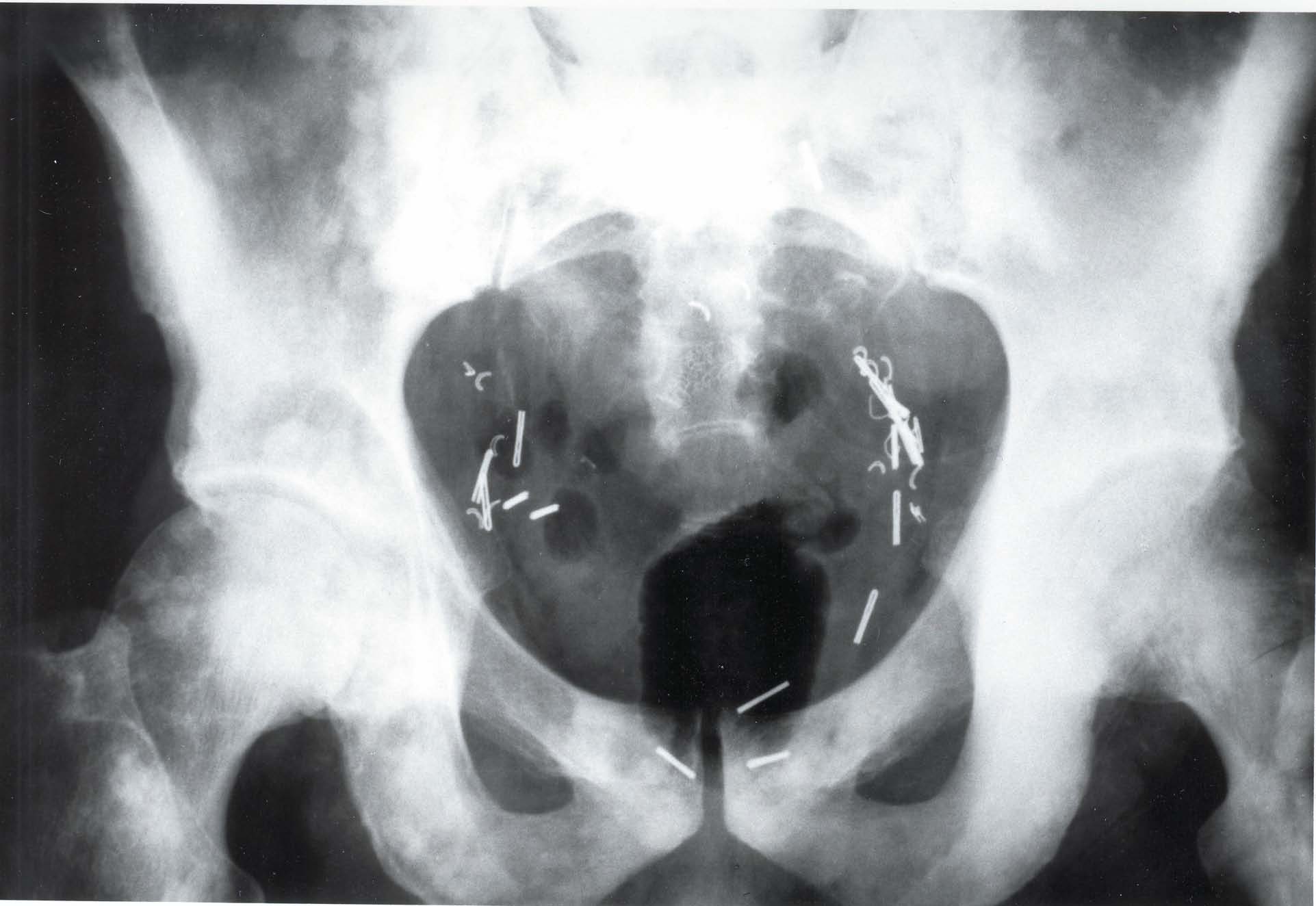
The first one, Sandy, came to me with a fusion of the lumbar vertebra with metal plates and screws. I knew there were contraindications, but this lady was persistent. Being a person of great love and caring for people, I decided to try to help her. Long story short, we treated her very slowly. The results were short of a miracle. After ten sessions, she went from having to have her husband and me pull her out of a chair to getting out of the chair herself, with ease. I have followed her case, and after two years she is still doing great. In fact, she speaks to prospective patients on the phone to answer any questions they may have about axial decompression and my office!

The second patient is Vivian. She had three spinal surgeries and, with the last one, a morphine drip was placed surgically inside her with a tube going into the disc. She also was in a great deal of pain. She also traveled quite a distance to see me, but her results were just as impressive as Sandy’s. Her neurosurgeon spoke with her and told her it would be okay for me to do axial decompression, and that I could even treat the disc in which the morphine drip was placed.

I was not very comfortable with that, so I chose another disc level and went to work. She responded extremely well to the care. However in her case, the treatment took longer than it would have for the average patient. Not unexpectedly, it took thirty-one treatment sessions to get her 85 percent improved. The patient went from hardly being able to walk to spending long hours on her husband’s motorcycle. Unfortunately, I won’t be able to track the longevity of her case, because twelve months after care, Vivian passed away. Vivian was struck by a blood clot that went from the lung into the heart. Her husband made a special and touching trip to my office to tell me how great Vivian’s last twelve months were without her crippling intense pain.

These two cases are the very reason I offer axial decompression in my office.

TAC: How do your patients pay?

Wilkins: Almost 98 percent of my decompression practice is cash. This is a great time to tell you why. If the patients have a vested interest in their health, they will naturally do much better with care than if someone else is responsible for it. But the biggest reason is what a patient said to me the other day, “If you had not made me pay for the service, I would probably have quit before I got the results I could have!”

I believe that having a great understanding of running a cash practice or a partial cash practice will be the difference of being in practice in the future, rather than not being in practice.

TAC: What single piece of advice would you give a new chiropractor just starting out?

Wilkins: In my heart, the answer to the question of advice for a new chiropractor just starting out would be two-fold, and in no particular order. The one that has always been with me came from Dr. Pierce and Dr. Gonstead; it was to be the master of one technique. By doing that, you are going to be proficient enough to treat 90 percent of the people coming in your door. You will have a reason for everything you do with regard to chiropractic. The second would be to sign up with a practice consultant.

TAC: What general advice would you give an established chiropractor whose practice might be struggling?

Wilkins: If you are looking for growth as a chiropractor, the easiest way I have found is to find a great coach. In my lifetime, I have had fantastic coaches. The first was Dr. Vern Pierce, who taught me a great adjustment technique and the philosophy of chiropractic.

The next coach that came into my life was Dr. Chuck Gibson. He taught me all the great procedures for running an office and managing patients—not in a clinical context, but in maintaining control, and educating the patients as to why they were there.

Another great coach was Dr. C. J. Mertz. He taught me how to do what Dr. Pierce and I had wanted to do for years and could not figure out how. That was how to care for the patients the way we wanted to—not the way the insurance companies wanted us to—and get paid for it.

When I became involved with axial decompression, I also needed a coach. For this, I researched and chose Freedom Awaits™. Dr. Richard E. Busch, III and Dr. Jack Ashton not only taught me how to use decompression properly, but also how to do it with love, passion, caring and ethics.

TAC: Any final words for our readers?

Wilkins: I can honestly say that there is nothing else in the world that I would rather be doing than chiropractic and axial decompression. Our profession is so very easy to do. You just have to have the right knowledge, and if you don’t have the right knowledge it is readily available; all you have to do is find the doctor that will teach you. I have mentioned five of them. Seek them out, and pick their minds. Don’t waste a precious moment of your life.There are too many patients that need you to help them achieve optimum health. These two quotes by Vince Lombardi sum it up best: “The difference between a successful person and others is not a lack of strength, not a lack of knowledge, but rather in a lack of will;” and, “The quality of a person’s life is in direct proportion to their commitment to excellence, regardless of their chosen field of endeavor.”

You may contact Dr. Wilkins at 724-547-5030, 724-547-3377, or email [email protected].

Caring for the Supermom

When I was thirteen, my two-year-old brother was suffering from a serious case of torticollis, which had been misdiagnosed as possible meningitis. After just one chiropractic treatment, he was virtually cured. At that moment, I knew that my contribution to healthcare would be in the chiropractic field.

Like any idealistic young chiropractic student, I was electrified by the story of Harvey Lillard getting his hearing back after one adjustment from D.D.—the first chiropractic adjustment in history. I expected that kind of result routinely but, too often, the reality didn’t live up to my hopes.

I now know that, in today’s modern world, innate intelligence can be compromised by things other than subluxations. The body’s inherent wisdom has the healing potential to repair anything, but sometimes it doesn’t. The body cannot meet all its challenges successfully without the proper genuine replacement parts in the form of nutritional support. If that realization sounds simple, getting there was anything but easy.

Being a perfectionist, I was continually beating myself up over the patients I wasn’t helping; I went to seminar after seminar in search of new answers, better solutions, and more reliable techniques.


The breakthrough came with learning not to diagnose, but simply to ask the body what was missing, and in what priority. The nutritional technique that I incorporated into my practice is a whole story in itself. Suffice it to say that I found the method I was looking for, and everything else fell into line, except for one thing: there were patients who really didn’t come to be helped, or who had gone down the medical road so long that they were beyond the reach of nutritional help. You have to let those go. When they walk into your office, check them out, and don’t take them on unless you can actually get a win by helping them. You actually help fewer people when your attention is fixated on a case that came to you too late. Don’t be a martyr. That was the advice of my greatest mentor, and I pass it along to anyone else who can relate to this story.

As a result of these breakthroughs in my practice, I was able to accept myself as an effective, valuable contributor to my community.

The majority of my practice consists of the “Supermoms,” females in the 35 to 55 age group. They need your help. And, since I am one of them, I know this for a fact. Getting into this profession, I never realized the challenges that would come with it as a female who also chose to have a family. It is one thing for the man to go off to work, but it is an entirely different game when mom goes off to work. Fifty years ago, running a home, taking care of the children and being the caretaker of the man was our full time job.

In today’s society, women are expected to do that “on the side,” so there is a real finesse to keeping it all going, not just as a chiropractor mom, but even just as a working mom, in general. When I tell people my life and my schedule, they always ask, “How in the world do you get it all done?”

So, here are the secrets. Number one is keeping myself and my family healthy. And, as I mentioned earlier, though we all get chiropractic adjustments, with the food sources today, it just isn’t enough. The key is what I have found in the nutritional realm. In addition to chiropractic adjustments to keep everyone well, nutrition is the thing that seals the deal. This is what allows me to keep up such a hectic schedule and get it all done. We don’t get sick and, if we do, it is very rarely, because, as we all know in chiropractic, an ounce of prevention is worth a pound of cure. To a working mother, it is probably more like a ton. When you are regularly adjusted and are on the correct nutritional program, you can nip this one in the bud for sure.

These women are the ones who have to keep things together at home with the husband and the kids while also working a full-time job, no matter how bad they feel. They need our help and no one is specializing in them and giving them the TLC they need. When you help mom, you have not only helped her but you have also helped the husband and the children. And, before you know it, she is bringing them in for care as well.

Her chief complaints tend to be stress, fatigue, weight issues and depression. But she can come with more symptoms that you can name. I don’t worry about or get caught up in her symptoms, because I don’t treat symptoms. I find the true cause of the dysfunction which, usually, stems from nutritional deficiencies. When these are properly handled, the body will heal itself and miracles will occur.

What is great about handling the true cause is that you can position yourself as the expert of any health problem. By tapping into the innate intelligence of the body, you let it tell you what is wrong and then you can fix it.


I have seen true miracles in my practice by making a so-called “incurable” disease process disappear, having bone density restored without drugs—as verified by medical diagnostics. I have resolved the hot flash case without natural or synthetic hormone therapy of any kind. I have resolved the gall bladder attack cases, even after the gall bladder was removed. Most cases have substantial improvement in energy levels in just one week and routinely experience weight loss as a great “side effect” of the program. I routinely resolve the chronic subluxation case that doesn’t budge, despite the fact that they are seeing my colleagues and receiving great adjustments.

Any time I hear of my patient going for chiropractic, physical therapy and/or other related musculo-skeletal treatments more than six visits, I then begin to do my detective work and find out what is really going on. D.D. Palmer, in his original writings, cites examples of serious illnesses completely resolving in three to six visits. I became curious as to how 36 visits became the norm in modern-day chiropractic. I found the answers in Nutrition Response Testing.

A mother brought me her four-year-old boy who presented with severe symptoms of autism. There were so many quirks and functional abnormalities that social workers were working with the boy three days a week. This had gone on without improvement for over two years.

I did a routine analysis. Interestingly enough, the priority of this child was his gall bladder. Based on my findings, I made some nutritional supplement recommendations and a few dietary modifications and within six weeks the child was substantially improved. The social workers told the mom it was a miracle—all of a sudden, their years of working with the little boy were finally paying off. They didn’t know yet that he was under my care.

Within three more weeks, the social workers pronounced him “cured.” There had been a complete resolution of all of the autistic symptoms—no more rocking of the body and head, no more flapping of the hands. His behavior was fully rational. The social workers decided the little boy should be considered normal after nine weeks of nutritional care.

I worked with a woman who had been suffering from Crohn’s disease for seven years. None of the medical treatments had done her any good, and she was doing an intense steroid drug therapy just to get through her day. Within three days of following the exact program I recommended, the patient was totally asymptomatic.

One of my most dramatic cases was a man who was on three different psychotropic medications. This poor gentleman was not doing well at all in life and really wasn’t functioning in society. He was 75 pounds overweight, with dark black circles under his eyes. He literally looked like the living dead. After doing the Nutrition Response Testing procedures, a year later he has all of the excess weight off, is off all three of the psychotropic drugs, holds a full time job, and—much to my surprise—is a very handsome and charming man; something that I surely had not seen a year earlier.

I feel very optimistic about the future of health care in this country, because of what I’ve learned and what I can do. This future starts by supporting the “Supermom” who is our most important patient. You just need great products, a great testing methodology, and a patient management system that keeps the practice stress-free. I feel truly blessed to have all three.


Dr. Lori Puskar lives in Clearwater Florida and is a member of the Florida Chiropractic Association. She is also married, the mother of a 5 and 7 year old, and a full time executive for Ulan Nutritional Systems, Inc. Dr. Puskar also delivers Nutrition Response Testing seminars locally and across the country and has spoken to thousands of doctors educating them on the need for real nutrition. She has built a highly successful nutritional cash practice in Hazleton, Pennsylvania, and still oversees the practice till this day. She is a stanch supporter of non-profit nutritional organizations preaching the truth in nutrition such as the Dr. Weston A. Price and Dr. Francis Pottenger Foundations. In addition she is active in groups regarding our children’s future such as fightforkids.org and Citizens Commission on Human Rights.

Thoracic Spine Compression Fractures

CASE HISTORY

This 70-year old female lost her balance and stepped off the curb and experienced severe lower thoracic pain.

DIAGNOSIS

Observe the compression fractures of the T7, T8 and T9 vertebral bodies. Note the osteoporosis which contributed to the weakening of the vertebrae allowing these fractures to occur with minimal trauma.

GENERAL CONSIDERATIONS


Radiographic Signs of Vertebral Compression Fracture. Radiographs of optimum quality are necessary in order to adequately demonstrate these fractures. Lateral radiographs best demonstrate fracture features. Radiographic signs of vertebral compression fracture include a step defect, wedge deformity, linear zone of condensation, displaced endplate, paraspinal edema, and abdominal ileus.

The Step Defect. Since the anterior aspect of the vertebral body is under the greatest stress, the first bony injury to occur is a buckling of the anterior cortex, usually near the superior vertebral endplate. This sign is best seen on the lateral view as a sharp step-off of the anterosuperior vertebral margin along the smooth concave edge of the vertebral body. In subtle compression fractures the “step” defect may be the only radiographic sign of fracture. Anatomically, the actual step-off deformity represents the anteriorly displaced corner of the superior vertebral cortex. As the superior endplate is compressed in flexion, a sliding forward of the vertebral endplate occurs, creating this roentgen sign.

Wedge Deformity. In most compression fractures, an anterior depression of the vertebral body occurs, creating a triangular wedge shape. The posterior vertebral height remains uncompromised, differentiating a traumatic fracture from a pathologic fracture. This wedging may create angular kyphosis in the adjacent area. The superior endplate is far more often involved than the inferior endplate. Up to 30 percent or greater loss in anterior height may be required before the deformity is readily apparent on conventional lateral radiographs of the spine. Normal variant anterior wedging of 10 to 15 percent or 1-3 mm is common throughout the thoracic spine most marked at T11 – L2.

In all compression fractures there should be clear differentiation from an underlying pathology that has produced the fracture. Key features of pathologic fractures may be identified by loss of the posterior body height, pedicle, and other sites of destruction, a paraspinal mass while on MR imaging abnormal marrow can be demonstrated.

Linear White Band of Condensation (Zone of Impaction). Occasionally, a band of radiopacity may be seen just below the vertebral endplate which has been fractured. The radiopaque band represents the early site of bone impaction following a forceful flexion injury where the bones are driven together. Callus formation adds to the density of the radiopaque band later, in the healing stage of the fracture injury. This radiographic sign is striking when present; however, it is an unreliable sign, since it is not present as often as might be expected. Its presence, however, denotes a fracture of recent origin (less than two months’ duration).

Disruption in the Vertebral Endplate. A sharp disruption in the fractured vertebral endplate may be seen with spinal compression fracture. This may be difficult to perceive on plain films and tomography; CT provides the definitive means to identification. The edges of the disruption are often jagged and irregular. The superior endplate is more commonly fractured than the inferior endplate.

Paraspinal Edema. In cases of extensive trauma unilateral or bilateral paraspinal masses may occur which represent hemorrhage. These are best seen in the thoracic spine on the anteroposterior projection but may occur adjacent to the lumbar spine, creating asymmetrical densities or bulges in the psoas margins.

Abdominal Ileus. This may occur with severe spinal trauma and is a warning sign to the observer that the trauma has been severe and the likelihood of fracture is great. Abdominal ileus is seen radiographically as excessive amounts of small or large bowel gas in a slightly distended lumen. It occurs as a result of disturbance to the visceral autonomic nerves or ganglia from pain, paraspinal soft tissue injury, edema or hematoma.

Dr. Terry R. Yochum is a second generation chiropractor and a Cum Laude Graduate of National College of Chiropractic, where he subsequently completed his radiology residency. He is currently Director of the Rocky Mountain Chiropractic Radiological Center in Denver, Colorado, and Adjunct Professor of Radiology at the Southern California University of Health Sciences, as well as an instructor of skeletal radiology at the University of Colorado School of Medicine, Denver, CO. Dr. Yochum can be reached at 1-303-940-9400 or by e-mail at [email protected].

Dr. Chad J. Maola is a 1990 Magna Cum Laude Graduate of National College of Chiropractic. Dr. Maola is a Chiropractic Orthopedist and is available for post-graduate seminars. He may be reached at 1-303-690-8503 or e-mail [email protected] 

REFERENCES

 1.      Yochum TR, Rowe LJ:  Essentials of Skeletal Radiology, 3rd ed., Williams & Wilkins, Baltimore, Maryland, p. 513, 2005.

Multiple Myeloma

HISTORY


This 60-year-old male patient presents with head and spinal pain.


DIAGNOSIS


Note the diffuse osteolytic “punched-out” lesions affecting the bony calvarium which has been called the “raindrop skull” of multiple myeloma.


DISCUSSION

Osteolytic Defects. The radiologic hallmark of multiple myeloma is the sharply circumscribed osteolytic defect. These radiolucent lesions have been historically referred to as punched-out lesions. They are multiple, round, and purely lytic. The most frequent sites are in bones with hematopoietic potential. This appearance is most common in the skull, pelvis, long bones, clavicles and ribs. The pattern of widespread lytic lesions of the skull has been referred to as the raindrop skull.1 Calvarial involvement may occasionally be differentiated from metastatic carcinoma by the more uniform size of the lytic lesions in myeloma. The coexistence of both large and small lesions is often the mode of presentation in metastatic disease.1,2


Spinal involvement. The lower thoracic and lumbar spine are usual sites; however no spinal region is exempt. Early osteoporosis may be the only radiographic sign. As the disease progresses, pathologic vertebral collapse is inevitable. This often takes the configuration of a vertebra plana compromising the posterior one third of the vertebral body, as well as its anterior two thirds. Pathologic fracture in the spine may be singular or multiple and has been called the wrinkled vertebra of myeloma. Demonstration of punched-out lesions in the spine is rare. The vertebral pedicles are involved much less frequently than the body. Jacobson, et al.,3 suggest that the paucity of red marrow in the pedicles may allow their preservation with vertebral involvement. This has been called the pedicle sign of multiple myeloma. Since Jacobson’s original paper in 1958, there have been numerous cases reported refuting this sign and demonstrating isolated pedicular or combined vertebral body and pedicular involvement in multiple myeloma. Therefore, the usefulness of this pedicle sign to differentiate multiple myeloma from osteolytic metastatic carcinoma appears doubtful.1,2

Pelvic and Long Bones. Diffuse osteolytic round or oval lesions predominate without any reactive sclerosis. Medullary bone destruction abuts the endosteal surface of the cortex. The diaphysis is an area frequently involved in the long bones, which is consistent with the anatomic distribution of active red bone marrow. The humerus and femur are favored sites. Widespread disease throughout the pelvis and sacrum creates diffuse lytic lesions, which are fairly symmetric.1,2


Roentgen Signs of Multiple Myeloma

Early: Normal radiographsGross osteoporosis

Late: Diffuse, punched-out lesions Uniform vertebral collapse (compromise of the posterior one-third of the body) Diaphyseal osteolytic lesions Rarely, sclerotic lesions (ivory vertebra) Pedicle sign (preservation of pedicles)


TREATMENT AND PROGNOSIS

In patients with multiple myeloma, the overall prognosis is poor.1,2 Over 90 percent die within three years. Treatment is usually palliative, with the aim of minimizing the patient’s suffering. The two major forms of treatment are radiotherapy and chemotherapy. Plasma cells are characteristically radiosensitive and radiotherapy is of established value in the control of localized symptomatic lesions, which typically transform to a blastic area. Of the chemotherapeutic agents presently available, Melphalan and Cytoxan are the two drugs most useful in attempted long-term management. The importance of ambulation and adequate hydration cannot be overemphasized. The constant threat of hypercalcemia, hypercalciuria, and hyperuricemia necessitate continual attention to these aspects of general care.1,2,3

Dr. Terry R. Yochum is a second generation chiropractor and a Cum Laude Graduate of National College of Chiropractic, where he subsequently completed his radiology residency. He is currently Director of the Rocky Mountain Chiropractic Radiological Center in Denver, Colorado, and Adjunct Professor of Radiology at the Southern California University of Health Sciences, as well as an instructor of skeletal radiology at the University of Colorado School of Medicine, Denver, CO. Dr. Yochum can be reached at 1-303-940-9400 or by e-mail at [email protected].

Dr. Chad J. Maola is a 1990 Magna Cum Laude Graduate of National College of Chiropractic. Dr. Maola is a Chiropractic Orthopedist and is available for post-graduate seminars. He may be reached at 1-303-690-8503 or e-mail [email protected].

References

1.        Yochum TR, Rowe LJ:  Essentials of Skeletal Radiology, 3rd ed., Lippincott, Williams & Wilkins, Baltimore, Maryland, 2005.

2.        Kyle RA: Multiple Myeloma—review of 869, Mayo Clin Proc 50:29, 1975.

3.        Murray RO, Jacobson HG: The Radiology of Skeletal Disorders. Ed 2, New York, Churchill Livingstone, 1977.

Adjusting the Holographic Body Part Four: Allergies

If the body is sick, the mind worries and the spirit grieves; if the mind is sick, the body and spirit will suffer from its confusion; if the spirit is sick, there will be no will to care for the body or mind. – J.R. Worsley

Posture of subluxation (POS)

In Parts One and Two, we discussed how some subluxations can only be accessed (and adjusted) when the patient is in a certain physical or emotional posture (the Posture of Subluxation—POS). In Part Three, we discussed locating and correcting the emotional POS.


There are many POS’s; a common one is allergies

The POS is not limited to physical or emotional states. Many diverse health conditions will exhibit a POS. For example, Koren Specific Technique (KST) doctors have discovered that allergies correlate to subluxation postures.


KST and allergies

After developing KST, I was often asked if it could help allergy sufferers. I’m happy to report that KST doctors have discovered a simple yet powerful way to help allergy sufferers break the allergy/symptom connection.


The analysis

First the patient is analyzed and adjusted so they are free of subluxations in neutral posture (i.e. lying on a table). Now put the patient in the allergy posture of subluxation.

How do you do that? It is rather simple to do. First, have the patient think of the allergy. While they are thinking of the allergy, the body will subluxate. Now they are in the allergy POS.

KST is a quick and accurate method of analysis utilizing a bio-indicator. Bio-indicators, used in Applied Kinesiology, DNFT/Activator/Truscott, Body Talk, Toftness and other healthcare modalities, are physiological responses to stressors. In KST, the bio-indicator we prefer is the occipital/mastoid drop (OD) because there is no patient muscle fatigue and there are no posture restrictions—you can test a patient in nearly any posture.

While the patient is thinking of the allergy, the practitioner will find that their subluxation-free patient is suddenly subluxated again! They will have a positive OD (biofeedback) and will be “locked up” or subluxated. In most cases, the subluxations are best accessed in the cranials.

Let’s say you have the patient think of their cat allergy. While they are in their “cat allergy” state of mind, their body will subluxate.


Now the correction

As the patient is thinking of the cat (allergen), the subluxations are adjusted. I like to use an adjusting instrument, as this permits the patient to stay in one position so the force/energy/information (adjustment) can be directed specifically.

Next, ask the patient to think of the allergen again. There should be no OD. If there is an OD, go through the analysis and adjustment again. You may have missed something.

Are you finished?

Not quite.

Now ask the patient to think about the allergy from a different angle. Tell the patient, “Imagine holding a cat.” Then test for an OD. If you get a positive response, analyze and adjust.

Please realize that we are not using the actual physical allergen to elicit this reaction (although we could do that as well); we are using the mental/emotional POS of the allergy.

Try other statements to defuse the allergy. Tell the patient to imagine how their symptoms feel when they have an allergic reaction. Have them think of petting a cat; have them think of the age they first experienced the allergy; have them think of the emotional stress that occurs when they have the allergy. Tell them to imagine breathing the allergen. Tell them to imagine clear sinuses.

Go through a number of these exercises until you simply can’t elicit an OD from the patient.


That’s it

It’s that simple. KST doctors are reporting very good results using this procedure.


What exactly are we doing?

As with other allergy elimination protocols, we appear to be breaking a psycho-neuro-immunological reflex that caused the patient to overreact to an allergen.

DD Palmer said that the causes of subluxations are emotions (auto-suggestion), toxins and trauma. With KST we are adjusting a patient when they are in the toxin (allergy) “posture of subluxation” or POS.

Using KST procedures, chiropractors can easily and quickly locate and correct subluxations as they reveal themselves in any posture—physical, emotional or chemical.

In Part Five, we’ll discuss the POS as it relates to weight loss, smoking, dyslexia and bad habits.

or call 1-800-537-3001. Write to Dr. Koren at [email protected].

Degenerative Spondylolisthesis

HISTORY


This 50-year-old female patient presents with a six-month history of lower back pain without peripheral radiation.

DISCUSSION

Degenerative spondylolisthesis (Type III) has been referred to as a “pseudospondylolisthesis” by Junghans, to differentiate spondylolisthesis with an intact neural arch from those with a true defect of the neural arch.1 Macnab2 prefers the phrase “spondylolisthesis with an intact neural arch,” which is a more accurate description. Thus, degenerative spondylolisthesis is another type of nonspondylolytic spondylolisthesis.

Degenerative spondylolisthesis is approximately ten times more common at L4 than at the L3 or L5 vertebrae and is six times more common in females sixty years of age or older, compared with males of the same age.3 Type III is rare in persons younger than fi fty years of age. Degenerative spondylolisthesis is three times more common in blacks than in whites, with no adequate explanations for these sexual and racial disparities. Finally, degenerative spondylolisthesis is four times more likely to be found in association with a sacralized fi fth lumbar vertebra.1,2

 Figure 1. Observe the 20 percent anterior
displacement of L4 upon L5 without pars defects.
There is underlying discogenic spondylosis and
posterior facet arthrosis. This represents a
degenerative spondylolisthesis (pseudospondylolisthesis).


The mechanisms of displacement are thought to involve a combination of zygapophyseal joint arthrosis, disc degeneration, and remodeling of the articular processes and pars.2 An increase of the “pedicle-facet angle” has been noted in the degenerative type of spondylolisthesis.1 This angle, formed by the long axis of the pedicle (or vertebral root) at its intersection with the long axis of the articular pillar, indicates the more horizontal alignment of the degenerative zygapophyseal joints as seen on the lateral radiograph and demonstrates the overriding of the articular surfaces.1

The Three F‘s of Degenerative Spondylolisthesis

                Female
                Four (L4)
        Above Forty years


Several explanations have been proposed for degenerative spondylolisthesis occurring with such great frequency at the L4 level. Allbrook has stated that the greater mobility of L4 due to the sagittal orientation of the facets at the L4/L5 level may explain the unusual frequency of degenerative spondylolisthesis at the L4 level. Additionally, the fi rmly attached, normal lumbosacral joint may place increased stress on the L4/L5 intervertebral joints, ultimately leading to hypermobility and degeneration of the articular triad.3 No greater than 25 percent anterior displacement of the L4 vertebral body occurs, and the majority involves 10 to 15 percent displacement.1,3


Dr. Terry R. Yochum is a second generation chiropractor and a Cum Laude Graduate of National College of Chiropractic, where he subsequently completed his radiology residency. He is currently Director of the Rocky Mountain Chiropractic Radiological Center in Denver, Colorado, and Adjunct Professor of Radiology at the Southern California University of Health Sciences, as well as an instructor of skeletal radiology at the University of Colorado School of Medicine, Denver, CO. Dr. Yochum can be reached at 1-303-940-9400 or by e-mail at [email protected].

Dr. Chad J. Maola is a 1990 Magna Cum Laude Graduate of National College of Chiropractic. Dr. Maola is a Chiropractic Orthopedist and is available for post-graduate seminars. He may be reached at 1-303-690-8503 or e-mail [email protected].

References:

1. Yochum TR, Rowe LJ: Essentials of Skeletal Radiology, 3rd ed., Lippincott, Williams & Wilkins, Baltimore, Maryland, 2005.

2. Macnab I: Spondylolisthesis With an Intact Neural Arch – So Called Pseudospondylolisthesis, J. Bone Joint Surg (BR) 32:325, 1950.

3. Rosenberg, MJ: Degenerative Spondylolisthesis, Clin Orthop 1976.117:112, 1976.

Prostate Osteoblastic Metastasis

HISTORY


This 60-year-old male patient presents with a history of prostate cancer with surgical removal. Note the metallic clips in the pelvic basin consistent with prostatectomy. There are multiple “snowball” areas of blastic metastasis scattered throughout the pelvis and proximal femora. This radiographic appearance is consistent with osteoblastic metastatic carcinoma from the prostate gland.


DISCUSSION


The sacrum and bones of the pelvis are involved in about 12% of skeletal metastases and may show either lytic or blastic lesions. Seeding from the viscera via Batson’s venous plexus explains this high incidence in the pelvis, as well as in the lumbar spine. Blowout lesions of renal and thyroid origin often affect the bony pelvis. Lesions located in the sacral ala or the posterior ilium are often difficult to perceive on standard radiographs. With the advent of CT scans, a wide variety of lesions involving the osseous pelvis can be more readily seen. The ability of CT to provide accurate measurements of tissue attenuation coefficients and to provide a cross-sectional scan for three dimensional viewing has made it a powerful tool in musculoskeletal diagnosis, with a profound influence on patient management. It provides information about the extent of the bony lesion, localization (for biopsy and radiation therapy), and relationships with other structures. As equipment improves, it seems probable that CT will assume a more primary role in diagnostic evaluation, particularly of the pelvis, where the complexity of bones and the overlying bowel content prevent ideal evaluation with conventional radiographs.


Occasionally, blastic lesions affecting the pelvic rim, especially from carcinoma of the prostate, exhibit an expansion of bone. This occurs as a result of cortical thickening from endosteal or periosteal apposition of bone. The bony enlargement may mimic the appearance of Paget’s disease. Usually, other skeletal lesions are present to assist in radiologic differentiation. Biopsy of the lesion may be necessary as a final step in diagnosis.


Dr. Terry R. Yochum is a second generation chiropractor and a Cum Laude Graduate of National College of Chiropractic, where he subsequently completed his radiology residency. He is currently Director of the Rocky Mountain Chiropractic Radiological Center in Denver, Colorado, and Adjunct Professor of Radiology at the Southern California University of Health Sciences, as well as an instructor of skeletal radiology at the University of Colorado School of Medicine, Denver, CO. Dr. Yochum can be reached at 1-303-940-9400 or by e-mail at [email protected].


Dr. Chad J. Maola is a 1990 Magna Cum Laude Graduate of National College of Chiropractic. Dr. Maola is a Chiropractic Orthopedist and is available for post-graduate seminars. He may be reached at 1-303-690-8503 or e-mail [email protected].


Multiple Myeloma

HISTORY

This 65-year-old female patient presents with acute symptoms of right-sided radiculopathy, paresthesia and pain in the right upper extremity. There has been no trauma.

DIAGNOSIS

Observe the gross osteoporosis about the entire cervical spine. There has been pathological collapse of the C5 vertebral body, which is uniform in configuration. This is very characteristic of a malignant pathological fracture. This patient’s radiculopathy occurred as a result of posterior compression on the spinal cord resulting from the pathological collapse. The compression created an extradural defect on the spinal cord.

General Considerations

Multiple myeloma occurs as a result of a malignant proliferation of plasma cells, which infiltrate the bone marrow. The first patient known to have multiple myeloma was seen in 1845, after severe recurrences of pain during a 17-month period. His urine contained unusual “animal matter” that became soluble when boiled and formed again when cooled. Although William MacIntyre recognized the effect of heat on the urine and outlined the clinical findings, it was the young physician and chemist Henry Bence Jones who described the protein in detail. In 1873, von Rustizky named and outlined in detail the clinicopathologic features of the this disease. Multiple myeloma is occasionally referred to as Kahler’s disease, after the clinician from Prague who lectured extensively on myeloma in the late nineteenth century. In more recent times, the diagnosis of myeloma was facilitated by Longsworth, et al., in 1939, with the development of electrophoretic techniques and with immunoelectrophoresis as described by Grabar and Williams.

Incidence

Multiple myeloma is the most common primary malignant tumor of bone and accounts for 27 percent of biopsied bone tumors. Together, myeloma and osteosarcoma account for almost half (46 percent) of the primary malignant tumors of bone. Multiple myeloma represents about one percent of all types of malignant disease and slightly less than ten percent of hematologic malignancies. In the last two decades, the death rate from multiple myeloma has increased; however, it is likely that these increases are related to earlier and more improved diagnosis rather than representing an actual rise in incidence.

Clinical Features

Age and Sex Distribution: Typically 75 percent of myeloma patients are between fifty and seventy years of age, with an average age of sixty. It is rarely seen before the age of forty, but a few cases have been reported before the age of thirty. There is a male to female ratio of 2:1.

Signs and Symptoms: The clinical picture of the disease comprises four types of abnormalities: anemia owing to replacement or alteration of the hematopoietic tissues by proliferating plasma cells, deossification of bones that house red marrow production of abnormal serum and urinary proteins, and renal disease. Pain is the cardinal initial symptom, often suggesting arthritis or neuralgia.

Initially, the bone pain is intermittent; in later stages, it becomes continuous. It is worse during the day and aggravated by exercise and weight bearing. The pain is often better at night with bed rest. Low back pain in myeloma patients is frequently misdiagnosed as disc or sciatic problems initially. A rapid onset of severe pain after slight strain or mild trauma usually indicates the development of a pathologic fracture. In the late stages of the disease, pathologic fractures occur in 20 percent of patients. Paraplegia may occur with vertebral collapse and is more common with a solitary presentation (plasmacytoma). As the disease progresses, the pain becomes more severe and prolonged, often requiring narcotics for relief.

SOLITARY PLASMACYTOMA

General Considerations

Solitary plasmacytoma represents a localized form of plasma cell proliferation. It is much less common than multiple myeloma. Approximately 50 percent of patients present before age fifty. Most commonly, patients complain of localized pain. Laboratory findings are occasionally normal, or the abnormal serum electrophoresis may disappear after tumor excision. The mandible, ilium, vertebrae, ribs and proximal femur and scapula are the favored sites. Pathologic fracture is common. Isolated cases have been reported in extramedullary sites affecting the soft tissues of the upper respiratory tract. Rarely, solitary plasmacytoma can present as an ivory vertebra. The typical roentgen appearance is a geographic radiolucent lesion, often highly expansile, with a soap bubble internal architecture. The radiographic differential diagnosis includes pseudo-tumor of hemophilia, hydatid disease of bone, fibrous dysplasia, giant cell tumor, brown tumor of hyperparathyroidism, and blow-out metastases from renal or thyroid origin.

Often, these lesions initially appear benign; however, 70 percent of patients who have what seems to be a solitary focus develop diffuse multiple myeloma and die within five years. Progression to multiple myeloma has been documented in cases up to twenty-three years after the initial presentation of solitary plasmacytoma, emphasizing the importance of long-term follow-up with these patients. The balance of the lesions remain localized and are treated quite successfully with local irradiation and/or surgical excision.


Dr. Terry R. Yochum is a second generation chiropractor and a Cum Laude Graduate of NationalCollege of Chiropractic, where he subsequently completed his radiology residency. He is currently Director of the Rocky Mountain Chiropractic Radiological Center in Denver, Colorado, and Adjunct Professor of Radiology at the Southern California University of Health Sciences, as well as an instructor of skeletal radiology at the University of Colorado School of Medicine, Denver, CO. Dr. Yochum can be reached at 303-940-9400 or by e-mail at [email protected].


Dr. Chad J. Maola is a 1990 Magna Cum Laude Graduate of National College of Chiropractic. Dr. Maola is a Chiropractic Orthopedist and is available for post-graduate seminars. He may be reached at 303-690-8503 or e-mail [email protected].


References

1. Yochum TR, Rowe LJ: Essentials of Skeletal Radiology, 3rd ed., Lippincott, Williams & Wilkins, Baltimore, Maryland, 2005.

2. Deutsch A, Resnick D: Eccentric cortical metastases to the skeleton from bronchogenic carcinoma, Radiology 137:49, 1980.

3. Yuh WTC, Zachar CK, Barloon TJ, et al.: Vertebral compression fractures: Distinction between benign and malignant causes with MRI Imaging. Radiology 172:215, 1989.

4. Shih TT, Huang KM, Li YW: Solitary vertebral collapse: Distinction between benign and malignant causes using MR patterns. J Magnetic Reson Imaging 9(5):635, 1999.